and G. N. C. Kenny2. 1Department of Anaesthesiology, Leiden University Medical Centre, Postbus 9600, 2300 RC Leiden, The. Netherlands. 2University ...
British Journal of Anaesthesia 82 (3): 333–9 (1999)
Effect-site modelling of propofol using auditory evoked potentials M. White1*, M. J. Schenkels1, F. H. M. Engbers1, A. Vletter1, A. G. L. Burm1, J. G. Bovill1 and G. N. C. Kenny2 1Department
of Anaesthesiology, Leiden University Medical Centre, Postbus 9600, 2300 RC Leiden, The Netherlands. 2University Department of Anaesthetics, Glasgow Royal Infirmary, Glasgow, UK *To whom correspondence should be addressed Auditory evoked potentials (AEP) were used to monitor central nervous system effects during induction and recovery from anaesthesia produced by infusion of propofol 30 mg kg–1 h–1 in 22 healthy male patients. Non-parametric and parametric modelling techniques were used successfully to calculate the parameter keo which linked pharmacokinetic with pharmacodynamic aspects of drug action in only 15 of the study patients. In the non-parametric analysis, keo was found to have a mean value of 0.2 (range 0.1–0.36) min–1. Estimation of keo allowed calculation of the effect-site concentration (Ce50) associated with 50% of AEP effect for the population (2.08 µg ml–1; 95% confidence limits 1.7–2.45). There were no significant differences between keo values calculated by non-parametric and individual parametric modelling techniques. During recovery, 50% of patients demonstrated evidence of waking at an effect-site concentration of 2.28 µg ml–1. Br J Anaesth 1999; 82: 333–9 Keywords: anaesthesia, depth; brain, evoked potentials; monitoring, evoked potentials; pharmacodynamics; pharmacokinetics, propofol; anaesthetics i.v., propofol Accepted for publication: September 27, 1998
The rational use of i.v. agents for induction and maintenance of anaesthesia requires an appreciation of both the pharmacokinetics and pharmacodynamics of the agent in use. Pharmacodynamic aspects of drug action can be linked to pharmacokinetic behaviour by extending a pharmacokinetic model to include a further ‘effect’ compartment.1 2 The only additional parameter required to describe drug behaviour in such a compartment is the parameter ‘keo’ which determines the equilibrium delay between the central compartment of the pharmacokinetic model and the effect compartment. Estimation of keo for a given drug allows calculation of the effect-site concentration (Ce) of the drug which has a much closer relationship in non-equilibrium situations to the intensity of drug effect than blood concentration (CP), as blood is not the site of anaesthetic drug effect. In this study, we have used auditory evoked potentials (AEP)3 to measure central nervous system (CNS) effects during infusion and recovery from propofol, with the objective of determining the value of keo for propofol in healthy male patients. Knowledge of the keo for propofol makes it possible to describe the relationship between observed AEP effect and effect-site concentration. We have compared a non-parametric analysis with a parametric analysis so that the values of keo derived in an analytically correct manner (but which has minimal practical or clinical use) are compared with those derived in an analytically
less ideal manner (but which facilitates simulations and predictions of relevance and value to clinical practice). We have calculated, where possible, values of keo for each individual, together with pooled data estimates for the study population. Calculation of keo in each instance allowed estimation of that effect-site concentration (Ce50) at which 50% of the maximal effect was observed.
Patients and methods After obtaining approval from our institute and informed consent, we studied 22 healthy (ASA I), unpremedicated, male patients undergoing elective general surgical or orthopaedic procedures. Mean age was 32.7 (range 19–51) yr and mean weight 83.4 (SD 10.6) kg. Exclusion criteria were concomitant medication, excessive alcohol intake (. 4 units daily), drug abuse, mental retardation, psychiatric disturbance and subjective hearing impairment. Before induction of anaesthesia, a 20-gauge radial arterial catheter was inserted under local anaesthesia to provide continuous haemodynamic monitoring. Arterial blood samples were obtained at 1-min intervals throughout the study. An i.v. 19-gauge catheter was inserted into a forearm vein on the contralateral side to the arterial line and was connected to an infusion system. A one-way valve was inserted at the site of the propofol administration system to prevent retrograde flow of the drug. Additional routine
© British Journal of Anaesthesia
White et al.
anaesthesia monitoring comprised continuous ECG monitoring, end-tidal capnography and pulse oximetry. The AEP monitor used, together with details concerning signal acquisition and AEP analysis, have been described elsewhere.4–6 The intensity of CNS effect was measured by the AEP index calculated by the AEP system. AEP index is a mathematical derivative which reflects AEP waveform morphology.7 8 A baseline AEP index was obtained during a 5-min period before induction of anaesthesia with the patient lying quietly with the eyes closed and breathing 100% oxygen via a face mask. When suitable baseline conditions had been achieved, a zero-order propofol infusion (30 mg kg–1 h–1) delivered by a Graseby 3400 infusion pump (Graseby Medical Ltd, Watford, UK) was commenced and continued until the patient was deemed clinically anaesthetized and the AEP index was unchanged over 3 consecutive minutes. Recovery from propofol infusion was thereafter monitored until the AEP index indicated impending patient arousal (AER index greater than 80% of baseline value) whereupon anaesthesia was subsequently deepened in preparation for surgery. The record of AEP index vs time for each patient was stored on computer disk for subsequent analysis.
Propofol assays Blood sampling and propofol assays were performed as described by Vuyk and colleagues.9 Sample volume was 5 ml and samples were stored at 0–4°C. Arterial blood samples were obtained within 10 s at 1-min intervals until each individual showed signs of emergence (duration of sampling period mean 20.2 (range 12–30) min). The coefficient of variation was ø7% in the concentration range encountered in the study and the lowest limit of detection was 5 ng ml–1. The inter-batch coefficient of variation was 5%.
Pharmacokinetic–pharmacodynamic analysis Calculation of Ce was performed in two stages.2 First, the blood concentration–time response to the drug input regimen was determined and, thereafter, effect-site concentrations were calculated on the basis of the blood concentration– time relationship for an optimized estimate of keo. Two different analytical approaches were used in the first stage of this process: (1) parametric and (2) non-parametric. A parametric estimation implies that effect-site concentrations of drug are calculated on the basis of plasma concentrations predicted by a given parametric pharmacokinetic model. Such parametric models have the advantage that each is described concisely by the model parameters. Such a model has a general utility in that the mathematical rules that govern the operation of the model can be combined with the model parameters to make predictions about the concentration–time relationship after a specified dose scheme. However, when used analytically, such models constrain against the fitting of the predicted blood concentration–time curve to the observed data and this can lead to
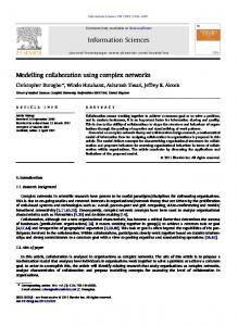
Fig 1 Non-parametric model. keo is the rate constant describing efflux of drug from the effect compartment.
inappropriately based calculations of effect-site concentrations when such fitting is poor.10 A non-parametric model has no predictive utility but is, nevertheless, analytically attractive because no assumptions are made regarding the physiological or pharmacokinetic nature of the model. The only assumption made is that the central compartment of an otherwise undefined pharmacokinetic model is linked to the effect-site by a first-order rate constant and that clearance of the drug from this effect compartment is also a first-order process (Fig. 1). Effectsite concentrations are calculated solely on the basis of the observed arterial concentrations of drug and no fitting procedure is involved. In a non-parametric analysis, subsequent determination of keo is then more accurately described as ‘semi-parametric’ rather than ‘non-parametric’ as first rate order processes are assumed in the link between the pharmacokinetic model and the postulated effect compartment. Nevertheless, for the purposes of this article, we retain, where appropriate, the term ‘non-parametric’.
Computer analysis Best fit individual pharmacokinetic models for the observed arterial concentrations of propofol were derived by PKOPT, a computer program written by one of the authors (M. W.). This program was written in GFABasic for Windows v37 (GFA Data Media (UK) Ltd) and runs in compiled form on an IBM Pentium PC under Windows95 (Microsoft Corporation). The program uses non-linear regression to minimize the extended least squares objective function (NONMEM objective function (–2 log likelihood (–2LL)).11 12 Data input for this program is completely automated and consists of pre-formatted files with details of measured propofol concentrations together with corresponding sampling times and details of the infusion administered to each patient. Population pharmacokinetic modelling was performed with the program NONMEM11 (University of California (San Francisco) version (IV); level (2.1)). keo values for each individual in the study were estimated (a)
334
Effect site modelling of propofol using AEP
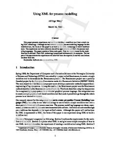
Fig 2 Arterial propofol concentrations and AEP index vs time in one patient. The prediction curve provided by the best fit individual pharmacokinetic two-compartment model calculated by the PKOPT program is superimposed on the raw data (– – –).
parametrically (on the basis of best individual parametric fit) and (b) non-parametrically, both by the program (Keo estimator). This program is based on that described by Fuseau and Sheiner13 and was also written by one of the authors (M. W.) in GFABasic for Windows. The program uses as automated input the propofol concentration data used by PKOPT together with the AEP index record file. The program estimates keo by iterating its value and selecting that value of keo at which hysteresis between the induction and recovery phases of the AEP index and effect site concentration (Ce) relationship is minimized.13 Sigmoid Emax curves were fitted from effect% vs effectsite concentration curves using WINNONLIN version 1.1 (1996) (Scientific Consulting Inc.,Cary, NC, USA). Effect% was calculated from the formula: Baseline AER index–observed AER index
Fig 3 AEP index vs time (A) for those individual patients for whom hysteresis curve collapse was successful and (B) and (C) for those in whom it was unsuccessful.
3 100 Baseline AER index–AER index at maximum effect
Table 1 NONMEM population two-compartment pharmacokinetic parameters for the study group
Statview for Windows (version 4.5) (1996) (Abacus Concepts Inc., Berkely, CA, USA) was used for statistical analyses. Probability values less than 0.05 were considered significant.
Parameter
Estimate
cv%
Volume (cpt1) Volume (cpt2) Clearance (cpt1) Clearance (cpt2)
104 (ml kg–1) 338 (ml kg–1) 33.7 (ml kg–1 min–1) 20.3 (ml kg–1 min–1)
7.9 2.05 5.99 8.22
Results Figure 2 shows the relationship between arterial blood propofol concentration and AEP index in a representative patient. For the non-parametric approach, the observed arterial concentrations of propofol were used directly to calculate Ce on the basis of a given estimate of keo, whereas for the parametric approach, the PKOPT program was used to derive the best fit two-compartment pharmacokinetic model for each individual and the model predictions of blood propofol concentrations were used to calculate Ce. The program NONMEM was used to calculate the estimates of a two-compartment population pharmacokinetic model for all 22 individuals in the study group on the basis of pooled data (Table 1). Both PKOPT and NONMEM failed to demonstrate any significant reduction in the respective objective functions when open three-compartment, rather
than two-compartment, modelling was used. For each of the three approaches, the relationship between Ce and effect (as determined by AEP index) for a given value of keo could then be determined for the course of the experiment. For both the non-parametric and the two parametric methods (individual and population), the value of keo was iterated by the computer program (Keo estimator) to determine the value at which hysteresis between the two limbs representing the induction and recovery phases of the experiment was minimized. Figure 3A, B and C demonstrate the time response of AER index during and after infusion of propofol. For seven patients in the study, the program was unable to estimate keo. In these cases, the record of AEP index either did not demonstrate a progressive increase in the value of AEP
335
White et al.
Table 2 Optimal keo values determined on the basis of various modelling techniques Patient Non-parametric Individual parametric Population parametric keo (min–1) keo (min–1) No. keo (min–1) 1 2 3 4 5 6 7 8 9 10 11 12 13 14 15 Median Mean
0.14 0.26 0.38 0.12 0.16 0.22 0.16 0.16 0.23 0.12 0.14 0.36 0.1 0.16 0.26 0.16 0.20
0.16 0.20 0.32 0.14 0.17 0.24 0.16 0.16 0.20 0.17 0.20 0.37 0.11 0.16 0.27 0.17 0.20
0.16 0.30 0.30 0.18 0.22 0.24 0.27 0.19 Failure 0.22 0.22 0.42 0.15 0.17 0.34 0.24 0.22
index during the recovery phase of the experiment but rather a flat response followed by a sudden increase only as the patient’s clinical level of arousal increased (five patients) (Fig. 3B) or, alternately, demonstrated unexplained periods of excitation during the experiment (two patients) (Fig. 3C). These patterns of response produced a Ce vs effect relationship which could not be efficiently minimized within the constraints set by the keo estimator program13 and therefore for each of these individuals a corresponding value of keo could not be calculated. Hysteresis between the induction and recovery limbs of the effect–Ce relationship was minimized at optimum keo. The optimum keo values calculated by the non-parametric and the individual and parametric analyses for those patients whose hysteresis curves were successfully collapsed are shown in Table 2. For the non-parametric analysis, the mean and median estimated values of keo were, respectively, 0.2 and 0.16 (range 0.1–0.36) min–1. The corresponding values for the individual parametric analysis were 0.2 and 0.17 (range 0.11–0.37) min–1 and for the population parametric analysis 0.22 and 0.24 (range 0.15–0.42) min–1. There was no significant difference between the non-parametric and individualized parametric sets of values on testing by the Wilcoxon ranked pair test. However, the corresponding difference between the values calculated from the nonparametric and population parametric analyses was significant. The relationship between effect and predicted Ce for each patient at the optimized value of keo was used to determine the effect% vs Ce relationship by fitting the effect%–Ce data to a sigmoid Emax curve14 15 of the nature: E5
Emax · Ceγ Ce50γ 1 Ceγ
where γ is a number (the Hill coefficient) which influences the slope of the curve and Ce50 is the effect-site concentration
at which 50% of maximum effect is obtained. Mean and median Ce50 values obtained in this way from the nonparametric analysis were 2.24 and 2.43 (range 0.52–3.76) µg ml–1. Figure 4A shows individual sigmoid Emax curves fitted by WINNONLIN from each individual’s optimally collapsed hysteresis curve. In Figure 4B, a population sigmoid Emax was constructed by fitting a curve to the pooled effect%–Ce data for all those patients for whom hysteresis could be successfully collapsed. Similar population sigmoid Emax curves were constructed from the effect%–Ce data derived using both the individual parametric and population parametric approaches (Fig. 4C). Estimates of Ce50 and γ for these three curves are tabulated in Table 3. The calculated effect-site concentration at which each individual began to demonstrate early clinical signs of arousal (movement or phonation) after termination of propofol infusion was noted and the percentage of aroused patients was plotted vs effect-site concentration (Fig. 5): a reversed sigmoid Emax curve was then fitted to the data. The EC50 for arousal was estimated to be 2.28 µg ml–1 (95% confidence limits 2.2–2.36; γ55.62).
Discussion We have used auditory evoked potentials as the determinant of the effect of propofol on the central nervous system in order to determine the parameter (keo) that dynamically characterizes the effect compartment for the drug in healthy male patients. The sets of keo values obtained when either non-parametric or individual parametric modelling techniques were used were not significantly different. However, there was a significant difference between the keo values calculated using the population parametric analysis compared with those obtained using the non-parametric analytical method. In these circumstances, the constraint of using population kinetic parameters to estimate blood concentrations of propofol and applying these parameters to individuals who are less representative of the population results in the calculation of keo values which differ from those calculated by the non-parametric method. Furthermore, the duration of sampling used in the study was relatively short (12–30 min) in relation to the terminal half-life of the model and this may also give rise to some inaccuracy in the estimation of the population model parameters. The mean value of keo obtained by the non-parametric method for the study group was 0.20 min–1. Hitherto, the only published keo for propofol is that reported in a congress abstract by Schuttler, Schwilden and Stoeckel16 (mean T 12 (keo)52.9 min: mean value keo50.24 min–1) in a group of six healthy male volunteers when EEG median frequency was used to determine the effect of propofol on the central nervous system and when parametric modelling techniques were used. We found that the range of estimated keo (using the non-parametric method) within our study group was considerable, varying between 0.1 and 0.36 min–1. This
336
Effect site modelling of propofol using AEP
Fig 4 A: Individual sigmoid Emax curves derived from non-parametrically derived effect%–Ce curves at optimized (minimal hysteresis) keo. B: Pooled data sigmoid Emax curve (thicker line) derived from non-parametrically derived effect%–Ce curves. The thick line is superimposed on the optimally collapsed hysteresis curves (thinner lines) for each of the individuals in the study group for whom such collapse was achieved successfully. C: Pooled data sigmoid Emax curves derived from non-parametric, individual parametric and population parametric (thin). Table 3 Estimates of Ce50 and γ derived by WINNONLIN for population curves of effect% vs Ce
Model
Parameter
Ce50 (µg ml–1) γ Parametric individual Ce50 (µg ml–1) γ Parametric population Ce50 (µg ml–1) γ Non-parametric
Estimate
Upper 95% CL
Lower 95% CL
2.08 1.77 2.10 1.75 2.17 1.76
2.45 2.41 2.48 2.32 2.53 2.30
1.7 1.12 1.72 1.19 1.81 1.22
Fig 5 Concentration–response curve for early arousal. The percentage of patients demonstrating arousal is plotted vs calculated effect-site concentration based on the non-parametric analysis.
variability in keo could not be ascribed purely to interpatient pharmacokinetic variability as use of both nonparametric and individualized parametric modelling techniques removed this factor from the overall modelling process. There is currently much interest in the use of auditory evoked potentials as a monitor of anaesthetic depth. The technique has the theoretical advantage that it derives its signal from a single neurological pathway in contrast with
unprocessed EEG which derives it signal from global central nervous system activity. A wide range of both inhalation and i.v. anaesthetic agents produce similar changes in the early cortical waves of the AEP signal. Furthermore, the AEP signal obtained during surgery appears to represent a balance between CNS depression caused by anaesthetic drugs and activation induced by noxious stimuli.3 Our study was performed in the total absence of noxious stimuli caused either by anaesthetist or surgeon. Under these circumstances, it was presumed that the changes in the AEP signal would be related directly to predicted concentration of drug in the brain. In each of the study patients, the AEP index decreased in a progressive manner after commencement of the propofol infusion until Emax was reached. However, in seven of 22 patients, it proved impossible to calculate a value of keo for the individual owing to the fact that it was not possible to collapse the hysteresis curve in an acceptable manner. In these individuals the inability to collapse the hysteresis curve was caused either by unexplained excitation during the experiment or by a flat response of the AEP index vs predicted Ce curve during the late stages of the recovery curve. In these latter instances, the AEP index returned to awake levels only when the patient had already began to demonstrate clinical signs of arousal. It appeared that, in these individuals, the process of awakening from propofol-induced anaesthesia was associated with an ‘on-off’ AEP index response rather than a gradual progression to awake levels. The observed data in these seven patients suggest that the proposed effect model was not applicable to all patients in the study. Doi and colleagues5 reported that during emergence from anaesthesia, the AEP index before eye opening correlated poorly with predicted blood propofol concentrations, whereas bispectral index and 95% spectral edge frequencies both correlated well with predicted blood concentrations of propofol. However, AEP
337
White et al.
index was demonstrated as being superior to EEG methods in distinguishing the conscious from the unconscious state. It may be that the pharmacodynamics of propofol may differ according to the monitoring technique used. AER, bispectral index and spectral edge frequency all require similar time periods (approximately 30 s) for complete update of their signal. In our study, the mean value of Ce50 was 2.08 µg ml–1 (95% confidence limits 1.7–2.45) using the pooled data from the non-parametric modelling analysis. It is of interest to relate these values to clinical end-points. Forrest and colleagues17 reported that, in an unpremedicated population of patients, an equilibrated blood propofol concentration of 3.1 (2.7–3.5) µg ml–1 was necessary to suppress 50% of maximum EEG median power frequency in the absence of external stimulation which implies that the AER index is more sensitive to the effects of propofol than unprocessed EEG. In the same study, it was found that, under steady state conditions, mean EC50 and EC95 values for loss of consciousness were 2.3 µg ml–1 and 3.1 µg ml–1 (where EC50 and EC95 are defined as those equilibrated blood concentrations of propofol at which 50% and 95%, respectively, of the study population lost consciousness). In a similar study using only clinical end-points, Vuyk and colleagues9 reported that the EC50 and EC90 values of propofol for loss of eyelid reflex in a group of 18 young female patients were 1.85 and 2.69 µg ml–1 whereas the corresponding values for loss of consciousness were 3.34 and 4.17 µg ml–1. Smith and colleagues18 reported that the EC50 (Cp50 in their terminology) and EC95 (Cp95) at which their study patients lost response to verbal command were, respectively, 3.3 and 5.4 µg ml–1. In a more recent study of Japanese subjects, Kazama, Ikeda and Morita19 found higher values for EC50 and EC95 for loss of consciousness (4.4 and 7.8 µg ml–1 ). The reason for the differences in the EC50 and EC95 values obtained in these studies is unclear. It may be that the level of the stimulus applied varied between studies, that the equilibration times were inadequate in some of the study patients, or that there are racial or cultural reasons for the observed differences. To avoid confusion in terminology, it should be borne in mind that EC50 and Ce50 are not equivalent and cannot be used in an interchangeable manner. EC50 (or Cp50) is that equilibrated plasma concentration at which 50% of individuals have reached a given non-continuous end-point (e.g. eye closure or loss of consciousness), whereas Ce50 denotes that effect-compartment concentration at which an individual or a ‘population-individual’ displays 50% of the recorded maximum effect. Figure 6 demonstrates this point. The EC50 at which 50% of patients started to show clinical evidence of arousal was 2.28 µg ml–1 whereas the Ce50 at which 50% of maximum suppression of AEP effect occurred was 2.08 µg ml–1. Knowledge of the keo value of a drug provides insight into its clinical use which is not apparent from pharmacokinetic modelling concepts alone. First, equilibration of the effect-
Fig 6 Non-parametrically derived population effect% vs Ce sigmoid Emax curve in relation to Ce50 and to EC50 (waking) for regain of consciousness.
site concentration with steady state blood concentrations takes 4–5 times T1/2 (keo) (where T1/2 (keo)50.693/keo). Thus if we were to request a certain blood concentration using a target controlled (TCI) delivery system,20 21 then we can predict that it would take approximately 15 min for equilibration between the effect and central compartments to occur if keo is taken to be 0.20 min–1. We have calculated a range of keo values between 0.1 and 0.36 min–1 for our patients. These results suggest that some studies which were designed to permit equilibration between blood and effect compartments did not allow adequate time for this process to occur in all patients since if the keo for a given individual is 0.1 min–1 then the time taken for equilibration between blood and the effect compartment is approximately 28 min. In order to achieve a given effect-site concentration in a shorter time, then the concept of overpressure (familiar from inhalation anaesthesia) can be used to force drug into the effect compartment at a faster rate, whereby a much higher blood concentration is initially achieved than the desired effect concentration. The mean value of keo of 0.20 min–1 derived in this study for propofol is substantially less than that of 0.58 min–1 derived for thiopental.22 This implies that propofol is taken up into the effect compartment less rapidly than that of thiopental and explains why propofol is a clinically less appropriate induction agent compared with thiopental when rapid intubation to secure the airway is the priority. We have calculated keo values for propofol using a nonparametric method but have demonstrated that the values obtained by a parametric method were similar. We have demonstrated that the values of keo thus obtained can be used in the context of a parametric model and that subsequently the values obtained have use with respect to pharmacokinetic model-based simulations. It is important to note that the estimated value of keo that correlates observed pharmacodynamics with pharmacokinetics varies according to the performance of a particular pharmacokinetic model in predicting experimentally observed blood concentrations of propofol and that the value of keo used in a simulation cannot be used in association with a
338
Effect site modelling of propofol using AEP
pharmacokinetic model other than that which was used in its estimation. The concept of the effect compartment, particularly when applied to anaesthetic and analgesic drugs, is especially useful because it allows the anaesthetist to relate drug dose to drug effect in a more sophisticated manner than is otherwise possible by consideration of blood concentrations alone, as such a concept provides an appreciation of the delays involved for equilibration between clinical effect and blood concentration. In our study, we reported the estimated values of keo for propofol in 15 of 22 healthy male patients and, on the basis of these values, described the relationship between AEP index and effect-site concentration of propofol. For the remaining seven patients, an individual value of keo could not be estimated.
Acknowledgement This work was supported by a research grant from Zeneca Pharma, Ridderkerk, The Netherlands.
References 1 Hull CJ, Van Beem HBH, McLeod K, Sibbald A, Watson MJ. A pharmacodynamic model for pancuronium. Br J Anaesth 1978; 50: 1113–22 2 Sheiner LB, Stanski DR, Vozeh S, Miller RD, Ham J. Simultaneous modeling of pharmacokinetics and pharmacodynamics: Application to d-tubocurarine. Clin Pharmacol Ther 1979; 25: 358–71 3 Thornton C. Evoked potentials in anaesthesia. Eur J Anaesthesiol 1991; 8: 89–107 4 Davies FW, Mantzaridis H, Kenny GNC, Fisher AC. Middle latency auditory evoked potentials during repeated transitions from consciousness to unconsciousness. Anaesthesia 1996; 51: 107–13 5 Doi M, Gajraj RJ, Mantzaridis H, Kenny GNC. Relationship between calculated blood concentration of propofol and electrophysiological variables during emergence from anaesthesia; comparison of bispectral index, spectral edge efficiency, median frequency and auditory evoked potential index. Br J Anaesth 1997; 78: 180–4 6 Gajraj RJ, Doi M, Mantzaridis H, Kenny GNC. Analysis of the EEG bispectrum, auditory evoked potentials and the EEG power
7
8
9 10
11 12 13
14 15
16 17
18
19
20
21
22
339
spectrum during repeated transitions from consciousness to unconsciousness. Br J Anaesth 1998; 80: 46–52 Mantzaridis H. Closed Loop Control of Anaesthesia. Doctoral thesis. Glasgow: Department of Bioengineering, University of Strathclyde, 1996; 117 Mantzaridis H, Kenny GNC. Auditory evoked potential index: a quantitive measure of changes in auditory evoked potentials during general anaesthesia. Anaesthesia 1997; 52: 1030–6 Vuyk J, Engbers FHM, Lemmens HJM, et al. Pharmacodynamics of propofol in female patients. Anesthesiology 1992; 77: 3–9 Shafer SL, Varvel JR, Gronert G. A semi-parametric method of estimating the equilibrium delay for neuromuscular blocking drugs. Anesthesiology 1988; 69: A518 Beal S, Sheiner LB. Nonmem User’s Guide Part 1. San Francisco: University of California, 1994 Gepts E, Shafer SL, Camu F, et al. Linearity of pharmacokinetics and model estimation of sufentanil. Anesthesiology 1995; 83: 1194–204 Fuseau E, Sheiner LB. Simultaneous modelling of pharmacokinetics and pharmacodynamics with a non-parametric pharmacodynamic model. Clin Pharmacol Ther 1984; 35: 733–41 Hill AV. The possible effects of the aggregation of the molecules of haemoglobin on its disociation curves. J Physiol 1910; 40: iv–vii Holford NHG, Sheiner LB. Understanding the dose–effect relationship: clinical application of pharmacokinetic– pharmacodynamic models. Clin Pharmacokinet 1981; 6: 429–53 Schuttler J, Schwilden H, Stoeckel H. Pharmacokinetic–dynamic modelling of Diprivan. Anesthesiology 1986; 65 (Suppl.): A549 Forrest FC, Tooley MA, Saunders PR, Prys-Roberts C. Propofol infusion and the suppression of consciousness: the EEG and dose requirements. Br J Anaesth 1994; 72: 35–41 Smith C, McEwan AI, Jhaveri R, et al. The interaction of fentanyl on the Cp50 of propofol for loss of consciousness and skin incision. Anesthesiology 1994; 81: 820–8 Kazama T, Ikeda K, Morita K. Reduction by fentanyl of the Cp50 values of propofol and hemodynamic responses to various noxious stimuli. Anesthesiology 1997; 87: 213–27 Alvis JM, Reeves JG, Spain JA, Sheppard LC. Computer assisted infusions of the intravenous analgesic fentanyl during general anaesthesia; an interactive system. Trans Biomed Eng 1985; 5: 323–9 Schuttler J, Kloos S, Schwilden H, Stoeckel H. TIVA with propofol and alfentanil by computer assisted infusions. Anaesthesia 1988; 43 (Suppl.): 2–7 Stanski DR, Maitre PO. Population pharmacokinetics and pharmacodynamics of thiopental: The effect of age revisited. Anesthesiology 1990; 72: 412–22