gDivision of Pharmacy and Optometry,Faculty of Biology, Medicine and Health, The University of. Manchester, UK. hIstituto Zooprofilattico Sperimentale del ...
Electronic Supplementary Material (ESI) for RSC Advances. This journal is © The Royal Society of Chemistry 2017
Electronic Supplementary Information (ESI) for RSC Advances.
Electrostatically driven scalable synthesis of MoS2-Graphene hybrid films assisted by hydrophobins AUTHORS: Jasneet Kaur,*a,b Alessandro Vergara,c,d,e Manuela Rossi,f Alfredo Maria Gravagnuolo,c,g Mohammadhassan Valadan,a Federica Corrado,h Mariarosaria Conte,i Felice Gesuele,a Paola Giardinac and Carlo Altucci*a
aDepartment bAkal
of Physics "Ettore Pancini”, University of Naples Federico II, Naples, Italy
College of Basic Sciences, Eternal University, Baru Sahib, Himachal Pradesh, India
cDepartment dCEINGE
of Chemical Sciences, University of Naples Federico II, Naples, Italy
Biotecnologie Avanzate scarl, Naples, Italy
eInstitute
of Biostructures and Biomaging, CNR, Naples, Italy
fDepartment gDivision
of Earth, Environment and Resources Sciences, University of Naples Federico II, Naples, Italy
of Pharmacy and Optometry, Faculty of Biology, Medicine and Health, The University of
Manchester, UK hIstituto iIRCCS,
Zooprofilattico Sperimentale del Mezzogiorno, Portici, Italy
SDN, Via E. Gianturco 113, 80143, Naples, Italy
Experimental
Extraction of Vmh2 from Pleurotus Ostreatus mycelia White-rot fungus, P. ostreatus (Jacq.: Fr.) Kummer (type: Florida; ATCC No. MYA-2306) was maintained at 4 °C through periodic transfer on potato dextrose agar (Difco) plates in the presence of 0.5% yeast extract. Mycelia were inoculated in 1L flasks containing 500 mL of potato-dextrose broth (24 g L−1 ) supplemented with 0.5% yeast extract, grown at 28 °C in shaken mode (150 rpm). After 10 days of fungal growth, mycelia were separated by filtration through gauze, treated twice with 2% sodium dodecyl sulfate (SDS) in a boiling water bath for 10 min, washed several times with water and once with 60% ethanol to completely remove the detergent. The residue was dried under nitrogen, grinded and treated with 100% trifluoroacetic acid (TFA) in a water bath sonicator (Elmasonic S30, Elma) for 30 min, and centrifuged (10 min at 3200 g). The supernatant was dried, and then lipids were extracted in a mixture of water–methanol–chloroform 2:2:1 v/v (5 min in bath sonicator). After centrifugation, proteins appeared as a solid aggregate at the interface
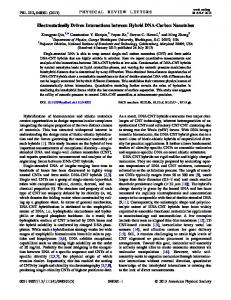
between the water–methanol and the chloroform phases. They were recovered by liquid phase removal. The aggregated protein was dried, treated with TFA for 30 minutes in bath sonicator, re-dried, and dissolved in 80% ethanol. The sample was centrifuged (90 min at 12 000 g) and ethanol was removed from the supernatant under vacuum at 40 °C using rotavapor and the material was freeze-dried, treated with TFA as above described, and dissolved again in 60% ethanol. We have optimized the procedure to remove non-protein contaminants (e.g. lipids and carbohydrates), as demonstrated by Attenuated Total Reflection Fourier Transform Infrared (ATR FT-IR) spectroscopy, and reported elsewhere.1 Only one protein, of the accurate mass of Vmh2 (UniProt2 Accession Number Q8WZI2; Vmh2-1, chain 25-111) was identified by matrix-assisted laser desorption/ionization mass spectrometry analysis.3 UV-Vis Analysis Optical extinction spectra were acquired on Jasco V-530 UV–VIS spectrophotometer using 1 cm optics quartz cuvettes. The extinction spectra of MoS2 dispersion was analyzed to determine the mean number of layers and mean lateral size of the nanosheets by metrics. 4,5 For graphene dispersion, the concentration of graphene flakes was determined by Lambert Beer law6,7, using the absorbance spectra shown in Fig. S1.
Fig. S1 Absorbance of bGr samples centrifuged at 620g (10 times dilution) and 2500g. ζ -potential and DLS measurements ζ- potential measurements were carried out on a Malvern Zetasizer Nano ZSP with irradiation from 633nm He-Ne laser. The samples were injected in capillary cells and the electrophoretic mobility (µ) was measured using a combination of electrophoresis and laser Doppler velocimetry techniques as reported elsewhere7. The ζ-potential was estimated from the electrophoretic mobility data through the Henry’s equation.
However, because of the particular solvent-sample relationship, the Henry’s function was approximated to Smoluchowsky limit to estimate the value of ζ-potential, in table 2. The specific monitoring of flakes in the mixture of free protein and protein-MoS2 mixture was possible because in the electrophoretic analysis, which is based on the combination of electrophoresis and laser Doppler velocimetry techniques,8 the signal is originated from the scattered laser light. Indeed, in the presence of the exfoliated flakes, which are more than one order of magnitude larger than proteins, the signal of the proteins is negligible and only the flakes are observed. All the measurements were conducted at 25°C at the natural pH of the MoS2, bGr and hybrid solutions. Optical Microscope Morphological and textural analyses of MoS2, graphene and hybrid samples were carried out by Axio Imager Am1 polarized and reflected light (Real Museo Mineralogico laboratory, Università degli studi di Napoli Federico II), Carl Zeiss Microscope. The observations on MoS2, graphene and hybrid structures were effectuated in reflected light configuration. In particular, the microscope is equipped with: reflector module, dark field and bright field ACR P&C for reflected light; reflector module DIC/Pol ACR P&C for reflected light. Moreover, the microscope is equipped with objectives to perform analyses in reflected light, in particular LD EC HC DIC M27 "Epiplan-Neofluar" 20x/0.22 and 50x/055. The microscope is equipped with the camera Axiocam ICC5 (D), with digital adaptor 60N-C 2/3x0.63. Finally, the software Zeiss was utilized to acquire the images is Axiovision SE64. Scanning Electron Microscopy with EDS Detailed morphological analyses and qualitative chemical analyses of MoS2, graphene and hybrid samples were performed with a scanning electron microscope (SEM) JEOL-JSM 5310 (CISAG, Università degli Studi di Napoli Federico II) equipped with energy dispersive X-Ray spectroscopy (EDS). The EDS spectrometer utilized a JEOL JSM-5310 SEM and an Oxford Instruments Microanalysis unit, equipped with an INCA X-act detector and operating at 15 kV primary beam voltage, 50-100 mA filament current, variable spot size, 20 mm WD and 40 s net acquisition real time. INCA X-act detector uses Energy software with XPP matrix correction scheme, developed by Pouchou and Pichoir (1991), and Pulse Pile up correction. The data were processed with INCA version 4.08 (Oxford Instruments, 2006). The samples were investigated previously without metallization for qualitative chemical analyses, later were metalized with gold by using a sputter coater, for morphological analyses and micrographs were acquired to 10000x and 15000x magnification. Transmission Electron Microscopy Samples were prepared for transmission electron microscopy (TEM) analysis by depositing a 10 μL drop on a standard carbon-coated copper grid (100 mesh) covered with a Formvar film, then the grid was dried in
air. Images were acquired using a FEI Tecnai 12 transmission electron microscope (FEI Company, Hillsboro, Oregon, U.S.A.) equipped with a Veleta CCD digital camera (Olympus Soft Imaging Solutions GmbH, Munster, Germany) and operating at 120 kV. Raman and Photoluminescence spectroscopy A confocal Raman microscope (Jasco, NRS-3100) was used to obtain Raman and photoluminescence spectra. The 514 nm line of an air-cooled Ar+ laser (Melles Griot, 35 LAP431 220), was injected into an integrated Olympus microscope and focused to a spot diameter of approximately 3 μm by a 20x objective with a final 4 mW power at the sample. A holographic notch filter was used to reject the excitation laser line. The Raman backscattering was collected using a 0.1 mm slit and a diffraction lattice of 1200 grooves/mm, corresponding to an average spectral resolution of 8 cm-1. Solutions were left evaporating on Si substrates, and it took 60 s to collect a complete data set by a Peltier-cooled 1024 x 128 pixel CCD photon detector (Andor DU401BVI). Raman measurements were at least triplicated for scope of reproducibility. Wavelength calibration was performed by using cyclohexane as a standard.
References
1
M. Portaccio, A.M. Gravagnuolo, S. Longobardi, P. Giardina, I. Rea, L. Cammarota and M. Lepore, Appl. Surf. Sci., 2015, 351, 673–680.
De Stefano, M.
2
The UniProt Consortium. (2014) Activities at the Universal Protein Resource (UniProt). Nucleic Acids Res. 42, D191-8.
3
A. Armenante, S. Longobardi, I. Rea, L. De Stefano, M. Giocondo, A. Silipo, A. Molinaro and P. Giardina, Glycobiology, 2010, 20, 594–602.
4
C. Backes, R. J. Smith, N. Mcevoy, N. C. Berner, D. Mccloskey, H. C. Nerl, A. O. Neill, P. J. King, T. Higgins, D. Hanlon, N. Scheuschner, J. Maultzsch, L. Houben, G. S. Duesberg, J. F. Donegan, V. Nicolosi and J. N. Coleman, Nat. Commun., 2014, 5, 4576.
5
J. Kaur, A. M. Gravagnuolo, P. Maddalena, C. Altucci, P. Giardina and F. Gesuele, RSC Adv., 2017, 7, 22400–22408.
6
A. M. Gravagnuolo, E. Morales-Narváez, C. R. S. Matos, S. Longobardi, P. Giardina and M. Arben, Adv. Funct. Mat., 2015, 25, 6084–6092.
7
M. Lotya, Y. Hernandez, P. J. King, R. J. Smith, V. Nicolosi, L. S. Karlsson, F. M. Blighe, S. De, W. Zhiming, I. T. McGovern, G. S. Duesberg and J. N. Coleman, J. Am. Chem. Soc., 2009, 131, 3611– 3620.
8
M. Lotya, A. Rakovich, J. F. Donegan and J. N. Coleman, Nanotechnology, 2013, 24, 265703– 265709.