TECHNICAL PAPER
RESEARCH ARTICLE 4555
Development 138, 4555-4564 (2011) doi:10.1242/dev.066779 © 2011. Published by The Company of Biologists Ltd
Evaluation and application of modularly assembled zincfinger nucleases in zebrafish Cong Zhu1,*, Tom Smith1,*, Joseph McNulty1, Amy L. Rayla1, Abirami Lakshmanan1, Arndt F. Siekmann1,†, Matthew Buffardi1, Xiangdong Meng1,‡, Jimann Shin2, Arun Padmanabhan3, Daniel Cifuentes4, Antonio J. Giraldez4, A. Thomas Look2, Jonathan A. Epstein3, Nathan D. Lawson1,§ and Scot A. Wolfe1,5,§
SUMMARY Zinc-finger nucleases (ZFNs) allow targeted gene inactivation in a wide range of model organisms. However, construction of target-specific ZFNs is technically challenging. Here, we evaluate a straightforward modular assembly-based approach for ZFN construction and gene inactivation in zebrafish. From an archive of 27 different zinc-finger modules, we assembled more than 70 different zinc-finger cassettes and evaluated their specificity using a bacterial one-hybrid assay. In parallel, we constructed ZFNs from these cassettes and tested their ability to induce lesions in zebrafish embryos. We found that the majority of zinc-finger proteins assembled from these modules have favorable specificities and nearly one-third of modular ZFNs generated lesions at their targets in the zebrafish genome. To facilitate the application of ZFNs within the zebrafish community we constructed a public database of sites in the zebrafish genome that can be targeted using this archive. Importantly, we generated new germline mutations in eight different genes, confirming that this is a viable platform for heritable gene inactivation in vertebrates. Characterization of one of these mutants, gata2a, revealed an unexpected role for this transcription factor in vascular development. This work provides a resource to allow targeted germline gene inactivation in zebrafish and highlights the benefit of a definitive reverse genetic strategy to reveal gene function.
INTRODUCTION The attributes of the zebrafish have established it as a powerful model for the study of vertebrate development. The accessibility of its externally fertilized embryos allows numerous manipulations, and the transparency of the zebrafish embryo permits direct serial visualization of biological processes in vivo (Beis and Stainier, 2006). These characteristics have enabled forward genetic screens for mutants affecting a wide range of developmental processes (Patton and Zon, 2001). However, the size of the zebrafish genome, the relatively long generation time, and the expense of maintaining large populations preclude forward genetic screening to saturation. Consequently, mutants exist for only a small fraction of zebrafish genes. Although modified antisense oligonucleotides can achieve targeted gene knockdown during embryogenesis (Nasevicius and Ekker, 2000), this technology is problematic with regard to persistence, specificity and penetrance (Robu et al., 2007; Eisen
1
Program in Gene Function and Expression, University of Massachusetts Medical School, Worcester, MA 01605, USA. 2Department of Pediatric Oncology, DanaFarber Cancer Institute, Harvard Medical School, Boston, MA 02115, USA. 3 Department of Cell and Developmental Biology, Perelman School of Medicine at the University of Pennsylvania, Philadelphia, PA 19104-6058, USA. 4Department of Genetics, Yale University School of Medicine, New Haven, CT 06510, USA. 5 Department of Biochemistry and Molecular Pharmacology, University of Massachusetts Medical School, Worcester, MA 01605, USA. *These authors contributed equally to this work Present address: Max-Planck Institute for Molecular Biomedicine, Laboratory for Cardiovascular Patterning, Roentgenstr. 20, 48149 Muenster, Germany ‡ Present address: Bio-Rad Laboratories, Gene Expression Division, 2000 Alfred Noble Drive, Hercules, CA 94547, USA § Authors for correspondence (
[email protected];
[email protected]) †
Accepted 3 August 2011
and Smith, 2008). Thus, definitive reverse genetic strategies are required, especially for the study of later developmental processes, analysis of adult physiology, and for the establishment of disease models. Zinc-finger nucleases (ZFNs) are chimeric fusions between a zinc-finger protein (ZFP) and the nuclease domain of FokI (Urnov et al., 2010). ZFNs have been employed to achieve heritable targeted gene disruption in the genomes of numerous plant and animal species, including zebrafish (Bibikova et al., 2002; Beumer et al., 2008; Doyon et al., 2008; Meng et al., 2008; Foley et al., 2009; Geurts et al., 2009; Cui et al., 2010; Mashimo et al., 2010; Meyer et al., 2010; Takasu et al., 2010). Gene disruption is achieved through imprecise repair of a ZFN-induced double-strand break within the coding sequence of a target gene (Urnov et al., 2010). ZFNs have also been utilized to achieve tailor-made genomic alterations in animal genomes through stimulation of homology-directed repair from an exogenous donor DNA (Beumer et al., 2008; Cui et al., 2010; Meyer et al., 2010). Although offtarget lesions in the genome can result from ZFN treatment, their frequency is significantly lower than at the target site and their location can be predicted based on ZFN specificity (Meng et al., 2008; Perez et al., 2008; Gupta et al., 2011). Together, these characteristics make ZFNs an ideal tool to facilitate reverse genetic studies in zebrafish and other organisms. Despite their utility, the widespread implementation of ZFNs is hindered by the difficulty in creating highly specific ZFPs. Two general approaches have been employed for this purpose: selectionbased methods, which identify ZFPs with a desired specificity from randomized libraries, and assembly-based methods, which utilize archives of predefined zinc fingers to construct ZFPs with a desired specificity. Each approach is hampered by limitations. Selectionbased approaches (Greisman and Pabo, 1997; Isalan et al., 1998;
DEVELOPMENT
KEY WORDS: gata2, Vascular development, Zebrafish, Zinc-finger nuclease
4556 RESEARCH ARTICLE
MATERIALS AND METHODS Fish lines
Zebrafish were handled according to established protocols (Westerfield, 1993) and in accordance with Institutional Animal Care and Use Committee (IACUC) guidelines of participating institutions (University of Massachusetts Medical School, University of Pennsylvania, Dana Farber Cancer Institute and Yale University). The Tg(kdrl:egfp)la116 line has been described elsewhere (Choi et al., 2007). Construction of a modular zinc-finger archive
An archive of zinc-finger modules originating from multiple sources (see Table S1 in the supplementary material and Results) was constructed by subcloning ZFPs into either a pBluescript or pCS2 vector using standard methods. Clones were sequence verified and arrayed into a 96-well plate to generate an address for each module. Plasmids from the archive are available through addgene.org. Modular assembly of ZFNs
To assemble three-finger ZFPs we first separately amplified modules for each of the three finger positions from plasmid templates by PCR. Distinct primer pairs were used to amplify modules for each position (fingers 1, 2 and 3) (see Table S2 in the supplementary material). PCR was performed using Advantage HF2 polymerase (Clontech) and 20 ng plasmid template. Finger 1 modules were amplified as follows: 94°C for 2 minutes, followed by 2030 cycles of 94°C for 30 seconds, 60°C for 30 seconds and 68°C for 20 seconds, with a final extension of 68°C for 5 minutes. For finger 2 or 3 modules: 94°C for 2 minutes, then 20-30 cycles of 94°C for 30 seconds, 57°C for 30 seconds and 68°C for 20 seconds, with a final extension of 68°C for 5 minutes. All initial PCR products were gel purified. Three-finger cassettes were constructed by overlapping PCR using 30 ng of each finger module in a 50 ml reaction with Advantage HF2 polymerase. The first five cycles were performed without primers: 94°C for 2 minutes, followed by five cycles of 94°C for 30 seconds, 55°C for 30 seconds and 68°C for 1 minute. We added 2 ml of 10 mM F1 forward and F3 reverse primer and performed the following steps: 24 cycles of 94°C for 15 seconds, 60°C for 30 seconds, 68°C for 1 minute, with a final extension of 68°C for 5 minutes. Final PCR products were gel purified, digested with Acc65I and BamHI and cloned into
the pCS2 nuclease backbone in frame with the EL/KK heterodimeric variants of FokI (Miller et al., 2007), such that the 5⬘ ZFP was fused to the EL nuclease and the 3⬘ ZFP was fused to the KK nuclease. In several cases, DD/RR nuclease variants (Miller et al., 2007; Szczepek et al., 2007) were used. All constructs were sequence verified. ZFN injections into zebrafish embryos
Synthesis and injection of ZFN mRNAs, dose optimization (see Table S3 in the supplementary material) and establishment and identification of germline lesions were performed as previously described (Meng et al., 2008; Gupta et al., 2011). Bacterial one-hybrid binding site selections
Sequence-verified ZFP cassettes were cloned into the 1352-UV2 expression vector using Acc65I and BamHI. Bacterial one-hybrid (B1H) binding site selections were typically carried out at 5 mM 3-amino-1,2,4triazole (3-AT), 10 mM IPTG and in the absence of uracil as previously described (Noyes et al., 2008; Gupta et al., 2011). For some ZFPs, lower stringency was required to achieve sufficient enrichment of recognition sequences above background. Illumina library preparation of B1H-selected binding sites
Preparation of selected binding sites for deep sequencing was performed as described (Gupta et al., 2011), except that amplicons were digested with either EcoRI or NotI prior to ligation of bar-coded adapters for the identification of sequences associated with each ZFP (see Table S2 in the supplementary material). Evaluation of the DNA-binding specificity of each ZFP and individual zinc-finger modules
Recognition motifs within the population of unique 28 bp sequences recovered from each binding site selection were identified using MEME (Bailey and Elkan, 1994). Position weight matrices (PWMs) were generated using the Log-Odds method (Hertz and Stormo, 1999) from the aligned sequences within the most statistically significant motif, weighting each sequence based on the number of counts within the Illumina dataset using the formula: 9
S= ∑ log2 1
fb,i pb,i
,
(1)
where S is the PWM score over the 9 bp target site s, b,i is the identity of base b at position i, fb,i is the normalized frequency of occurrence of base b at position i, and pb,i is the probability of observing the same base at the same position in a background model, which is assumed to be an equal distribution. Each ZFP was evaluated by calculating the score for its target site from its derived PWM. Each individual finger module was evaluated by calculating the score for its corresponding 3 bp recognition element (assuming canonical recognition) from this PWM. When data were available for multiple identical modules at identical positions, the scores for these modules were averaged for an overall assessment of the quality of the finger module. Modules with (average) scores that were two standard deviations below the average score of all modules at that position (i.e. finger 1, finger 2 or finger 3) were considered ‘questionable’ and flagged. Analysis of somatic lesion frequency
Somatic lesion frequency was determined by Illumina sequencing as previously described (run #1) (Meng et al., 2008; Gupta et al., 2011). A second round of sequencing was performed for a subset of ZFNs from an independent set of injections (run #2). To avoid cross-contamination with samples from the first analysis, a unique pair of PCR primers was designed for each target site that would not amplify the PCR products from the first trial. These primers encoded the Illumina P1 and P2 sequences within each primer pair (see Table S2 in the supplementary material). In this case, PCR products for each ZFN target site were gel purified and amplified using Illumina genomic DNA primers (1.1 and 2.1). The PCR products for each target were then pooled at equal molar ratio, sequenced at 7 pM on a HiSeq 2000 (Illumina), and analyzed in a manner identical to run #1.
DEVELOPMENT
Maeder et al., 2008; Meng et al., 2008), although effective in generating specific ZFPs, can be time-consuming and technically challenging (Kim et al., 2010). Assembly-based approaches are more straightforward (Carroll et al., 2006; Kim et al., 2009) but are dependent on the quality of the available zinc-finger archive and the difficulty in predicting context-dependent effects between neighboring zinc fingers (Desjarlais and Berg, 1993; Greisman and Pabo, 1997; Wolfe et al., 1999). Although assembly-based, or modular, ZFNs have suffered from relatively low success rates (Ramirez et al., 2008), continued characterization of available archives and investigation of context dependence (Carroll et al., 2006; Kim et al., 2009; Sander et al., 2009), along with the generation of new archives with expanded specificity, will make this a more viable approach. In this study we evaluated an assembly-based approach for creating ZFNs for targeted gene disruption in the zebrafish genome. We constructed an archive of ZFP modules recognizing 27 different triplet sequences and characterized the DNA-binding specificity for three-finger ZFP cassettes assembled from this archive. In parallel, we tested ZFNs constructed from these ZFPs for their ability to induce both somatic and germline lesions in zebrafish embryos. To facilitate the application of this technology in the zebrafish community, we developed a publicly accessible database that assists users in modular ZFN construction using our archive. Finally, we characterized the phenotypes in zebrafish embryos bearing a ZFN-induced deletion in the gata2a gene, revealing its role in vascular morphogenesis.
Development 138 (20)
RESEARCH ARTICLE 4557
Calculation of the occupancy probability for a ZFN pair at its target site
The probability of a ZFP occupying its 9 bp target site was calculated using statistical thermodynamic free energy derived from a modified PWM score based on its B1H-determined DNA-binding specificity: 9
ΔG = – RT • ( ∑ ln 1
fb,i pb,i
),
(2)
where fb,i and pb,i are defined as in (1), R0.001987 kcal/(mol.K) and T301.5K (28.5°C). All 9mers within the zebrafish genome (Zv8) were extracted considering both strands then the free energy of binding each 9mer by each ZFP was calculated using the scoring function shown in formula (2). These energies were then used to calculate the probability (P) for on-target binding by the ZFP within the zebrafish genome using: –
P=
e l
ΔGs RT –
∑ ni e
ΔGs,i RT
,
(3)
1
where DGs is the free energy of binding to the 9mer matching the desired ZFP target site, DGs,i is the free energy of binding of the ith 9mer, ni is the occurrence of the ith 9mer in the genome, and l is total number of 9mers. A threshold for DGs,i was set such that DGs,i–DGs≤4.0 to simulate the nonspecific binding affinity of each ZFP. The occupancy probability of each ZFN pair was calculated as the product of the occupancy probabilities of the two ZFPs (3⬘ ZFP and 5⬘ ZFP). Sequence and statistical analyses
All sequence analyses, PWM score calculations, ZFP occupancy calculations and lesion frequency calculations were performed using customized Perl scripts. All statistical analyses, plots and DNA-binding logos were generated using R (http://www.r-project.org). Analysis of gata2aum27 mutant embryos
For phenotypic analysis, embryos were obtained from individual incrosses of heterozygous carriers. To analyze vascular development, gata2aum27 was crossed into the Tg(kdrl:egfp)la116 background. Vascular morphology and circulatory function were observed by confocal microscopy, confocal microangiography and video microscopy as described elsewhere (Covassin et al., 2006); Quantum Dots were from Invitrogen. Whole-mount in situ hybridization was performed as previously described using antisense riboprobes against kdrl, hey2, efnb2a, vegfaa, flt4, tal1 and gata1 (Lawson et al., 2001; Hart et al., 2007). Following phenotypic analysis or in situ hybridization, DNA was isolated from selected individual embryos as described elsewhere (Roman et al., 2002). The presence of the 10 bp um27 deletion was determined by PCR amplification (see Table S2 in the supplementary material for primer sequences), followed by gel electrophoresis or analysis on a QiaXL system (Qiagen). A rescue construct was made by amplifying the gata2a coding sequence by PCR using primers containing the Gateway attB1 and attB2 sites (see Table S2 in the supplementary material), followed by BP cloning into pDONR221 (Invitrogen). The resulting plasmid, pME-gata2a, was used in an LR reaction with pCSDest (Villefranc et al., 2007) to generate pCSgata2a, which was linearized with NotI and used as a template to synthesize mRNA. Embryos derived from crosses between gata2aum27 heterozygous carriers were injected at the 1-cell stage with 200 pg gata2a mRNA, or a comparable amount of mcherry mRNA, and were scored for the presence or absence of trunk circulation at 48 to 55 hours post-fertilization (hpf). Following phenotypic scoring, embryos were genotyped as above.
RESULTS Overall rationale and experimental approach To assess ZFNs constructed via modular assembly, we developed the following approach (see Fig. S1 in the supplementary material). We generated a collection of plasmids encoding zinc-finger modules recognizing 27 different triplet sequences. In parallel, we constructed
a database of sites within zebrafish protein-coding genes (Zv7) that could be targeted by ZFNs assembled from this archive. From this database, we chose target genes and generated the corresponding ZFP cassettes. We characterized the specificity of these ZFP cassettes and determined their function in ZFNs by assessing their ability to induce somatic and germline lesions in vivo. Finally, we created zebrafish bearing a truncation allele in gata2a and characterized the phenotypes associated with this mutation. A zinc-finger archive for modular assembly We compiled an archive of zinc fingers recognizing 27 different triplet sequences in the three finger positions of the Zif268 backbone that can be rapidly assembled by PCR into three-finger ZFPs (see Table S1 in the supplementary material). This archive comprises modules from previously defined finger archives (Segal et al., 1999; Liu et al., 2002; Carroll et al., 2006) or that have been designed based on previously described recognition principles (Isalan et al., 1998; Segal et al., 1999; Wolfe et al., 1999; Dreier et al., 2001; Dreier et al., 2005). We also generated 14 new modules by bacterial one-hybrid (B1H) selection (Meng et al., 2008). In some cases, distinct recognition helices were utilized at different finger positions for a common triplet for the B1H-generated fingers. In general, the archive was focused around GNN recognition elements (see Table S1 in the supplementary material) because of their reliable functional properties (Ramirez et al., 2008). Nine HNG modules with a preference for either G or T at the neighboring 3⬘ position due to an RSD motif at positions –1, 1 and 2 of the recognition helix were included as they should synergistically recognize a composite sequence (NNGGNN) with GNN modules. Finally, AGA and TGT modules were included to expand the set of triplets that could be specified. Target gene selection We identified zebrafish genes that could be targeted using ZFNs constructed from the modular archive. In this analysis, each ZFN monomer consists of a three-finger cassette recognizing a 9 bp sequence. Therefore, we searched all coding exons (including the 10 bp flanking each exon) for potential ZFN sites that constitute adjacent, appropriately oriented 9 bp recognition sites compatible with our archive and separated by a 5 or 6 bp gap. Nearly 75% of all annotated zebrafish protein coding genes contained accessible ZFN sites. To generally assess the archive, targets sites were chosen in more than thirty genes (see Table S4 in the supplementary material) by four different laboratories, in which the corresponding ZFNs were constructed. The target genes are involved in multiple biological processes and are located randomly on 19 different chromosomes throughout the zebrafish genome. We restricted target sites to those that should yield a loss-of-function allele (i.e. those in the 5⬘ half of the coding sequence or in a known functional domain) in the event of a ZFN-induced lesion. We gave preference to targets in which a restriction enzyme site overlapped the sequence gap between the ZFP binding sites to allow straightforward lesion identification in founder fish. Binding specificity of modularly assembled ZFPs To assess the recognition properties of three-finger ZFPs, we determined their DNA-binding specificities by interrogating each ZFP against a 28 bp randomized library using the B1H system (Noyes et al., 2008). The majority of selections (56 out of 76; see Table S5 in the supplementary material) were successful (i.e. yielded a significant increase in colonies over background) under standard stringency (5 mM or 10 mM 3-AT). Selections were successful for
DEVELOPMENT
A zebrafish knockout kit
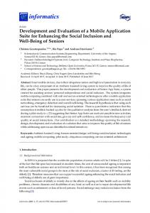
Fig. 1. DNA-binding specificity of modular zinc-finger proteins (ZFPs). (A-C)Sequence logos and position weight matrix (PWM) scores determined for 5⬘ (5p) or 3⬘ (3p) target sites for zebrafish (A) gata2a, (B) drosha and (C) nf1b. (D)Box plot depicting meta-analysis of PWM scores for each target site across all ZFPs for which bacterial one-hybrid (B1H) selections were successful. PWM scores for the previously described kdrl ZFPs are shown for reference. Whiskers indicate the largest (smallest) datum still within 1.5 interquartile range (IQR) of the upper (lower) quartile, where outliers are indicated as open circles.
19 of the remaining 20 ZFPs at lower stringency (≤2.5 mM 3-AT). For each ZFP a recognition motif (sequence logo) (Schneider and Stephens, 1990) and position weight matrix (PWM) were generated based on binding sites recovered following selection. In general, determined recognition motifs resembled the desired target motif, although there was variability in the overall quality of the PWM score of the target site across all ZFPs (see Fig. S2 in the supplementary material). For example, the 5p ZFP targeting gata2a properly specified all 9 bp and displayed among the highest PWM scores, suggesting excellent specificity for its target (Fig. 1A). Most ZFPs exhibited more modest specificity for their target, similar to the drosha (rnasen – Zebrafish Information Network) 3p ZFP (Fig. 1B), whereas several displayed weaker preference, such as the nf1b 5p (Fig. 1C). Overall, the distribution of PWM scores for the modular ZFP cassettes was similar to the engineered ZFPs successfully used to target the zebrafish kdrl locus (Fig. 1D) (Meng et al., 2008; Gupta et al., 2011), indicating that the ZFPs constructed using this archive generally display good recognition properties. We further analyzed the B1H binding site selection data to assess the quality of individual modules, allowing us to identify those with poor or context-dependent specificity. Overall, most modules displayed a strong preference for their recognition triplet based on individually derived PWM scores (Fig. 2A), although modules at fingers 1 and 3 display lower median scores than those at finger 2.
Development 138 (20)
This is likely to be due to fraying effects at the edges of the proteinDNA complex that reduce the specificity of the determinants at these positions (Choo, 1998). Comparison of PWM values across all individual modules revealed that those targeting purine-rich triplets were among the most robust, regardless of their finger position. For example, the GAG module consistently displayed excellent specificity in multiple finger positions in most ZFPs (Fig. 2B), as did modules targeting GAT, GGG and GGT (see Tables S5 and S6 in the supplementary material). We also identified four modules within the archive that displayed poor specificity at a specific position or in a context-dependent manner. For example, the TTG module displays context-dependent alterations in specificity. We originally utilized the TTG module at finger 3 in ZFPs targeting the zebrafish kdrl locus, where it displays moderate preference (NtG) for its triplet (Meng et al., 2008; Gupta et al., 2011), similar to the efnb2a 5p ZFP in this study (Fig. 2C). However, when it is placed at the finger 2 position adjacent to a Cterminal module bearing an RSD motif at positions –1, 1 and 2, the specificity shifts to a strong preference for a GNG binding site [Fig. 2C, zgc:66439 (clec14a – Zebrafish Information Network), 3p ZFP]. This dramatic alteration in specificity is not observed when the neighboring C-terminal module lacks an RSD motif (Fig. 2C, kif1b, 5p ZFP). Overall, the majority of analyzed modules have favorable recognition properties in multiple contexts, although several might specify incorrect triplets in particular contexts. In vivo activity of modular ZFNs We evaluated the ability of ZFNs constructed from our modular archive to induce lesions at a target site within the zebrafish genome. Twenty-nine pairs of ZFNs targeting 28 genes (see Table S7 in the supplementary material) were constructed from 76 ZFPs described above (see Table S5 in the supplementary material) by fusing them to engineered heterodimeric FokI nuclease domains (Miller et al., 2007; Szczepek et al., 2007). We assessed lesion frequency by deep sequencing the target region in normal embryos at 24 hpf following injection of mRNAs encoding ZFNs at an optimal dose (see Table S3 in the supplementary material). Eight of the 29 ZFNs displayed lesions frequencies of 1% or more in at least one set of injections (see Table S7 in the supplementary material). The previously characterized kdrl ZFNs were injected as a positive control and generated an in vivo lesion frequency of ~7%, similar to our previous observations using Illumina sequencing (Gupta et al., 2011). Germline transmission of ZFN-induced mutant alleles Deep sequencing is likely to underestimate the frequency of ZFNinduced lesions because of short read lengths, and assessment of somatic lesions in embryos may not reflect the frequency of
Fig. 2. Assessing the quality and behavior of individual ZFP modules. (A)Box plot depicting meta-analysis of PWM values for individual ZFP modules in each of the three positions in the Zif268 backbone. 5p (red diamonds) and 3p (blue circles) scores for fingers in the kdrl ZFPs are shown for reference. Whiskers indicate the largest (smallest) datum still within 1.5 interquartile range (IQR) of the upper (lower) quartile, where outliers are indicated as open circles. (B) Sequence logos for ZFP monomers recognizing GAG in each finger position. (C)Sequence logos for ZFPs containing TTG modules at the indicated finger position.
DEVELOPMENT
4558 RESEARCH ARTICLE
A zebrafish knockout kit
RESEARCH ARTICLE 4559
Table 1. Somatic lesion frequency and germline transmission rate of ZFN-induced mutagenic alleles ZFN ID
ZFNv1_68779 ZFNv1_53751 ZFNv1_27773 ZFNv1_5254 ZFNv1_5253 ZFNv1_87654 ZFNv1_19302 ZFNv1_94061 ZFNv1_83611 ZFNv1_37185 ZFNv1_53765 ZFNv1_25601
Gene
Somatic lesion frequency (%)
Frequency of founders (%)
chd5 kif1b braf nf1b nf1b gata2a nf1a ago2 cxcr4a numb-like kif1b gata3