human prostate cancer cells (LNCaP, DU145, and PC3) in relation to the expression of messenger RNAs (mRNAs) for 17ÃHSD types 1â4. These cell systems ...
0013-7227/97/$03.00/0 Endocrinology Copyright © 1997 by The Endocrine Society
Vol. 138, No. 11 Printed in U.S.A.
Expression of Different 17b-Hydroxysteroid Dehydrogenase Types and Their Activities in Human Prostate Cancer Cells* LUIGI A. M. CASTAGNETTA, GIUSEPPE CARRUBA, ADELE TRAINA, ORAZIA M. GRANATA, MONIKA MARKUS, MICHELE PAVONE-MACALUSO, CHARLES H. BLOMQUIST, AND JERZY ADAMSKI Institute of Oncology (L.A.M.C., G.C.) and Institute of Urology (M.P.-M.), Policlinico, University Medical School; and Experimental Oncology and Molecular Endocrinology Units, Palermo Branch of the National Cancer Institute of Genoa (L.A.M.C., A.T., O.M.G.), Cancer Hospital Center, Palermo, Italy; Max Planck Institute for Experimental Endocrinology (M.M.), Hannover, Germany; and GSFNational Research Center, Institute of Mammalian Genetics (J.A.), Neuherberg, Germany; and the Department of Obstetrics and Gynecology, Healthpartners for Environment and Health, St. PaulRamsey Medical Center (C.H.B.), St. Paul, Minnesota 55101-2595 ABSTRACT The 17b-hydroxysteroid dehydrogenase (17bHSD) enzyme system governs important redox reactions at the C17 position of steroid hormones. Different 17bHSD types (no. 1– 4) have been identified to date in peripheral human tissues, such as placenta, testis, and breast. However, there is little information on their expression and activity in either normal or malignant prostate. In the present work, we have inspected pathways of 17b-oxidation of either androgen or estrogen in human prostate cancer cells (LNCaP, DU145, and PC3) in relation to the expression of messenger RNAs (mRNAs) for 17bHSD types 1– 4. These cell systems feature distinct steroid receptor status and response to hormones. We report here that high expression levels of 17bHSD4 were consistently observed in all three cell lines, whereas even greater amounts of 17bHSD2 mRNA were detected solely in PC3 cells. Neither 17bHSD1 nor 17bHSD3 mRNAs could be detected in any cell line. From a metabolic standpoint, intact cell analysis showed a much lower extent of 17b-oxidation of both androgen [testosterone (T)] and estrogen [estradiol (E2)] in LNCaP and DU145 cells compared to PC3 cells, where a greater precursor degradation and higher formation rates of oxidized derivatives (respectively, androstenedione
and estrone) were observed. Using subcellular fractionation, we have been able to differentiate among 17bHSD types 1– 4 on the basis of their distinct substrate specificities and subcellular localization. This latter approach gave rise to equivalent results. PC3 cells, in fact, displayed a high level of microsomal activity with a low E2/T activity ratio and approximately equal apparent Km values for E2 and T, suggesting the presence of 17bHSD2. Dehydrogenase specific activity with both E2 and T was also detected, although at lower levels, in LNCaP and DU145 cells. No evidence for reductase activity could be obtained in either the soluble or microsomal fraction of any cell line. As comparable expression levels of 17bHSD4 were seen in the three cell lines, 17bHSD2 is a likely candidate to account for the predominant oxidative activity in PC3 cells, whereas 17bHSD4 may account for the lower extent of E2 oxidation seen in both LNCaP and DU145 cells. This is the first report on the expression of four different 17bHSD types in human prostate cancer cells. It ought to be emphasized that for the first time, analysis of different 17bHSD activities in either intact or fractionated cells harmonizes with the expression of relevant mRNAs species. (Endocrinology 138: 4876 – 4882, 1997)
T
efficiency as the reduction of E1 (3, 4). 17bHSD2 is a microsomal enzyme of 387 amino acids that slightly favors the oxidation over the reduction of either androgen or estrogen and is expressed at high levels in the human placenta (5, 6). The microsomal 17bHSD3, which is exclusively expressed in human testis, consists of 310 amino acids and is responsible for the reduction of estrogens and androgens (7). Recently, cDNAs encoding for human, mouse, and porcine 17bHSD4 have been identified (8 –11). They share 85% amino acid identity and metabolize estrogens and androgens very efficiently, displaying a 400-fold preference for steroid oxidation. A 3-kb messenger RNA (mRNA) codes for peroxisomal 80-kDa (737 amino acids) protein, featuring domains that are absent in the other 17bHSDs. We have shown that both the 80-kDa and its N-terminal 32-kDa (amino acids 1–323) fragment are able to perform the dehydrogenase reaction not only with steroids at the C17 position, but also with 3hydroxyacyl coenzyme A (12). Evidence for key steroid enzymes in human prostate tis-
HE REDOX reactions at position C17 of the steroid molecule represent a key step in both biosynthesis and metabolism of gonadal steroids, either androgen or estrogen (1, 2). In particular, the 17b-hydroxysteroid dehydrogenase (17bHSD) enzyme system presides over important steroid interconversions, including that of testosterone (T), androstenedione (D4Ad), estradiol (E2), and estrone (E1). To date at least four different human 17bHSDs have been identified, their complementary DNAs (cDNAs) cloned, and their amino acid sequences deduced. The soluble 17bHSD1, consisting of 327 amino acids, was originally isolated from human placenta and performs the oxidation of E2 at the same Received March 7, 1997. Address all correspondence and requests for reprints to: Dr. Luigi A. M. Castagnetta, Istituto di Oncologia, Universita` di Palermo, Via Marchese Ugo 56, 90141 Palermo, Italy. * This work was supported in part by the Italian Association for Cancer Research, the National Research Council (Special Project Aging, Contract 95.01017.PF40; to M.P.-M.), and Deutsche Forschungsgesellschaft Grant AD127/1–1 (to J.A.).
4876
17bHSD TYPES IN HUMAN PROSTATE CANCER
4877
TABLE 1. Primers used for PCR amplification Enzyme
Position
Forward primer
Position
Reverse primer
17bHSD1 17bHSD2 17bHSD3 17bHSD4
55–74 112–131 119–139 95–122
GGCCTGCACTTGGCCGTACG GGATCTGCCTGGCTGTCCCC GCGTGAGATTCTTCCAGATGTG GAATAATAGATGTTGTGG
604– 624 697–717 699–720 674– 695
CAGCACCTCCTCTGGGCTGCC CTTTGGATTTTCTAAGAAGAG CTGGATGATGACTTCTTTTGC AAGCCAAAGGACAAGAGGTGC
Primer sequences are given 59339.
sues has been repeatedly reported (13, 14). However, little is known of the expression and function of the different 17bHSD types in either normal or malignant prostate gland. Most previous studies have used crude extracts of homogenized tissues to compare the activities of steroid enzymes in normal, hyperplastic, and carcinomatous human prostate (15). Although this in vitro characterization of enzymes (as either purified or expressed proteins) may represent a versatile tool in understanding their potential physiological roles, in vivo conditions (including subcellular localization, pH, concentrations of cofactors and substrates, feedback mechanisms, etc.) may well drive the reactions with different kinetics and oxidation/reduction preferences. This is also reflected in the discrepancies often emerging between estimates of intratissue amounts of individual steroids and the extent of relevant enzyme activities, as measured by crude extract methods. After optimization of liquid chromatographic procedures, we established an original intact cell analysis that allows measurement of several enzyme activities of steroid metabolism in living cultured cells (16 –18). This in vivo approach, while retaining cells in a physiological microenvironment, enables to use close to physiological amounts of steroid precursors and preserves conversion rates and direction of steroid metabolism, which could be otherwise disturbed by tissue homogenization or simplification to few enzymes in vitro. In the present work, we have used both intact cell analysis and subcellular fractionation assays to inspect pathways of 17b-oxidation of androgens and estrogens in human prostate cancer cell lines that feature distinct steroid receptor contents and responses to hormones; these are used as helpful model systems for either hormone-sensitive or refractory prostate cancer. We compared the metabolic conversion rates of cells in culture with the expression of mRNAs for the four types of 17bHSD identified to date to verify which enzyme(s) is responsible for the observed reactions. The results of these studies are reported herein. Materials and Methods Chemicals All chemicals were of analytical grade. [a-32P]deoxy-CTP was obtained from Hartmann Analytic (Braunschweig, Germany).
Cell culture LNCaP.FGC (passage 19), DU145 (passage 59), and PC3 (passage 17) human prostate cancer cells were obtained from the American Type Culture Collection (Rockville, MD). For routine maintenance, cells were grown in RPMI 1640 medium, supplemented with 10% FCS, 2 mm l-glutamine, and antibiotics (100 IU/ml penicillin, 100 mg/ml streptomycin, and 0.25 mg/ml amphotericin B), at 37 C in a humidified atmosphere of 5% CO2 in air. Cells were periodically tested for mycoplasma contamination. For all experiments, the passage number of cells in cul-
FIG. 1. Extent of androgen and estrogen 17b-oxidation in PC3 (closed bars), LNCaP (dotted bars), and DU145 (open bars) human prostate cancer cells. Cells were incubated for 24 h in the presence of 1 nM tritiated T or E2, and the formation of relevant oxidized derivatives (D4Ad or E1, respectively) was measured by means of reverse phase HPLC and on-line radioactive detection. Values represent the average percent conversion 6 SD from three different experiments performed in triplicate. ture was kept in a narrow range (LNCaP, 22–25; DU145, 63– 67; PC3, 19 –23).
Steroid metabolism The methodological procedures used to assess patterns of steroid metabolism in in vitro systems were extensively described previously (19 –21). Cells (0.5–2 3 106) were plated onto 60-mm cell culture dishes and left undisturbed for 24 – 48 h. After two washes in PBS-A (170 mm NaCl, 3.4 mm KCl, and 2 mm Na2PO4, pH 7.2), the medium was substituted with FCS-free, phenol red-free RPMI medium to avoid interfering factors that might modify the metabolic ability of the cells. After an additional 24 h, medium was replaced with the same medium containing 1 nm radioactive T ([1,2,6,7-3H]T; SA, 92.1 Ci/mmol; DuPont de Nemours Italiana, Milan, Italy) or E2 ([6,7-3H]E2; SA, 48 Ci/mmol; DuPont) used as precursors. Both steroids were periodically checked and purified using HPLC before experimental use. After 24-h incubation, medium was transferred to sterile plastic tubes (Costar, Cambridge, MA) and stored at 280 C until analysis. For time-course experiments, triplicate dishes (5 3 105 cells/dish) were incubated in the presence of 1 nm labeled T for 30 min and 2, 8, and 24 h under exactly the same experimental conditions. Medium and cells were thus processed as described below.
Extraction procedures Extraction of both conjugate and free steroids was carried out, as previously described (19, 20), from the incubation medium as it has been shown to contain proportionally greater amounts of radioactive metabolites than those present in the cells (22, 23). Briefly, medium aliquots (1 ml) were transferred to scintillation vials to assess the total radioactivity. Before any sample manipulation, all glassware was coated with 4 mg of the same radioinert steroid to minimize radioactivity losses. Extracts of either conjugate or hydrolyzed steroids were finally dried and stored at 220 C until chromatographic analysis.
17bHSD TYPES IN HUMAN PROSTATE CANCER
4878
Endo • 1997 Vol 138 • No 11
TABLE 2. Testosterone metabolism in human prostate cancer cells after 24-h incubation
PC3 LNCaP DU145
T
DHT
3adiol
3bdiol
D4Ad
5aAdione
A
EpiA
11.4 6 0.9 816.6 6 33.4 728.6 6 22.1
ND 8.2 6 0.3 111.4 6 6.4
ND 8.8 6 0.2 28.6 6 1.9
ND ND 6.8 6 0.2
653.2 6 47.1 44.1 6 2.1 6.6 6 0.3
217.4 6 19.9 ND 4.6 6 0.1
4.2 6 0.3 ND 3.4 6 0.1
13.6 6 0.9 ND ND
Data represent average (6SD); (femtomoles per ml) values obtained from four different experiments, performed in triplicate, after correction for equal cell numbers. ND, Not detectable.
Chromatographic analysis
RNA blotting
The dried extracts were resuspended in 30 ml acetonitrile (androgens) or in a mixture consisting of 10 ml acetonitrile, 10 ml acetic acid (0.2 m), and 10 ml equilin (estrogens), with the latter used as internal standard. Twenty microliters of the resulting solution were used for HPLC analysis, while 5 ml were measured in a b-counter to quantify the radioactivity extracted for each steroid fraction; the extraction efficiency was calculated as reported previously (19). Extracted steroids were separated under isocratic conditions on HPLC in the reverse phase mode and quantified using an on-line Flo-One/Beta (500TR) three-channel radioactive detector (Camberra Packard, Meriden, CT). All of the chromatographic procedures used have been previously established and optimized in our laboratories (18). Precursor degradation and formation of metabolic products were expressed either as a percentage of the conversion rates or as femtomoles per ml. Data were normalized for total radioactivity and/or corrected for equal cell numbers when appropriate.
Term placenta tissue was collected after normal delivery, and testicular tissue was obtained from patients orchidectomized for prostate cancer. Extraction of mRNA was performed directly from tissues or PBS-washed cells (3 3 107) using oligo(deoxythymidine) Dynabeads (Dynal, Hamburg, Germany) according to the manufacturer’s instructions. Size fractionation of polyadenylase-enriched RNA was achieved by electrophoresis through a 1% (wt/vol) agarose gel, followed by capillary blotting to a Hybond-N membrane (Schleicher and Schuell, Dassel, Germany) (9). Blotting procedures were performed in duplicate, using two different sets of cells for any cell line. Loading of mRNA (10 mg from cell lines, 3 mg from testes, and 5 mg from placenta) was checked using ethidium bromide staining and subsequent UV visualization. The membranes were prehybridized in a solution containing 100 mg/ml denatured salmon sperm DNA, 50% formamide, 5 3 Denhardt’s solution, 0.3% SDS, and 6 3 SSPE (0.1 3 SSPE 5 15 mm NaCl, 1 mm sodium phosphate, and 0.1 mm EDTA, pH 7.4; for 12 h at 42 C), and hybridization (in the same solution except without Denhardt’s solution) was carried out with a-32P-labeled probes for 16 h at 42 C. Blots were sequentially hybridized with probes for human glyceraldehyde-3-phosphate dehydrogenase (GAPDH; Clontech, Heidelberg, Germany), four different types of 17bHSD, and again for GAPDH. The membranes were then washed to a final stringency of 0.1 3 SSPE containing 0.3% SDS at 42 C. The blots were exposed to x-ray X-Omat AR film (Eastman Kodak, Rochester, NY) with an intensifying screen for 12 h after quantification of signals by a Fuji BAS 1000 phosphoimager. Before every hybridization the membranes were stripped of bound radioactive DNA three times in 0.001 3 SSPE-0.3% SDS (wt/vol) at 80 C. The efficiency of stripping was checked by phosphoimager. Signals for GAPDH remained unchanged after five hybridizations.
Subcellular fractionation Cell monolayers were washed twice with PBS-A. Cells were then collected by centrifugation, suspended in 0.04 m potassium phosphate buffer (pH 7.0) containing glycerol (20% vol/vol) and 10 mm EDTA, and gently homogenized by hand in a glass Dounce homogenizer (Kontes Co., Vineland, NJ). Cell homogenates were centrifuged at 1,000 3 g for 10 min to remove cell debris and then at 105,000 3 g at 4 C for 40 min. The resulting supernatant was saved as the cytosol. The pellet, designated microsome, was suspended in 0.04 m potassium phosphate (pH 7.0) containing 10 mm EDTA but lacking glycerol. Both fractions were stored at 4 C until analysis.
17bHSD activity 17bHSD activity was assayed under conditions that differentiate among type 1, 2, 3, and 4 enzymes, as described previously (24). Briefly, 10-ml aliquots of cytosols or microsomes were combined with 10-ml aliquots of the reaction mixture to give 0.5 mm NAD, 1.0 mm [3H]E2 or [3H]T, and 0.1 m bicine (pH 9.0) for the dehydrogenase assay and 0.5 mm NADH, 1.0 mm [3H]E1 or [3H]D4Ad and 0.1 m HEPES (pH 7.2) for the measurement of reductase activity. For the determination of apparent Km (Kmapp) and apparent maximum velocity (Vmapp), 0.1 m HEPES, pH 7.2, was used for both dehydrogenase and reductase assays. Reactions were run at 37 C and stopped by transferring the reaction mixture to the preadsorbent layer of a TLC plate (Silica Gel HL, Analtech, Newark, DE). After the addition of 30 ml of an unlabeled carrier steroid (4.0 mg/ml) in ethanol, the spots were allowed to dry, and the plate was developed in benzene-acetone (4:1, vol/vol). Substrate and product were located by a light spraying with water. After drying, the spots were scraped into 10 ml Ecolumn (ICN Research Products Division, Costa Mesa, CA) for liquid scintillation counting. Specific activity (nanomoles per mg protein/30 min) was calculated from the counts per min recovered in product as a percentage of the total counts per min recovered in substrate and product, as reported previously (25).
Protein determination Protein content was measured by the method of Markwell et al. (26). BSA was used as the standard.
Data analysis The Kmapp and Vmapp values were estimated according to the graphic method of Cornish-Bowden and Eisenthal (27).
Hybridization probes Specific DNA probes for 17bHSDs of approximately 600 bases were PCR amplified using cDNA of 17bHSD1 (provided by Dr. J. Simard), 17bHSD2 and 17bHSD3 (provided by Dr. S. Andersson), and 17bHSD4 (10) as templates. The sequences of the primers used are given in the Table 1. Pfu DNA polymerase (Stratagene, Heidelberg, Germany), an annealing temperature of 60 C, and 35 cycles were used. To quantify the amounts of mRNA on the blots, a human GAPDH probe (Clontech) consisting of a 1.1-kilobase (kb) EcoR I/XhoI fragment of GAPDH cDNA was employed. The probes were labeled with a random hexanucleotide Prime-It RmT Kit (Stratagene) and had specific activities greater than 109 cpm/mg DNA.
Results
Inspection of 17bHSD oxidative activity was firstly carried out using intact cell analysis through incubation of cultured cells with physiological amounts of a labeled steroid precursor. The reliability of the data was strengthened by the high extraction efficiency and recovery of radioactivity values (.90% in either case). The results obtained revealed that either androgen (T) or estrogen (E2) oxidation was remarkably and consistently different in the three cell lines studied (see Fig. 1). As far as T metabolism is concerned (Table 2), PC3 cells showed large precursor degradation (,2% unconverted T after 24 h) to
17bHSD TYPES IN HUMAN PROSTATE CANCER
4879
TABLE 3. Estradiol metabolism in human prostate cancer cells after 24-h incubation
PC3 LNCaP DU145
E2
E1
16aOHE1
2MeOE2
28.6 6 1.36 574.8 6 5.17 648.9 6 11.2
613.8 6 7.51 92.4 6 3.48 46.4 6 5.62
25.9 6 1.83 16.7 6 0.71 ND
ND 8.39 6 0.27 ND
Data represent average (6SD); (femtomoles per ml) values obtained from three different experiments, performed in triplicate, after correction for equal cell numbers. ND, Not detectable.
FIG. 2. The time course of T metabolism in PC3 (A), LNCaP (B), and DU145 (C) human prostate tumor cells. Cells (5 3 105) were incubated in the presence of 1 nM [3H]T for 0.5, 2, 8, 12, and 24 h. Each data point represents the mean 6 SD of duplicate experiments, performed in triplicate, after correction for equal cell numbers. For abbreviations, see text. E, [3H]T; F, D4Ad; M, DHT; ‚, 3a-diol; f, 5aAdione; Œ, androsterone (A); , epiandrosterone (EpiA).
yield marked amounts (.70%) of D4Ad and its derivatives of the 17-keto series, 5a-androstenedione (5aAdione), androsterone, and epiandrosterone (in total, ;25%). In contrast, both LNCaP and DU145 cells gave rise to limited T conversion rates into D4Ad, with the latter never exceeding 5% of all radioactive androgens. Peculiarly, the formation of dihydrotestosterone (DHT) and its derivatives 3a- and 3bandrostenediols (3a/3b-diols) varied greatly in the three cell lines. In time-course experiments, PC3 cells did not show measurable DHT or 3a/3b-diol production at any incubation time, whereas the prevalence of the oxidative pathway leading to D4Ad formation was confirmed (see Fig. 2A). In fact, T was increasingly converted into D4Ad; an appreciable amount (.8%) of this metabolite was found after only 30-min incubation. A proportional increase in 5aAdione was also seen at 8 and 24 h. Formation of D4Ad plus 5aAdione was inversely and significantly related to the proportion of metabolized T (r 5 20.9706; P , 0.03, by Spearman correlation test). In contrast, the time course of T metabolism in both LNCaP and DU145 cells revealed low T degradation rates (,8% and ,18%, respectively). Overall, D4Ad formation did not exceed 5%, whereas relatively greater amounts of both DHT and 3a/3b-diols were found in DU145 cells (in total, nearly 15%) with respect to LNCaP cells (only 2%; see Fig. 2, B and C). As shown in Table 3, oxidation of E2 to E1 was strikingly greater in PC3 cells (nearly 92% by 24 h) than in LNCaP (14%) or DU145 (7%) cells. The proportion of undegraded precursor (E2) at 24 h ranged from 81–94% in LNCaP and DU145 cells, whereas it remained below 4% in PC3 cells. A small proportion of 16a-hydroxy-E1 was also seen in either LNCaP (2.4%) or PC3 (3.8%) cells, whereas small amounts of 2methoxy-E2 were found in LNCaP cells only. Likewise for T, time-course experiments reinforced 24-h data on estrogen metabolism; an increasingly greater extent of E2 oxidation into E1 was seen in PC3 cells over time (not shown). Subcellular fractions were assayed for 17bHSD activity under conditions that differentiate among 17bHSD types 1, 2, 3, and 4 (28). The highest level of activity with both E2 and T was detected in microsomes from PC3 cells. The E2/T activity ratio was approximately 1 (see Table 4). The Kmapp and Vmapp values were comparable, as were the Kmapp/ Vmapp ratios (Table 5). 17bHSD activity with E2 and T was detected in the other cell lines as well, but at a lower level. Dehydrogenase specific activity for both E2 and T was highest in the microsomal fraction from LNCaP cells. The cytosolic and microsomal specific activities of DU145 cells were comparable, being the lowest among the three cell lines. Expression of transcripts for 17bHSD types 1– 4 was scrutinized using Northern blot analysis of cellular mRNA under
17bHSD TYPES IN HUMAN PROSTATE CANCER
4880
Endo • 1997 Vol 138 • No 11
TABLE 4. 17bHSD specific activities with E2 and T of cytosols and microsomes from human prostate cancer cells 17bHSD SA (nmol/mg proteinz30 min) T3D4Ad
E23 E1
PC3 LNCaP DU145
Cytosol
Microsome
Cytosol
Microsome
0.108 6 0.030 0.034 6 0.037 0.054 6 0.004
1.110 6 0.026 0.130 6 0.027 0.041 6 0.018
0.190 6 0.052 0.025 6 0.021 0.018 6 0.008
1.112 6 0.032 0.102 6 0.032 0.084 6 0.035
The values are the mean (6SE) of duplicate assays. Reaction mixtures containing 0.08 substrate were incubated at 37 °C for 60 min (microsome) or 180 min (cytosol). TABLE 5. Kinetic parameters for 17bHSD dehydrogenase activity of PC3 microsomes with E2 and T
E23 E1 T3D4Ad
Kmapp (mM)
Vmapp (nmol/mg proteinz30 min)
Vm/Km
2.7 3.2
1.6 1.9
0.6 0.6
Reaction mixtures contained 0.08 M HEPES (pH 7.2), 0.5 mM NAD, and 1.0, 3.0, 6.0, 11.0, or 21.0 mM steroid substrate. Assays were run at 37 °C for 150 min.
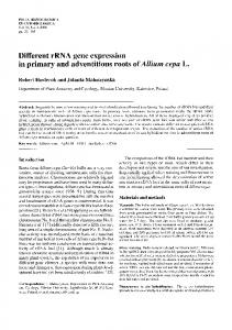
stringent conditions. Cells were kept under exactly the same experimental conditions as those used to measure 17bHSD activity and then subjected to mRNA isolation. The fast isolation procedure and the relatively high amounts of mRNA (10 mg) used ensured low detection limits; meanwhile, equal lengths of hybridization probes allowed for a direct comparison of the expression levels of the mRNAs. Human testis and placenta tissues provided mRNA for positive controls. As shown in Fig. 3A, remarkable expression levels of 17bHSD2 mRNA were seen in PC3 cells, whereas no signal for 17bHSD2 was revealed in either LNCaP or DU145 cells; this could not be due to uneven loading of mRNAs on the blots, as excluded by either UV visualization of ethidium bromide-stained gels or quantification at laser densitometry of GAPDH signals. In addition, all three cell lines expressed comparable and significant amounts of a 2.9-kb mRNA species for 17bHSD4. In the same experiments, substantial amounts of 17bHSD1 and 17bHSD3 were detected in placenta and testes, respectively. However, neither 17bHSD1 nor 17bHSD3 mRNAs could be detected in any cell line, not even after exposure of blots for 1 week (not shown). Duplicate experiments gave rise to the same results. Densitometric estimates of relative signal intensities after normalization for GAPDH are illustrated in Fig. 3B. As can be seen, LNCaP cells expressed abundant 17bHSD4 mRNA, whereas lower levels were found in DU145 and, especially, PC3 cells. In the latter, the amounts of 17bHSD2 mRNA were nearly twice as high as those observed for 17bHSD4. Discussion
Data from previous studies on 17bHSD expression in human prostate tissues and cells have been sparse and even inconsistent. In a recent paper, Pylkkanen and colleagues (29) reported that both newborn and adult human prostate weakly stained for 17bHSD1, and that its NADPH-dependent reductase activity was present in cell-free homogenates of prostate tissues. By contrast, other investigators failed to detect 17bHSD1 mRNA in primary cultures of benign prostatic hyperplasia and prostate cancer tissues (30). The re-
M
bicine (pH 9.0), 0.5 mM NAD, and 1.0 mM steroid
searchers found that prostate cultured epithelial cells and, to a lesser extent, fibroblasts express appreciable amounts of 17bHSD2 transcript, whereas it was undetectable in prostate tissue homogenates. Equally, the presence of a 2.2-kb mRNA species coding for 17bHSD2 has been previously reported in the human prostate (31). This observation is also in accordance with the results of Wu et al. (5). We have investigated the expression and activity of 17bHSDs in cultured human prostate cancer cells (PC3, LNCaP, and DU145). These cell lines have been previously characterized in our laboratories for their growth response to both androgen and estrogen as well as for their respective receptor contents. In particular, LNCaP cells contain both androgen and estrogen receptors; their proliferative activity is significantly stimulated by either steroid (32, 33). In contrast, androgen receptor-negative PC3 cells and DU145 cells fail to respond to androgens; this despite DU145 cells retain high affinity sites and apparently intact capacity for androgen binding (32). In the present work we have seen remarkable expression of 17bHSD2 in androgen-nonresponsive PC3 cells, whereas no specific mRNA could be detected in DU145 or in hormone-responsive LNCaP cells. On the other hand, no specific transcript for 17bHSD1 (1.3 or 2.3 kb) or 17bHSD3 (;1.3 kb) was found in any cell line, even after long exposure of blots, thus confirming previous reports. The authenticity of these observations is proven by strong parallel hybridization signals in placenta (17bHSD1) and testes (17bHSD3). Although 17bHSD1 and -2 could be detected in many tissues (6, 7, 31), 17bHSD3 was observed only in testis (5). High levels of 17bHSD4 were seen in all three cell lines studied; the amounts of mRNA were comparable to those we previously observed in kidney and much greater than those in placenta or normal prostate (10). Recent studies on 17bHSD4 expression in porcine testis have localized the enzyme in peroxisomes of Leydig cells (34). As the expression levels of 17bHSD4 are comparable in the three cell lines, 17bHSD2 is a likely candidate for the predominant oxidative activity seen in PC3 cells. 17bHSD4 may be of minor importance in this respect, but it may account for the lesser extent of E2 oxidation observed in both LNCaP and DU145 cells. As the latter cell lines did not express detectable 17bHSD2 mRNA, the enzymatic activity responsible for T oxidation remains to be identified. From a metabolic standpoint, striking differences emerged in the extent of 17b-oxidation of both androgen (T) and estrogen (E2) using intact cell analysis; the extent of reaction was, in fact, significantly greater in PC3 cells, lower in LNCaP cells, and very low DU145 cells. This evidence was repro-
17bHSD TYPES IN HUMAN PROSTATE CANCER
FIG. 3. Distribution of mRNAs for human 17bHSD. A. Northern blotting analysis. Polyadenylated RNA isolated from human tissues (testis, 3 mg; placenta, 5 mg) and prostate cancer cell lines (10 mg) was subjected to electrophoresis in the presence of formaldehyde and blotted to a nylon membrane that was sequentially hybridized as indicated to the left. Specific 32P-labeled probes of 600 bp, corresponding to the most N-terminal parts of 17bHSD1 to -4 enzymes, were amplified with PCR. Human GAPDH probe, consisting of a 1.1-kb EcoR I/HindIII fragment of GAPDH cDNA, was used to quantify the amounts of mRNA on the blots. The molecular mass of the observed RNA is given to the right. B. Signal quantification. After each hybridization, the membranes were subjected to quantification of signals using a Fuji BAS 1000 phosphoimager. Signal intensities are given after normalization to GAPDH levels.
ducible and consistent for both T and E2 metabolism. It is worth mentioning that the large E2 oxidation seen in PC3 cells combines with the formation of 16a-hydroxy-E1, exactly as we have observed in other hormone-unresponsive human cancer cell lines, in which production of this estrogen derivative is much enhanced by FCS or its major component albumin (35).
4881
The assay conditions used in this study for estimating 17bHSD activity on cell homogenates differentiate among the four 17bHSD types on the basis of their differing substrate specificities and subcellular localization (28). 17bHSD1 is a cytosolic enzyme with an E2/T activity ratio of approximately 100 (25). In contrast, 17bHSD2 is a microsomal enzyme with an E2/T activity ratio of approximately 1 (5). For PC3 cells, the high level of microsomal activity compared with cytosol, the low E2/T activity ratio, and the approximately equal Kmapp values for E2 and T are consistent with the presence of type 2 17bHSD. Although less than that of PC3 cells, a significant level of activity with E2 and T was detected in microsomes from LNCaP cells. The low E2/T activity ratio is suggestive of the presence of 17bHSD2. However, the absence of type 2 17bHSD mRNA and the detection of mRNA for the type 4 isoform suggest that this low ratio may be misleading. It is noteworthy in this regard that 17bHSD4 is highly specific for E2, with little or no affinity for T (10). The presence of a high level of 17bHSD4 mRNA in LNCaP and DU145 cells suggests that the microsomal dehydrogenase activity found with E2 may be due to the type 4 enzyme. A comparable level of microsomal activity with T in the absence of 17bHSD2 mRNA raises the interesting possibility of the presence in these cancer cells of an as yet unidentified enzyme reactive with T. To our knowledge, this is the first report on the expression of the four different 17bHSD types in human prostate cancer cells. Furthermore, for the first time, in vivo measurement of 17bHSD activity harmonizes with the amounts of relevant 17bHSD transcripts, as demonstrated by Northern blot analysis. The apparent lack of both type 1 and 3 enzyme transcripts, precluding any reductive activity by 17bHSD enzymes, deserves a deeper scrutiny, using more sensitive assays for specific mRNAs and proteins. All of this evidence would imply that distinct 17bHSDs may be differently regulated in cells with different sensitivities to sex steroids, eventually leading to a differential accumulation of biologically active hormones. Taking into account the multifarious response to estrogen we have observed in these prostate tumor cells (33, 36), this could be relevant to both growth and progression of human prostatic carcinoma. Further studies should ascertain whether a prevalent 17b-oxidation driven by 17bHSD2 and/or 17bHSD4 associates with hormone refractory prostate cancer, in vivo. Acknowledgments We thank Dr. J. Simard, CHUL (Quebec, Canada), for providing cDNA for 17bHSD1, and Dr. S. Andersson, University of Texas (Dallas, TX), for cDNA for 17bHSD2 and -3. We thank Dr. I. Khalifa, Kreiskrankenhaus (Peine, Germany) for providing human placenta, and Dr. S. Pfliege-Bruss, Zentrum for Dermatologie und Andrologie (Giessen, Germany), for human testis tissues. The authors are grateful to the Centro Interdipartimentale di Ricerca in Oncologia (CIROC) for its continuing support.
References 1. Andersson S 1995 17b-Hydroxysteroid dehydrogenases: isozymes and mutations. J Endocrinol 146:197–200 2. Reed MJ 1991 Oestradiol 17b-hydroxysteroid dehydrogenase: its family and function. J Endocrinol 129:163–165 3. Peltoketo H, Isomaa V, Ma¨entausta O, Vihko R 1988 Complete amino acid
4882
4.
5.
6. 7.
8. 9.
10. 11. 12.
13. 14. 15. 16. 17. 18. 19.
17bHSD TYPES IN HUMAN PROSTATE CANCER
sequence of human placental 17b-hydroxysteroid dehydrogenase deduced from cDNA. FEBS Lett 239:73–77 Luu-The V, Labrie C, Zhao HF, Coue¨t J, Lachance Y, Simard J, Leblanc G, Coˆte´ J, Be´rube´ D, Gagne´ R, Labrie F 1989 Characterization of cDNAs for human estradiol 17b-dehydrogenase and assignment of the gene to chromosome 17: evidence of two mRNA species with distinct 59-termini in human placenta. Mol Endocrinol 3:1301–1309 Wu L, Einstein M, Geissler WM, Chan HK, Elliston KO, Andersson S 1993 Expression cloning and characterization of human 17b-hydroxysteroid dehydrogenase type 2, a microsomal enzyme possessing 20a-hydroxysteroid dehydrogenase activity. J Biol Chem 268:12964 –12969 Casey ML, MacDonald PC, Andersson S 1994 17b-Hydroxysteroid dehydrogenase type 2: chromosomal assignment and progestin regulation of gene expression in human endometrium. J Clin Invest 94:2135–2141 Geissler WM, Davis DL, Wu L, Bradshaw K, Patel S, Mendonca BB, Elliston KO, Wilson JD, Russell DW, Andersson S 1994 Male pseudohermaphroditism caused by mutations of testicular 17b-hydroxysteroid dehydrogenase 3. Nat Genet 7:34 –39 Adamski J, Husen B, Marks F, Jungblut PW 1992 Purification and properties of oestradiol 17b-dehydrogenase extracted from cytoplasmic vesicles of porcine endometrial cells. Biochem J 288:375–381 Leenders F, Husen B, Thole HH, Adamski J 1994 The sequence of porcine 80 kDa 17b-estradiol dehydrogenase reveals similarities to the short chain alcohol dehydrogenase family, to actin binding motifs and to sterol carrier protein 2. Mol Cell Endocrinol 104:127–131 Adamski J, Normand T, Leenders F, Monte´ D, Begue A, Ste´helin D, Jungblut PW, de Launoit Y 1995 Molecular cloning of a novel widely expressed human 80 kDa 17b-hydroxysteroid dehydrogenase IV. Biochem J 311:437– 443 Normand T, Husen B, Leenders F, Pelczar H, Baert J-L, Begue A, Flourens A-C, Adamski J, de Launoit Y 1995 Molecular characterization of mouse 17b-hydroxysteroid dehydrogenase IV. J Steroid Biochem Mol Biol 55:541–548 Leenders F, Tesdorpf JG, Markus M, Engel T, Seedorf U, Adamski J 1996 Porcine 80 kDa protein reveals intrinsic 17b-hydroxysteroid dehydrogenase, fatty acyl-CoA-hydratase/dehydrogenase and sterol transfer activities. J Biol Chem 271:5438 –5442 Bartsch W, Klein H, Schiemann U, Bauer HW, Voigt KD 1990 Enzymes of androgen formation and degradation in the human prostate. Ann NY Acad Sci 595:53– 66 Klein H, Bressel M, Kastendieck H, Voigt KD 1991 Biochemical endocrinology of prostate cancer. In: Voigt KD, Knabbe C (eds) Endocrine-Dependent Tumors. Raven Press, New York, pp 131–163 Krieg M, Bartsch W, Janssen W, Voigt KD 1979 A comparative study of binding, metabolism and endogenous levels of androgens in normal, hyperplastic and carcinomatous human prostate. J Steroid Biochem 11:615– 624 D’Agostino G, Mitchell F, Castagnetta L, O’Hare MJ 1984 Solvent optimization for RP-HPLC of polar adrenal steroids using computer predicted retention for a modified COF procedure. J Chromatogr 305:13–26 D’Agostino G, Castagnetta L, Mitchell F, O’Hare M 1985 Computer-aided mobile phase optimization and chromatogram simulation in high-performance liquid chromatography. J Chromatogr 338:1–23 Castagnetta L, Granata OM, Lo Casto M, Calabro` M, Arcuri F, Carruba G 1991 Simple approach to measure metabolic pathways of steroids in living cells. J Chromatogr 572:25–39 Castagnetta L, Granata OM, Polito L, Blasi L, Cannella S, Carruba G 1994 Different conversion metabolic rates of testosterone are associated to hormonesensitive status and -response of human prostate cancer cells. J Steroid Biochem Mol Biol 49:351–357
Endo • 1997 Vol 138 • No 11
20. Castagnetta L, Granata OM, Farruggio R, Cannella S, Montesanti A, Oliveri G, Sorci C, Mesiti M, Carruba G 1995 Oxidative and reductive pathways in hormone responsive and non-responsive human breast cancer cells in vitro. J Steroid Biochem Mol Biol 53:367–374 21. Castagnetta L, Montesanti A, Granata OM, Oliveri G, Sorci C, Amodio R, Liquori M, Carruba G 1995 17b-Hydroxysteroid dehydrogenase activity in endometrial cancer cells: different metabolic pathways of estradiol in hormone-responsive and non-responsive intact cells. J Steroid Biochem Mol Biol 55:573–579 22. Castagnetta L, Granata OM, Lo Casto M, Miserendino V, Calo` M, Carruba G 1986 Estrone conversion rates by human endometrial cancer cell lines. J Steroid Biochem 25:803– 809 23. Adams JB, Phillips NS, Hall R 1988 Metabolic fate of estradiol in human mammary cancer cells in culture: estrogen sulfate formation and cooperativity exhibited by estrogen sulfotransferase. Mol Cell Endocrinol 58:231–242 24. Blomquist CH, Leung BS, Zhang R, Zhu Y, Chang P 1995 Properties and regulation of 17b-hydroxysteroid dehydrogenase oxidoreductase of OVCAR-3, CAOV-3 and A431 cells: effects of epidermal growth factor, estradiol and progesterone. J Cell Biochem 59:409 – 417 25. Blomquist CH, Bealka D, Hensleigh HC, Tagatz G 1994 A comparison of 17b-hydroxysteroid oxidoreductase type 1 and type 2 activity of cytosol and microsomes from human term placenta, ovarian stroma and granulosa-luteal cells. J Steroid Biochem Mol Biol 49:183–189 26. Markwell MK, Haas SM, Tolbert NE, Bieber LL 1981 Protein determination in membrane and lipoprotein samples: manual and automated procedures. Methods Enzymol 72:296 –303 27. Cornish-Bowden A, Eisenthal R 1978 Michaelis constant and maximum velocity from the direct linear plot. Biochim Biophys Acta 523:268 –272 28. Blomquist CH 1995 Prediction of multiple forms of 17b-hydroxysteroid dehydrogenase. J Steroid Biochem Mol Biol 55:515–524 29. Pylkkanen L, Santti R, Salo L, Maentausta O, Vihko R, Nurmi M 1996 Immunohistochemical localization of estrogen-specific 17b-hydroxysteroid oxidoreductase in the human and mouse prostate. Prostate 25:292–300 30. Delos S, Carsol J-L, Ghazarossian E, Raynaud J-P, Martin P-M 1995 Testosterone metabolism in primary cultures of human prostate epithelial cells and fibroblasts. J Steroid Biochem Mol Biol 55:375–383 31. Luu-The V, Labrie C, Simard J, Lachance Y, Zhao H-F, Coue¨t J, Leblanc G, Labrie F 1990 Structure of two in tandem 17b-hydroxysteroid dehydrogenase genes. Mol Endocrinol 4:268 –275 32. Carruba G, Leake RE, Rinaldi F, Chalmers D, Comito L, Sorci C, PavoneMacaluso M, Castagnetta L 1994 Steroid-growth factor interaction in human prostate cancer. I. Short-term effects of transforming growth factors on growth of human prostate cancer cells. Steroids 59:412– 420 33. Castagnetta L, Miceli MD, Sorci C, Pfeffer U, Farruggio R, Oliveri G, Calabro` M, Carruba G 1995 Growth of LNCaP cells is stimulated by estradiol via its own receptor. Endocrinology 136:2309 –2319 34. Carstensen JF, Tesdorpf JG, Kaufmann M, Markus MM, Husen B, Leenders F, Jacob F, de Launoit Y, Adamski J 1996 Characterization of 17b-hydroxysteroid dehydrogenase IV. J Endocrinol [Suppl] 150:S3–S12 35. Bradlow H, Arcuri F, Blasi L, Castagnetta L 1995 Effect of serum albumin on estrogen metabolism in human cancer cell lines. Mol Cell Endocrinol 115:221–225 36. Carruba G, Pfeffer U, Fecarotta E, Coviello DA, D’Amato E, Lo Casto M, Vidali G, Castagnetta L 1994 Estradiol inhibits growth of hormone non responsive PC3 human prostate cancer cells. Cancer Res 54:1190 –1193