ABSTRACT. To facilitate the purification of wild type p53 protein, we established a recombinant p53 vaccinia viral expression system. Using this efficient ...
Nucleic Acids Research, Vol. 20, No. 13 3435-3441
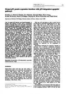
Expression of wild-type and mutant p53 proteins by recombinant vaccinia viruses Dvora Ronen, Yael Teitz, Naomi Goldfinger1 and Varda Rotterl,* Department of Human Microbiology, Sackler School of Medicine, Tel Aviv University, Ramat Aviv, Tel Aviv 69978 and 'Department of Cell Biology, The Weizmann Institute of Science, Rehovot 76100, Israel Received February 27, 1992; Revised and Accepted June 12, 1992
ABSTRACT To facilitate the purification of wild type p53 protein, we established a recombinant p53 vaccinia viral expression system. Using this efficient eukaryotic expression vector, we found that the expressed p53 proteins retained their specific structural characteristics. A comparison between wild type and mutant p53 proteins showed the conservation of the typical subcellular localization and the expression of specific antigenic determinants. Furthermore, wild type p53 exhibited a typical binding with large T antigen, whereas no binding was detected with mutant p53. Both wild type and mutant p53 proteins were highly stable and constituted 5-7% of total protein expressed in the infected cells. These expression recombinant viruses offer a simple, valuable system for the purification of wild type and mutant p53 proteins that are expressed abundantly in eukaryotic cells.
INTRODUCTION Wild type p53 was shown to be a growth regulator that functions as a suppressor gene (1-3). Inactivation of this gene through deletion or mutation, which probably allows a cell to escape normal growth control, plays a critical role in the development of malignant transformation. In human colorectal carcinoma the transition from the benign to the malignant state correlates with the loss of both wild type alleles for p53, indicating a tumor suppressor function for p53 (4, 5). Allelic degeneration of p53 was also found in human carcinoma of the lung (6) and in a number of commonly occurring types of carcinoma (7-9). In most of the carcinomas mutant p53 protein forms are overexpressed, whereas in other malignancies, mostly those of myeloid origin, the p53 gene is rearranged and no p53 protein is evident (10-13). The concept that p53 indeed functions as a suppressor gene was proven by several experimental approaches. Wild type p53 expression inhibited malignant transformation of primary embryonic rat fibroblasts induced by co-transfection with activated oncogenes (14-16). Furthermore, expression of wild *
To whom correspondence should be addressed
type p53 induced direct growth arrest of proliferating cells in vitro (17-20) and in vivo (20-23). There are indications that wild type p53, that is spatially regulated during the cell cycle, functions as the cell advances from the GO/GI to the S phase (17, 19, 24-27). The molecular mechanism by means of which the wild type p53 protein functions is unclear as yet; however, there are some indications as to its activities. It was suggested that the protein plays a direct role in DNA replication. Indeed, p53 protein competed with DNA ca polymerase in the replication of SV40 large T antigen (28-31) and was found to co-stain with Herpes viral origin of replication (32). Furthermore, it was shown that the protein functions as a transcription factor (33, 34). In both these functional assays, mutant p53 proteins were inactive. Comparisons between wild type and mutant p53 protein forms, showed variations in their DNA binding affinities. Wild type p53 protein exhibited a high DNA binding activity, whereas mutant p53 protein forms showed low DNA binding capacities (35, 36). Although clusters of mutations were found in the p53 gene isolated from different tumors (3, 9) inactivation of wild type p53 protein was not restricted to a specific region. This suggests that the intact tertiary structure of the protein is critical for its activity, and loss of function may result from a variety of modifications occurring in the wild type protein. To study the function of the wild type protein it is critical to establish a suitable source for the purification of the protein, that will retain its authentic natural conformation. The vaccinia virus produces large amounts of the proteins that undergo proper physiological posttranslational modifications occurring in eukaryotic cells (37 -39). Therefore, it was chosen as a suitable system for the expression of wild type p53 protein. In the present study we describe the construction of recombinant vaccinia viruses containing the cDNAs coding for the wild type p53 and mutant p53 proteins. Characterization of the proteins expressed in the vaccinia system showed that the virus yielded high levels of the authentic protein expressing antigenic determinants corresponding in their conformation to both wild type p53 and mutant p53 proteins. Furthermore, the differential ability of the wild type p53 recombinant protein to form a complex with the large T antigen and the typical subcellular localization in the cell were conserved.
3436 Nucleic Acids Research, Vol. 20, No. 13
MATERIALS AND METHODS Cells and Virus TK- 143, a TK_ human osteosarcoma cell line (40), the cell lines RK13, HeLa and Cos were all grown in Dulbecco's modified Eagle's medium (DMEM) containing 10% fetal bovine serum. The wild type vaccinia virus used for recombination was the WR strain.
Monoclonal Antibodies The following anti-p53 antibodies were used: monoclonal antip53 Pb-240, PAb-242, PAb-246 (41, 42); PAb-421 (43). PAb-419 is an anti large T antigen monoclonal antibody (43). The antibodies were either supernatants of growing cell lines or ascitic fluids obtained from the peritoneal cavity of hybridomabearing syngeneic mice, spun to remove tissue debris, diluted in PBS, and used without further purification. Construction of Recombinant Vaccinia Viruses The previously described plasmids pSVL-CD, coding for the wild type p53 protein, and pSVL-M8, coding for the mutant p53 protein (20, 44), were used as sources for construction of p53 recombinant vaccinia viruses. The two cDNA inserts were isolated by digestion with Bam HI and subcloned into the Bam HI site of vaccinia virus vector, pgpt-ATA-18 (kindly provided by Dr. H. Stunnenberg of EMBL), carrying a xanthine guanine phosphoribosyl transferase gene (gpt) for selection (45, 46). Transfer of the p53 genes from the recombinant plasmids to vaccinia virus WR was achieved by using a modification of the standard homologous recombination method (47). Briefly, TK143 cells were infected with wild type vaccinia virus WR at a multiplicity of infection (M.O.I.) of 0.1 PFU per cell. At 2 h post-infection, the cells were transfected, by the calcium phosphate precipitation procedure (48), with 1 jig of pgpt-ATA-18 plasmid DNA carrying either p53CD or p53M8 coding sequences. Two days later, recombinant viruses were collected from the infected cells and plaque-purified on RK13 cells overlaid with 1 % agarose in growth selectable medium (DMEM supplemented with 10% FCS and containing 20 jtg/ml mycophenolic acid, 150 yg/ml xanthine and 15 ,ug/ml hypoxanthine). Individual recombinant plaques were purified by 3 x plaque purification and amplified on RK13 cells. Virus recombinant stocks carrying either the wild type p53 cDNA, designated vaccinia p53WT (containing the p53CD coding for the wild type p53 protein), or the mutant p53 cDNA, designated vaccinia p53mut (containing the p53M8 coding for mutant p53 protein), were prepared from the amplified cultures and titrated for level of virus PFU before use.
Preparation of Infected Cell Lysate: Imunoprecipitation and hmmunoblotting Monolayer cultures were infected with vaccinia p53WT, vaccinia pS3mut or parental vaccinia virus WR vaccinia vector at a M.O.I. of 5 PFU per cell of each virus. 24 h post-infection, the cells were metabolically labelled with 0.125 mCi of 35S-methionine (Amersham) for 1 h at 37°C in methionine-deficient (met-) Eagle's modified medium supplemented with 10% heatinactivated dialyzed fetal calf serum. Cells were lysed in lysis buffer (50 mM Tris pH 7.5; 150 mM NaCl; 0.5% NP40; 0.5% deoxycholate; 0.01 % SDS; 2 mM PMSF) and pre-cleared with 10% fixed Staphylococcus aureus. Equal amounts of TCAinsoluble radioactive material were reacted with specific antibodies for 2 h at 4°C. The immune complexes were
precipitated with 10% fixed Staphylococcus aureus and washed 3 x in PLB buffer (10 mM NaH2HPO4 pH 7.5; 100 mM NaCl; 1% Triton X100; 0.5% sodium deoxycholate; 0.1% sodium dodecyl sulfate). The immune complexes were separated on SDS-PAGE (49). For immunoblotting, cells were lysed in sample buffer and subjected to PAGE, as above. The fractionated proteins were electrotransferred to nitrocellulose membranes and the proteins were detected using the protoblot western blot Ap system (Promega). Indirect Immunofluorescent Staining Cells were plated on sterile cover slides and infected with recombinant vaccinia viruses, as described above. Twenty-four hours after infection, the cells were fixed with 3% formaldehyde in PBS and washed twice with PBS, permeabilized for 5 min in acetone at -10°C and again washed twice with PBS. Subsequently the slides were incubated for 30 min with an appropriate specific antibody, washed with PBS and incubated with a rhodamine-conjugated goat anti-mouse IgG antibody. The slides were washed extensively in PBS rinsed with water and mounted on objective microscope slides with Gelvatol.
RESULTS Construction of Wild Type and Mutant p53 Recombinant Vaccinia Viruses The two cDNAs representing the wild type p53 and mutant p53 proteins were previously defined by sequence analysis and biological activities (14, 50, 51). pS3CD cDNA representing the wild type p53 protein was isolated from a cDNA library prepared from a mitogen-stimulated normal T cell library (51). The protein coded by this cDNA lacks any transforming ability upon cotransfection into rat embryo fibroblasts with activated human H-ras oncogene (14). Conversely, it inhibited the appearance of transformed foci upon co-transfection with mutant p53 and the H-ras oncogene (44). p53M8 cDNA representing mutated p53 protein was isolated from a chemically transformed MethA cDNA library (50). This cDNA contains a single point mutation at amino acid position 132 (cys to phe). In addition, the C-terminus was altematively spliced, resulting in a shorter rnslaon prduct (51). The protein coded by this cDNA clone possesses a transforming activity, as determined in the cooperation assay (44). p53CD and p53M8 cDNAs were isolated and cloned into the pgpt-ATA-18 vaccinia virus recombinant vector. The resulting constructs vaccinia p53WT and vaccinia p53mut were then transfect TK- 143 cells (40), previously infected with wild type vaccinia virus (WR strain). Homologous recombination between sequences of the gpt-ATA-18 vector and the wild type vaccinia virus genome resulted in the recombination of the p53CD or p53M8 sequences into vaccinia viral genome. Recombinant viruses were drug selected. Plaques were picked from the third round of purification and propagated in RK13 cells. The putative recombinant virus DNAs were isolated from the infected cells and tested for proper gene insertion. Expression of Wild Type and Mutant p53 Proteins by Recombinant Vaccinia Viruses In order to examine the p53 proteins expressed by the recombinant viruses, we chose the HeLa cells, that almost totally lack detectable endogenous p53 protein (52), as a convenient cell system for measuring the expression of p53 protein introduced by viral infection. We infected HeLa cells with vaccinia p53WT,
Nucleic Acids Research, Vol. 20, No. 13 3437 vaccinia p53M8, or vaccinia virus vector devoid of any foreign DNA. At 24 h post-infection, the cultures were labelled for 1 h with 35S-methionine, and equal amounts of Trichloroacetic acid (TCA)-insoluble radioactivity of cell lysates were reacted with PAb-242 (41) anti-p53 monoclonal antibodies which
A
co) LO C~~~~aa
OL
a 0
.U
Cb
ia_
~~~E
LO
>
>
r
-~
HB
o U 0)
b
1
a
.M-
-
0
aZ
b
IIa b-I
1
w -Op 4-
4b
a
B
281
4h
6h
p53
i"
(data not shown).
_t
811 1 01h 24[i Ul VV\ Nil
CO)
U)
CZ 4-
. .
p53
_.........
C)
.
~~~~~~~Ark
0~
> on U3 C')
an
recognize both wild type p53 and mutant p53 proteins. Figure IA represents cellular and viral specific proteins synthesized in the cells infected with the various recombinant viruses. Lanes a (Figure 1A) show the pattern of total proteins expressed in the cells. At that level of resolution it is clear that cells infected by vaccinia p53WT or vaccinia p53mut expressed an additional band of the expected p53 product. Upon immunoprecipitation with specific anti-p53 antibodies, the expected corresponding p53 proteins were immunoprecipitated only from cultures infected with recombinant vaccinia viruses, and no specific immunoprecipitation occurred from cultures infected with the vaccinia virus vector alone. The prbteins expressed by the recombinant viruses represent the authentic sizes of the expected p53 proteins. The mutant p53 protein translated from the p53M8 cDNA is shorter than the wild type p53 protein and exhibits a slightly faster migration (Figure IA, lanes b). In order to detect the earliest time point for maximal production of recombinant p53 proteins expressed after infection, we measured the rate of p53 synthesis at various time intervals after infection. HeLa cells were infected with recombinant wild type p53 or mutant p53 vaccinia viruses. At times indicated in Figure 1B, the cells were harvested, cell lysates were analyzed by the Western technique using PAb-242 specific anti-p53 antibodies. The synthesis of both the recombinant wild type p53 and the recombinant mutant p53 proteins was detected as early as 8 h after infection (Figure iB). Thirty hours after infection, the level of p53 proteins plateaued and remained as after 24 h
-
p53
4Z;-
Figure 1. Expression of p53 protein by recombinant vaccinia viruses. A. HeLa cells were infected for 60 min, at M.O.I. 5 PFU per cell, with vaccinia, the wild type vaccinia virus (strain WR) vector, with p53WT, the vaccinia recombinant coding for wild type p53 (p53CD), or with p53mut, the vaccinia recombinant coding for mutant p53 protein (p53M8). Twenty-four hours after infection, the cells were metabolically labelled with 3 S-methionine for lh, after which protein content was measured. Total cell lysates, 5 x 10⁵ cpm, prior to immunoprecipitation (a) were compared to specific immunoprecipitated product binding the PAb-242 monoclonal antibodies (b). B. Western blot analysis. HeLa cells were infected with either vaccinia p53WT or vaccinia p53mut and at indicated time points. Cells were harvested and reacted with the anti-p53 monoclonal antibody PAb-242. UI, cell lysates of uninfected HeLa cells; VV, cell lysates of HeLa cells 24 h post-infection with WT vaccinia virus. M, markers indicating the positions of molecular weight standards.
Evaluation of Wild Type p53 Protein Levels in Vaccinia p53WT Infected HeLa Cells Infection of cells with vaccinia virus shuts off the expression of most cellular proteins and induces the predominant expression of the viral encoded proteins (45, 46). As expected, infection of HeLa cells with the vaccinia pS3WT showed that, in addition to the viral encoded proteins, the cells produce as expected the p53 protein (compare the patterns of total proteins expressed in HeLa cells infected with vaccinia vector or with vaccinia p53WT expression vector in Figures 1A and 2A). These specific patterns of protein expression suggested that HeLa cells infected with p53 vaccinia vectors were a suitable source for p53 purification. It was important, however, to measure the levels of p53 protein in the infected cells. We performed densitometric analyses and found that p53 constitutes about 5-7% of the total protein expressed in the virally infected HeLa cells. These levels are about 100 x higher than those found in any cells expressing wild type p53 protein. For further estimation of the cellular levels of wild type p53 expressed in the HeLa infected cells, we compared the level of p53 protein to the level of tubulin, a structural protein expressed in cells in rather high amounts (about 5-7% of total protein content). Equal protein samples of either uninfected or vaccinia p53WT infected HeLa cells were exposed to a Western blot analysis probed with anti tubulin or anti-p53 PAb-421 monoclonal antibodies. As can be seen in Figure 2B, uninfected HeLa cells expressed the cx and ,3 forms of tubulin. As expected, HeLa cells infected with the various vaccinia vectors expressed almost no tubulin (see Figure 2B). However, Western analysis of equivalent protein amounts, showed that HeLa cells infected with vaccinia p53WT expressed p53 protein levels comparable to the tubulin levels expressed in uninfected cells (5-10%). This further supports the conclusion that p53 protein levels in infected cells
3438 Nucleic Acids Research, Vol. 20, No. 13
Figure 4. Antigenic epitope analysis of wild type and mutant p53 proteins derived
:4m
from cells infected with recombinant vaccinia viruses. HeLa cells were infected with vaccinia vector (a), vaccinia p53WT (b), or vaccinia p53mut (c). At 24 h post-infection, cells were labelled with 35S-methionine for 1 h. Equal amounts of TCA-insoluble radioactive material were immunoprecipitated with the indicated anti-p53 monoclonal antibodies.
Figure 2. Evaluation of p53 protein levels in HeLa cells infected with vaccinia p53WT. A. Equal amounts of radiolabelled protein obtained from HeLa cells infected with vaccinia p53WT or with vaccinia vector were compared. B. Equal amounts of protein obtained from uninfected or vaccinia p53WT infected HeLa cells were exposed to Western blot analysis to measure the expression of (x and 13 tubulin (anti-tubulin) or p53 (PAb-421).
4
am-
_
_
.5
Figure 3. Stability of recombinant p53 vaccinia virus expressed proteins. HeLa cells were infected by recombinant p53 vaccinia viruses under conditions as in Figure IA. Twenty-four hours post-infection, the cells were pulse-labelled with 35S-methionine for 45 min. The labelled cells were either lysed immediately after pulse time or chased with excess of cold methionine for times indicated. Cell lysates were prepared and immunoprecipitated with PAb-242 and analyzed on 10% SDS-PAGE.
are relatively high and therefore this expression system can be used as a convenient source for the purification of wild type p53
protein.
Stability of Wild Type and Mutant p53 Proteins Expressed by Recombinant Vaccinia Viruses Previous studies have shown that wild type p53, which is expressed at low molar concentrations, is a labile protein with a short half life (53). Thus, it is difficult to purify the protein
from normal eukaryotic cells. In subsequent experiments we evaluated the stability of the p53 proteins expressed by the vaccinia virus. It was important to determine whether the primary structure of the protein or the cellular environment influence their turnover (54). We performed a series of pulse chase experiments with wt and mut p53 vaccinia virus recombinant p53 proteins. At 24 h post-infection, the cells were pulse labelled with 35Smethionine for 1 h and then chased with an excess of cold methionine for selected times. Figure 3 shows that both wild type p53 and mutant p53 proteins are highly stable and are detectable even after 30 h of chase time. The fact that both wild type and mutant p53 proteins exhibit a similar turnover in the same cellular environment suggests that p53 protein stability is dictated by the cellular milieu rather than by the primary structure of the protein.
Epitope Analysis of Recombinant Wild Type and Mutant p53 Proteins Expressed by Vaccinia Virus The above Western analysis (Figure IB) indicated that both wild type and mutant p53 recombinant proteins express the PAb-242 antigenic epitope that maps at the N-terminus of p53 protein (41). To further investigate the authenticity of the recombinant proteins expressed by vaccinia viruses, we compared the specific antigenic phenotype of the virally expressed p53 products. To that end we used a battery of well characterized monoclonal anti-p53 antibodies. HeLa cells were infected with either wild type or mutant p53 recombination viruses. At 24 h post-infection, the cells were pulse-labelled with 35S-methionine for lh and cell lysates were prepared and immunoprecipitated with appropriate antibodies. Wild type p53 protein and mutant p53 protein forms can be differentially distinguished by their antigenic determinants. Wild type p53 expresses the PAb-246 determinant whereas mutant p53 protein forms have lost this determinant and express the specific PAb-240 determinant instead (41, 42). As expected, the PAb-246 antibodies reacted specifically with the epitope displayed by the
Nucleic Acids Research, Vol. 20, No. 13 3439 at the C-terminus of the molecule). Therefore, it represents a protein with a lower nuclear localization capacity (55). In the next experiment we have analyzed the subcellular localization of the viral expressed p53 proteins. Figure 5 shows a typical subcellular distribution of the vaccinia expressed proteins. Wild type p53 exhibited a strict nuclear localization visualized by immunofluorescent stalning with either PAb-242 (B) or PAb-246 (C). As expected, the mutant p53 protein encoded by p53M8 showed a less intensive nuclear staining, accompanied by cytoplasmic staining (E stained with PAb-242 or F stained with PAb-240). Control HeLa cells transfected with the wild type vaccinia virus exhibited no specific staining (A stained with PAb-242 and D stained with 242). This suggests that the subcellular localization of the viral p53 proteins is recognized and regulated by authentic cellular mechanisms.
n'
u.
E.
F.
Figure 5. Subcellular localization of p53 proteins encoded by p53 recombinant vaccinia viruses. HeLa cells were infected with vaccinia vector, vaccinia p53WT or vaccinia p53mut, as in legend to Figure IA. At 24 h post-infection, cells were fixed and processed for indirect immunofluorescent staining with different p53 monoclonal antibodies. Panels A and D: Cells infected with vaccinia vector and stained with PAb-242. Panel B: Cells infected with vaccinia p53WT stained with PAb-242. Panel C: Cells infected with vaccinia p53WT stained with PAb-246. Panel E: Cells infected with vaccinia p53M8 stained with PAb-242. Panel F: Cells infected with vaccinia p53mut stained with PAb-240.
wild type p53 recombinant protein, while no precipitation occurred when they were reacted with the mutant p53 recombinant protein (Figure 4). Alternatively, the PAb-240 antibodies directed against the mutant conformation clearly precipitated the mutant p53 recombinant protein, while only slight precipitation occurred with the recombinant vaccinia virus wild type p53 protein (compare intensities of bands in lanes b immunoprecipitated with PAb-240 to those detected by PAb-246, 242 or 421). This might have been due to the fact that the protein immunoprecipitated with PAb-240 of the wild type p53 protein had low binding affinity or, alternatively, that a certain percentage of the p53 protein population that were denatured under the experimental conditions provided and retained a mutant specific phenotype. The PAb-421 antibodies (43) directed against the Cterminal region of the protein recognized only the wild type recombinant protein and not the mutant p53 protein. Lack of this antigenic determinant in this mutant p53 protein was due to the fact that the p53M8 represents an alternatively spliced product that does not code for the PAb-421 determinant (50, 51). Subcellular Localization of Wild Type and Mutant p53 Proteins Expressed by Vaccinia Virus Another typical feature of the p53 protein is its nuclear localization. Previous studies showed that nuclear localization is essential for wild type p53 protein activity (27, 44). We found that wild type p53 protein encoded by pS3CD yielded an intact protein that expressed three nuclear localization signals which were responsible for nuclear localization (55). p53M8 codes for a protein that contained only a single nuclear localization signal (the other two were lost as a result of the alternative splicing
Complex Formation Between Large T Antigen and Vaccinia Virus Expressed p53 Proteins The capacity to form complexes with the viral large T antigen of SV40 is a typical feature of the wild type p53 protein only (56, 57). In our subsequent experiments we examined complex formation of p53 vaccinia expressed proteins and large T antigen expressed in Cos cells. Cells were infected with the various vaccinia viruses and the different cell lysates were immunoprecipitated with anti p53 (PAb-246, 240 and 421) and anti large T antigen monoclonal antibodies (PAb-419). As shown in Figure 6, wild type p53 formed a complex with the T antigen. Indeed large T antigen was detected when cell lysates were immunoprecipitated with PAb-246, the wild type specific p53 protein. Some of the wild type p53 protein bound the PAb-240 (Figure 4), however, this product did not form a complex with the large T antigen, as is the case with the mutant p53 protein. This supports our conclusion that at least a certain fraction (about 10%, assayed by autoradiogram intensity) lost the typical wild type features. Cos cells infected with the mutant coding virus expressed substantial levels of of the mutant p53 protein (binds PAb-240) but no complex with large T antigen was detected (Figure 6; compare lanes PAb-240 and PAb-419 of vaccinia-p53 mutant). Cos cells infected with vaccinia vector alone expressed the endogenous p53 as well as the large T antigen (Figure 6).
DISCUSSION The fact that the p53 suppressor gene functions at low molar concentrations (50) greatly complicates the direct search for modulation of p53 protein activity in normal cells. The finding that in most systems induction of p53 expression usually causes a dramatic cessation of growth, precluding any further analysis of its function in growing cells, coupled with the fact that wild type p53 is a short lived product (53), makes it essential to establish sources other than the normal cell for the purification of this product. The activity of wild type p53 seems to be highly controlled by the intact conformation of this protein. Therefore it is critical that the protein expressed undergo all he structural processing which occurs in the normal eukaryotic cell. For that reason, we adapted the vaccinia expression system, which is a very high expression system in eukaryotic cells (37 -39). With this system we detected expression of both wild type and mutant p53 proteins as early as 8h after infection, before cytopathic changes became evident, and the proteins produced were of the expected sizes and were both transported to the nucleus. Our conclusion that the p53 proteins expressed by vaccinia
3440 Nucleic Acids Research, Vol. 20, No. 13
40 4.
Figure 6. Complex formation between large T antigen and p53 vaccinia viral expressed proteins. Cos cells were infected with the various vaccinia viruses, and the different cell lysates were immunoprecipitated with anti p53 (PAb-246, 240 and 421) and anti large T antigen monoclonal antibodies (PAb-419). NI, cell lysates immunoprecipitated with non-immune serum.
viruses exhibit authentic structure and function was assessed on the basis of the following parameters: (a) Nuclear localization. p53 is a nuclear protein that is known to be spatially regulated in the cytoplasm. As the cell moves from the resting GI/Go phase into the S phase, newly synthesized protein moves from the cytoplasm to the nuclear compartment (44, 59). Nuclear localization is controlled by three nuclear signals located at the C-terminus of the molecule (55, 60, 61). In the present study, we compared the wild type p53 protein containing three intact p53 signals to a mutant p53 form that contains a single nuclear localization signal (55). As expected, the wild type p53 exhibited a strict nuclear localization, while the mutant product showed occasionally a combined nuclear and cytoplasmic pattern. On the basis of these subcellular localization patterns, we concluded that the products expressed were regulated by mechanisms found in the eukaryotic cell. (b) Expression of antigenic determinants. Our antigenic analysis of p53 recombinant proteins indicates that both proteins adopt a native conformation. Both proteins display several epitopes characteristic of wild type and mutant p53 proteins. The wild type p53 protein and the mutant p53 protein, which are identical in their amino terminal structure, are both recognized by PAb-242 p53 antibodies directed against the epitope located at this region of the molecule. As expected, the mutant p53 encoded by p53M8 lacks the PAb-421, a C-terminal specific epitope. The wild type p53 efficiently binds the PAb421. The differential expression of PAb-246 and PAb-240 in wild type and mutant p53, respectively, is a convenient tool to distinguish between the two. In the case of the vaccinia viral products, it is clear that the PAb-246 is typical of the wild type p53 product; no binding was evident with the mutant p53 product. The results with PAb-240 showed efficient binding with the mutant p53 protein; nevertheless a certain level of binding was also evident with the vaccinia wild type p53 protein. This could mean that the wild type p53 contains a fraction (about 10%) of molecules that have undergone denaturation under our experimental conditions or, alternatively, this could be an inherent property of the wild type p53 protein that, because of lower resolution conditions in other expression systems, was heretofore
undetectable. A third possibility is that the PAb-240 population represents the minor endogenous mutant p53 protein levels expressed in HeLa cells that was significantly stabilized following complex formation with the exogenous viral p53 products (52). To examine this last possibility, we intend to study viral vectors in strictly p53 non producer cells lines. (c) Differential complex formation. Wild type and mutant p53 proteins were shown to vary in their binding affinities to large T antigen. Wild type p53 forms a stable complex with the large T antigen, whereas mutant p53 does not (56, 57). It was suggested that formation of such complexes accounts for the inactivation of the p53 gene (3). In the vaccinia expression system, we found that expressed mutant p53 had no binding affinity for the large T antigen. The wild type product, immunoprecipitating with the wild type-specific monoclonal antibodies (PAb-246), bound large T antigen. However, the fraction of molecules of the wild type population binding the PAb-240 (mutant-specific) did not complex with the large T antigen. This further confirms, by a different criterion, i.e., binding of the large T antigen, the assumption that a fraction of the molecules expressed by the wild type p53 cDNA exhibits a mutant phenotype. A potential cellular target protein that may be involved in complexing with mutant p53 protein is the heat shock 70 protein (hsp-70) which was shown previously to be able to complex only with the mutant p53 protein (30, 57, 62-64). Indeed, our immunoprecipitation results (Figure 4) show a band which may correspond to the putative hsp-70 that results from complexing only with the mutant p53 protein (Figure 4, Panel C, immunoprecipitation with PAb-242 and PAb-240) and not with PAb-246. No such bands was detected in the wild type p53 product. (d) Stability of p53 protein. One of the important prerequisites for purification of the wild type p53 is that the produced protein be stable. Our results show that the wild type p53 which is expressed has a high protein stability, comparable to that of the mutant p53 product. Recent in vitro experiments demonstrated that the p53 proteins, like other oncoproteins, are the most rapidly degraded proteins and have a very short turnover (54). This short half-life was shown to be contributed to the ubiquitin-mediated proteolytic system. However, examination of p53 turnover in vivo in various cell lines revealed that this cellular proteolytic process can be overcome, resulting in an increased half-life of the p53 protein. This was suggested to be the case when the wild type p53 protein formed a stable complex with the large T antigen (53, 64, 65), or when the mutant p53 either retained an altered conformation (63, 64) or formed a complex with the cellular hsp-70, increasing its half-life (63, 64). The markedly increased half-life turnover of both vaccinia virus p53 expressed proteins demonstrated here may be explained by the fact that the vaccinia virus vector causes an almost total shut-off of protein synthesis, including the total cessation of production of the natural proteolytic system of the cell. Thus, the viral p53 encoded protein is able to escape physiological cell proteolysis and increase its metabolic turnover. This supports the idea that the short life span of wild type p53 is not an inherent characteristic feature dictated by the protein's primary structure but rather a feature dominated by cellular proteins expressed in the specific host cells. In conclusion, the results presented here describe an efficient system for producing authentic p53 proteins which can be purified and used to study p53 protein structure and function.
Nucleic Acids Research, Vol. 20, No. 13 3441
ACKNOWLEDGMENTS We thank Dr. H. G. Stunnenberg (European Molecular Biology Laboratory, Heidelberg, Germany) for providing the vaccinia
virus recombination vector and for his guidance in constructing the recombination viruses. This work was supported in part by grants from the Leo and Julia Forchheimer Center for Molecular Genetics. V.R. is the incumbent of the Norman and Helen Asher Professorial Chair in Cancer Research at the Weizmann Institute and the recipient of a Career Development Award from the Israel Cancer Research Fund (ICRF). Ms. M. Baer prepared and edited the manuscript.
REFERENCES 1. Lane, D., and Benchimol, S. (1990). Genes and Development 4, 1-8. 2. Vogelstein, B. (1990). Nature 348, 681-682. 3. Levine A. J., Momand, J., and Finlay., C.A. (1991). Nature 351, 453 -456 4. Baker, J.S., Fearon, E.R., Nigro, J.M., Hamilton, S.R., Preisinger, A.C., Jessup, J.M., van Tuinen, P., Ledbetter, D.H., Barker, D.F., Nakamura, Y., White, R., and Vogelstein, B. (1989). Science 244, 217-221. 5. Nigro, J.,M.,Baker, J.S, Preisinger A.,C,Jessup J.M., Hostetter, K., Cleary, K., Bigner S.H, Davidson, N., Baylin S. Devilee P., Glover, T., Collins F.S. Weston A. Modali R. Harris C.C., and. Vogelstein, B. (1989). Nature
342, 705-708. 6. Takahashi, T., Nau, M.M., Chiba, I., Birrer, M.J., Rosenberg, R.K., 7.
8.
9. 10. 11. 12.
13. 14. 15.
16. 17.
18. 19. 20.
21. 22.
23. 24.
25.
26. 27.
28. 29.
Vinocour, M., Levitt, M., Pass, H., Gazdar, A.F., and Minna, J.D. (1989). Science 246, 491-494. Baker S.J., Markowitz, S., Fearon, E.R., Willson, J.K.V. and Vogelstein, B. (1990). Science 249, 912-915. Rodrigues, N.R., Rowan, A., Smith, M.E.F., Kerr, I.B., Bodmer, W.F., Gannon, J.V. and Lane, D.P. (1990). Proc. Natl. Acad. Sci. USA, 87, 7555-7559. Hollstein, M., Sidransky, D., Vogelstein, B., and Harris, C., (1991). Science 253, 49-53. Wolf, D., and Rotter V., (1985). Proc Nat Acad Sci 82, 790-794. Prokocimer, M., Shaklai, M., Ben-Bassat, H., Wolf, D., Goldfmger, N., Rotter, V., (1986) Blood 68, 113-118. Kelman, Z., Prokocimer, M., Peller, S., Kahn, Y., Rechavi, G., Manor, Y., Cohen A., and Rotter, V. (1989). Blood 74, 2318-2324. Ahuja, H., Bar-Eli, M., Advani, S.H., Benchimol, S., and Cline, M.J. (1989). Proc. Natl. Acad. Sci. USA 86, 6783-6787. Eliyahu, D., Goldfinger, N., Pinhasi-Kimhi, O., Shaulsky, G., Skumik, Y., Arai, N., Rotter, V. and Oren, M. (1988). Oncogene 3, 313-321. Eliyahu, D., Michalovitz, D., Eliyahu, S., Pinhasi-Kimhi, 0. and Oren, M. (1989). Proc. Natl. Acad. Sci USA 86, 8763-8767. Finlay, C.A., Hinds, P.W., and Levine, A.J. (1989). Cell 58, 1083-1093. Mercer, W.E. Amin, M. Sauve, G. J. Appella, E. Ullrich, S. J. and Romano J. W. (1990). Oncogene 5, 973-980 Michalovitz, D. Halevy O., and Oren M. (1990). Cell 62, 671-680 Diller, L., Kassel, J., Nelson, C.E., Gryka, M.A., Litwak, G., Gebhardt, M., Bressac, B., Ozturk, M., Baker, S.J., Vogelstein, B. and Friend, S.H. (1990). Mol. Cell. Biol. 10, 5772-5781. Shaulsky, S., Goldfmger, N., Peled A., and Rotter, V. (1991). Proc. Nat. Acad of Sci. 88, 8982-8986. Shaulsky, G., Goldfinger, N., and Rotter, V. (1991). Cancer Research 51, 5232-5237. Chen, P.-L, Chen, Y., Bookstein, R., and Lee, W.-L. (1991). Science, 250, 1576-1580. Chen, Y., Chen, P.L., Arnaiz N., Goodrich, D., and Lee, W.H., (1991). Oncogene 6, 1799-1805. Shaulsky, G., Goldfinger, N., Peled, A., and Rotter V., (1991). Cell Growth and Differentiation. 2, 661-667. Deppert, W., Buschhausen-Denker, G., Patschinsky, T., and Steinmeyer, K. (1990). Oncogene 5, 1701-1706. Ginsberg, D., Michalovitz, D., Ginsberg, D,. and Oren, M. (1991). Mol. Cell. Biol. 11, 582-585. Martinez J., Georgoff, I., Martinez J., and Levine, A. J. (1991). Genes and Development 5, 151-159. Braithwaite, A.W., Sturzbecher, H.W., Addison, C., Palmer, C., Rudge, K. and Jenkins, J.R. (1987). Nature, 329, 458-460. Gannon, J.V. and Lane, D.P. (1987). Nature 329, 456-458.
30. Sturzbecher, H.W., Brain, R., Maimets, T., Addison, C., Rudge, K., and Jenkins, J.R. (1988). Oncogene 3, 405-413. 31. Wang, E.H., Friedman, P.N., and Prives, C., (1989). Cell 57, 379-392. 32. Wilcock, D. and Lane, D. P. (1991). Nature 349, 429-431. 33. Fields, S. and Jang, S. K. (1990). Science 249, 1046-1049. 34. Raycroft, L., Wu, H. and Lozano, G. (1990). Science 249, 1049-1051. 35. Kern, S.E., Kinzler, K.W., Baker, S.J., Nigro, J.M., Rotter, V., Levine, A.J., Friedman, P., Prives, C. and Vogelstein, B. (1991). Oncogene 6, 131- 136. 36. Shohat-Foord O., Bhattacharya, P., Reich, Z., and Rotter, V., (1991) in Nucleic Acid Research.19, 8982-8986. 37. Flexner, C., Broyles, S., Earl, P., Chakrabarti, S., and Moss, B., (1988). virology 166, 339-349. 38. Eduards, R.H., Selly, M,Y., Mobley, W.C., Weinrich, S.L., Hruly, D.E., and W.J., Rutter (1988). Mol. Cell. Biol. 8, 2456-2464. 39. Hu, S.L. Plowman, G.D., Sridher, P., Stevenson, U.S., Brown, J.P., and Estin C.D. (1988). J. Virol. 62, 176-180. 40. Rhim, J.S., Cho, H.Y., and Huebner, R.J. (1975). Int. J. Cancer 15, 23-29. 41. Yewdell, J.W., Gannon, J.V. and Lane, D.P.(1986). J. Virol. 59, 444-452. 42. Gannon, J.V., Greaves, R., Iggo, R., and Lane, D.P. (1990). EMBO J. 9, 1595-1602. 43. Harlow, E., Crawford, L.V., Pim, D.C., and Williamson, N.M. (1981). J. Virol. 39, 861-869. 44. Shaulsky, G., Goldfinger, N., Tosky, M.S., Levine, A.J. and Rotter, V. (1991). Oncogene 6, 2056-2066. 45. Hanggi, M., Bannworth, W., and Stunnenberg H., (1986). EMBO J. 5, 1071-1076. 46. Stunnenberg, H., Lange, H., Philipson L., Van Miltenberg R., and Van der Viliet, P., (1988). Nucleic Acid Research 16, 2431-2444. 47. Mackett, M., G.L. Smith and Moss, b., (1985). In: DNA Cloning, Volume II (D.M. Glover, ed.), IRL Press, Oxford/Washington, D.C., pp. 191-211. 48. Graham, F.L., and Van der Eb, A.J. (1973). Virology 52, 456-467. 49. Laemmli, U.K. (1971). Nature 227, 680-685. 50. Wolf, D., Harris, N., Goldfinger, N., and Rotter, V. (1985). Mol. Cell. Biol. 5, 127-132. 51. Arai, N., Nomura, D., Yokota, K., Wolf, D., Brill, E., Shohat, O., and Rotter V. (1986). Mol. Cell. Biol. 6, 3232-3239. 52. May, E., Jenkins J.R., and May P., (1991). Oncogene 6, 1363- 1365.51. 53. Oren, M., Maltzman, W., and Levine, A.J. (1981). Mol. Cell. Biol. 2, 443-449. 54. Ciechanover, A., Diguiseppe, J.A., Bercovich, B., Orian, A., Richter, J.D., Swartz, A.L., and Brodeur, G.M., (1991). Proc. Natl. Acad. Sci. USA 88, 139-143. 55. Shaulsky, G., Goldfinger, N., Ben-Ze'ev, A., and Rotter, V. (1990). Mol. Cell. Biol. 10, 6565-6577. 56. Tan, T.H., Wallis, J. and Levine, A.J. (1986). J. Virol. 59, 574-583. 57. Hinds, P.W., Finlay, C.A., Frey, A.B., and Levine, A.J. (1987). Cell. Biol. 7, 2863-2869. 58. Rotter, V., Boss, M.A, and Baltimore, D., (1981). J Virol 38, 336-346. 59. Shaulsky, G., Ben-Ze'ev, A., and Rotter, V. (1990). Oncogene 5, 1707-1711. 60. Dang, C.V., and Lee, W.M.F., (1989). J. Biol. Chem. 264, 18019-18023. 61. Addison, C., Jenkins, J.R., and Sturzbecher, H.W. (1990). Oncogene 5, 423-426. 62. Pinhasi-Kimhi, O., Michalovitz, D., Ben-Ze'ev, A., and Oren, M. (1986). Nature 320, 182-185. 63. Finlay, C.A., Hinds, P.W., Tan, T.H., Eliyahu, D. Oren, M., and Levine, A.J. (1988). Mol. Cell Biol. 8, 531-539. 64. Halevy, O., Hall A. and Oren M. (1989). Mol. Cell. Biol. 9, 3385-3392. 65. Jia-Yuh Lin and D.T. Simmons (1990). Virology 176, 302-305.