IOP Conference Series: Materials Science and Engineering
PAPER • OPEN ACCESS
Fabrication of Ag-Loaded TiO2 Nanotube Arrays on Ti-6Al-7Nb for Dental Implants Application To cite this article: A R Murnandityas et al 2018 IOP Conf. Ser.: Mater. Sci. Eng. 395 012008
View the article online for updates and enhancements.
This content was downloaded from IP address 178.171.38.28 on 30/08/2018 at 18:09
7th Nanoscience and Nanotechnology Symposium (NNS) IOP Publishing IOP Conf. Series: Materials Science and Engineering 395 (2018) 012008 doi:10.1088/1757-899X/395/1/012008 1234567890‘’“”
Fabrication of Ag-Loaded TiO2 Nanotube Arrays on Ti-6Al7Nb for Dental Implants Application A R Murnandityas1, M Ibadurrohman1, B M Bachtiar2 and Slamet1,3 1
Department of Chemical Engineering, Faculty of Engineering, Universitas Indonesia, Depok 16424 2 Department of Oral Biology and Oral Science Research Center, Faculty of Dentistry, Universitas Indonesia, Jakarta 10430 3 Author to whom any correspondence should be addressed E-mail:
[email protected] Abstract. In the present study, Ag-loaded Titanium Nanotube Arrays (TNTAs) were fabricated on the surface of non-toxic dental material, Ti-6Al-7Nb. A layer of TNTAs were introduced on Ti-6Al-7Nb surface, hence extending its effective area and inducing antibacterial properties for biofilm inhibition. TNTAs were generated on the surface of Ti-6Al-7Nb by an anodization method in an electrolyte solution containing glycerol, water and NH 4F. This was followed by depositing Ag via a photo-assisted deposition method to obtain Ag-loaded TNTAs. Surface morphology and crystallinity were evaluated by FESEM-EDX and XRD, respectively. FESEM-EDX analysis showed that Ag-loaded TNTAs were formed and well aligned on the surface of Ti-6Al-7Nb, while XRD confirmed the presence of anatase and Ag crystals. Keywords: Dental implants; Ti-6Al-7Nb; Silver; TNTAs
1. Introduction Dental implantation is considered as reliable treatment option for teeth restoration, owing to its prominent features in terms of strength, effectiveness and durability. Its application, however, is by no means without side-effects. Biocompability of dental implant material and risk of peri-implantitis have become concern in dental implantation. Ti-6Al-4V as titanium alloys is the most common material for dental implants, due to its preeminent mechanical properties. However, biocompability of Ti-6Al-4V has been called to account, due to its vanadium content that can cause local adverse tissue reactions and immunological responses [1, 2]. Potentially toxic elements such as vanadium, copper and tin should be avoided completely from biomedical material applications [3]. Therefore, vanadium-free titanium alloys, such as Ti-6Al-7Nb have been developed as implant materials [4]. Although application of Ti-6Al-7Nb may have solved toxicity problems, peri-implantitis has been identified as another risk in dental implants application. Peri-implantitis has been recognized to be inflicted by the formation of bacterial biofilms on dental implant surface [5]. In fact, Peri-implantitis was identified in 28% of subjects and 43% of implant sites [6]. Peri-implantitis is an inflammatory reaction with loss of supporting bone in the tissues surrounding an implant, hence causing failure in osseointegration process that leads to implant failure [7]. Bacterial biofilm formation occurs due to incapability of dental implants to inhibit bacterial biofilm growth [8]. Therefore, modifications of dental implant material are needed to prevent peri-implantitis.
Content from this work may be used under the terms of the Creative Commons Attribution 3.0 licence. Any further distribution of this work must maintain attribution to the author(s) and the title of the work, journal citation and DOI. Published under licence by IOP Publishing Ltd 1
7th Nanoscience and Nanotechnology Symposium (NNS) IOP Publishing IOP Conf. Series: Materials Science and Engineering 395 (2018) 012008 doi:10.1088/1757-899X/395/1/012008 1234567890‘’“”
One way to prevent peri-implantitis is through modification of dental implant material surface morphology, makes it more resistant to bacteria adhesion or to incorporate antibacterial agent on its surface [9]. Those two functions can be achieved by fabricating TiO2 nanotube arrays (TNTAs) on the surface of Ti-6Al-7Nb as titanium alloys. TiO2 (Titanium Oxide) is known for its photocatalytic properties and wide use in disinfection, and was reported to cause oxidative damage in absence of photooxidation [10, 11]. Formation of natural oxide Titanium layer (TiO2) on the surface of Ti-6Al-7Nb provides implant osseointegration [12, 13]. Natural oxide Titanium layer also enhanced antibacterial activity, since TiO2 is known for its photocatalytic properties and has been widely utilized in disinfection [14]. In addition, TiO2 was also reported to cause oxidative damage in the absence of photooxidation [11]. However, natural oxide Titanium layer can be easily broken and does not perform adequate protective functions in the working condition of a biological environment [12,13,14]. Consequently, modification of the surface morphology is needed to induce implant antibacterial activity and osseointegration process. Synthesis of TiO2 nanotube arrays (TNTAs) is one way to generate TiO2 layer on the surface of Ti-6Al-7Nb as dental implant alloys. TiO2 in nanotube arrays will maximize the spesific surface area for catalytic reaction in disinfection process [15]. TiO2 nanotube arrays (TNTAs) is generated on Titanium-based dental implant surface, hence extending its effective area, which constitutes more sites for Ag occupation during Photo-assisted Deposition (PAD) process as well as more reactive sites for disinfection process. Silver (Ag) also has been utilized as an antibacterial agent with low toxicity in the human body [16]. Silver (Ag) may also enhance photocatalytic efficiency, by preventing recombination of electrons and holes produced upon photo-driven excitation [17]. In the presence of Ag, mechanism of biofilm adhesion inhibition is expected to be driven by the anti-bacterial features of Ag. However, it should be noted that anti-bacterial properties of Ag nanoparticles have not fully understood yet. According to Zheng, et al (2012), silver nanoparticles could damage the microbial cell-wall structure or cause cell separation, and inhibit the adhesive abilities by preventing gene expression. Also, the nanostructure of materials may also be effective in reducing related gene expression, possibly by damaging the cell wall, effecting cell separation, or altering the organism’s adhesive ability [18]. Without the presence of Ag nanoparticles, the mechanism of biofilm inhibition is most likely driven by the reactive oxygen species (ROS) generated from photocatalytic activities. These species, which include superoxide anion radicals, hydroxyl radicals, hydrogen peroxide, or atomic oxygen radicals are expected to damage DNA, RNA, proteins, and lipids of the bacteria in aqueous media. This subsequently leads to bacterial cell malfunction or destruction of the cell wall, extracting cell liquids and causing dehydration [19]. Among all the methods for preparation of TiO2 nanotube arrays, such as sol-gel [20, 21], hydrothermal [22, 23] and chemical vapor disposition, the electrochemical anodization [24-28] has the advantages of low cost, simple equipment, and easy process [29, 30]. On the other hand, this method has been recognized as an efficient approach to produce integrative, vertically and highly ordered nanotube arrays with controllable dimensions of nanotubes [26]. Hence, electrochemical anodization is suitable for creating nanotubular oxide layers for biological applications, as it also offers an excellent strength of layer adhesion as compared to those fabricated by other techniques [13, 15]. In the present work, a two fold strategy to produce advanced implant materials with capability of preventing peri-implantitis were implemented. Anodization of Ti-6Al-7Nb plate in a NH4F containing electrolyte successfully generated a nanotubular layer of TiO2 on the surface, which is followed by photo-assisted deposition of Ag nanoparticles. Further study about the modified samples capability to prevent biofilm is also essential. Influence of surface modification and concentration of Ag should be studied in order to optimized parameters in this research. Biofilm assay to measure the capability of the as-synthesized Ag-loaded TiO2 nanotubes is currently in progress and will be reported in the future paper. This work was done to further develop potential applications of TiO2 nanotubes on modification of Ti-6Al-7Nb with similar methods in previous study by Rafieerad et al (2015) [13]. Moreover, this study is expected to contribute to the advancement of highly functioning and biocompatible implant materials. 2
7th Nanoscience and Nanotechnology Symposium (NNS) IOP Publishing IOP Conf. Series: Materials Science and Engineering 395 (2018) 012008 doi:10.1088/1757-899X/395/1/012008 1234567890‘’“”
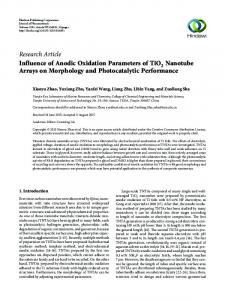
2. Methods Ti-6Al-7Nb plate (200 x 110 x 6 mm3, Baoji Chuangxin Metal Materials Co. Ltd China) were used as substrate. The plate was cut into 40 x 10 mm pieces. Prior to anodic oxidation, the Ti-6Al-7Nb samples were ground with abrasive SiC papers up to 1500 CW, and then wet-polished in a mixture of HF, HNO3 and demineralized water. The samples were subsequently cleaned by sonication in water for 5 min. Anodization was carried out in a two-electrode electrochemical cell in which a platinum sheet and the samples were connected to cathode and anode electrodes, respectively. The anodization process was carried out using a direct current (DC) power source at a constant potential of 50 V for 2 h. Optimized electrolyte mixture of glycerol, demineralized water and NH4F (with stirring at room temperature) were used. After anodization, all of the samples were washed with demineralized water, and then annealed at 500 C for 3 h in compressed air atmosphere to obtain crystalline phases. The annealed samples (TNTAs/Ag/0) were soaked in AgNO3 solutions (with ethanol as solvent) with three different concentrations (0.05 M, 0.1 M and 0.15 M) and irradiated with UV light for 3 hours while stirred. These samples were denoted as TNTAs/Ag/0.05, TNTAs/Ag/0.1 and TNTAs/Ag/0.15, respectively. The obtained Ag-loaded TNTAs Ti-6Al-7Nb samples were washed thoroughly using DI water, and cleaned ultrasonically afterwards. Micro-imaging of the samples was accomplished by a Field Emission Scanning Electron Microscope (FESEM, JEOL JIB-4610F) operated at EHT of 15 kV. The microscope is also equipped with a Thermo Noran spectrometer for EDX elemental probing. Xray diffraction patterns of the prepared samples were recorded using an X-ray Diffractometer (PANalytical Empyrean), in order to elucidate their crystallite properties. 3. Results and Discussions 3.1. Surface morphology of modified material Anodization of Ti-6Al-7Nbplates in the condition presented in this report results in formation of a TiO2 nanotube layer on the plate surface as depicted in the FESEM micrographs (Fig. 1a). The resultant nanotubular structure is vertically aligned and highly ordered across the surface, although variation in terms of tube height and diameter can be observed from 3D image in figure 1b. The average internal diameter of the nanotubes is ca. 130 nm with wall thickness of ca. 30 nm. A slight irregularity of the tube height might be originated from the original roughness of the plate or from two different phases (α+β) that might exist on the surface of Ti-6Al-7Nb. Nb and Al were added onto Ti to make an alloy with improved mechanical strength, considering the fact that Ti in α-phase has relatively low mechanical strength. Accordingly, the presence of Nb and Al assisted Ti to transform into its β-phase to some extent, resulting a stronger metallic structure. This observation is in agreement with that reported by Macak (2005) who also investigated the formation of TNTAs on the surface of Ti-6Al-7Nb in an Ethylene-Glycol-contained electrolyte [31]. (a)
(b)
Figure 1. Surface morphology of modified Ti-6Al-7Nb (a) top view (b) tilted view.
3
7th Nanoscience and Nanotechnology Symposium (NNS) IOP Publishing IOP Conf. Series: Materials Science and Engineering 395 (2018) 012008 doi:10.1088/1757-899X/395/1/012008 1234567890‘’“”
Fig. 2 showed the morphology of TNTAs layer after the deposition of Ag, revealing that silver nanoparticles (white spots) with particle size in the range of 40–60 nm were well dispersed in the superstructure of TNTAs. These particles are spread across the surface, situated on top of TNTAs, as well as on both the inner and outer sides of the tube walls (Fig. 2). These results imply that the presence of nanotubular TiO2 on the surface of Ti-6Al-7Nb provides extensive sites for Ag particles to occupy during photo-assisted deposition. Ultimately, both factors, i.e. higher surface area and adequate amount of deposited Ag particles, are expected to play significant roles in bactericidal activities and inhibition of biofilm formation.
Figure 2. Ag-loaded TNTAs on modified Ti-6Al-7Nb. Fig. 3 shows FESEM images of the modified samples, while fig. 4 shows the elemental (Ti, O and Ag) EDX mapping that were based on figure 2. From figure 3, addition of various concentration of Ag on the surface of TNTAs results in different distribution of Ag. Fig. 4 illustrates the dispersion of Ag particles, scanned across the surface of the TNTAs by energy dispersive X-ray (EDX) elemental mapping. The results confirm that Ag particles were successfully deposited on TNTAs, with the intensity of Ag signal (yellow spots) being positively correlated to the concentration of AgNO3 during PAD operation. It is also inferred from Figure 3 that the homogeneity of the particle dispersion deteriorates as the concentration become higher. Future tasks with regards to the operating conditions of PAD will be exerted in order to improve the uniformity of the resultant particle dispersion. (a)
(b)
(c)
(d)
Figure 3. FESEM images of modified Ti-6Al-7Nb samples (a) TNTAs/Ag/0 (b) TNTAs/Ag/0.05 (c) TNTAs/Ag/0.1 (d) TNTAs/Ag/0.15. Depositing Ag nanoparticles on a support with a nanotubular structure, as described in this report, is believed to offer advantageous features in the context of bacteria disinfection. Akhavan et al. [32] stipulated two key points that ensure the effectiveness of Ag as an anti-bacteria agent. Firstly, Agbased materials should be loaded on a nanostructured support which provides a high surface-area-tovolume ratio (enormous active sites). By imposing higher interfacial area between Ag particles and their bacteria target, microbial activities are expected to be significantly enhanced without the need to increase the amount of deposited silver. Considering the fact that silver is a high-value metal, this strategy offers economic benefits in addition to technical advantages. Secondly, immobilization of Ag nanoparticles in a nanostructured and porous host would ensure their durability and long-term 4
7th Nanoscience and Nanotechnology Symposium (NNS) IOP Publishing IOP Conf. Series: Materials Science and Engineering 395 (2018) 012008 doi:10.1088/1757-899X/395/1/012008 1234567890‘’“”
antimicrobial activities, because immobilized particles are more stable against washing which might otherwise give rise to material losses.
(a)
(b)
(c)
(d)
(a)
(b)
(c)
(d)
(b)
(c)
(d)
Figure 4. EDX mapping of Titanium (top), Oxygen (middle), and Silver (bottom) of modified Ti-6Al7Nb samples (a) TNTAs/Ag/0 (b) TNTAs/Ag/0.05 (c) TNTAs/Ag/0.1 (d) TNTAs/Ag/0.15. The XRD patterns of all samples are depicted in fig. 5. As shown in figure 5, the diffraction peaks appeared at 2θ = 25.4°, 37.86°, and 48.1° could be assigned to TiO2 anatase (JCPDS#01-0711166). From XRD results, it can be concluded that TiO2 anatase phase is obtained in all samples. Presence of TiO2 rutile wasn’t detected in the XRD profile, since rutile usually obtained via high temperature (greater than 800°C) calcination of anatase or in an acidic hydrothermal process. Presence of Ag (JCPDS#04-0783) can be found where the diffraction peaks at 2θ value of 44.42°. The other diffraction peaks at about 2θ = 35°, 38.4°, 40.3º and 53.1º can be indexed to the titanium substrate (JCPDS#44-1294). From figure 5 modified sample of Ti-6Al-7Nb, with Ag-loading concentration of 0.15 M and 0.05 M shows peaks at 44.42°, while it is not detected in 0.1 M Agloaded TNTAs. These may occur due to Ag deposited in TNTAs/Ag/0.05 existed in small amount, as confirmed with EDX mapping analysis. Using Scherrer equation, the size of TiO2 anatase crystalline can be obtained, with value 44.1 nm. The photocatalytic activity of the anatase form of TiO 2 is approximately 1.5 times higher than that of the rutile form. Accordingly, with XRD analysis confirms presence of anatase, photocatalytic activity may be enhanced.
5
7th Nanoscience and Nanotechnology Symposium (NNS) IOP Publishing IOP Conf. Series: Materials Science and Engineering 395 (2018) 012008 doi:10.1088/1757-899X/395/1/012008 1234567890‘’“”
Figure 5. XRD patterns of modified Ti-6Al-7Nb samples (a) TNTAs/Ag/0 (b) TNTAs/Ag/0.05 (c) TNTAs/Ag/0.1 (d) TNTAs/Ag/0.15. 4. Conclusions In this study, Ag-loaded TiO2 nanotubes layer was successfully generated on Ti-6Al-7Nb surface with a regularly aligned nanotubular structure, hence extending its effective surface area. A photo-assisted deposition technique was evidently an effective way to deposit Ag nanoparticles on TiO2 surface, as an electron trapper during activation and/or an antibacterial agent. Thus, some parameters of this research still need to be optimized in order to obtain the best strategy for the advancement of dental implant technology. In addition, further biofilm assay to measure capability of modified samples to inhibit bacterial biofilm growth is still in progress, and will be reported in a forthcoming paper. 5. References [1] Okazaki Y, Gotoh E 2005 Comparison of metal release from various metallic biomaterials in vitro Biomaterials 26 11-21 [2] Elias CN, Lima JHC, Valiev R, Meyers MA 2008 Biomedical applications of titanium and its alloys Jom 60 46-9 [3] Dominguez COaA. Titanium as a Biomaterial for Implants. In: Fokter SK, editor. Recent Advances in Arthroplasty: CC BY 3.0 license; 2012. p. 149 - 62. [4] Manfred F. Semlitsch HW, Robert M. Streicher and Rolf Sch6n 1992 Joint replacement components made of hot-forged and surface-treated Ti-6Al-7Nb alloy Biomaterials 13 [5] Dhir S 2013 Biofilm and dental implant: The microbial link J Indian Soc Periodontol 17 5-11 [6] Zitzmann NU, Berglundh T 2008 Definition and prevalence of peri-implant diseases J Clin Periodontol 35 286-91 [7] Prathapachandran J, Suresh N 2012 Management of peri-implantitis Dent Res J (Isfahan) 9 51621 [8] Yoshinari M, Oda Y, Kato T, Okuda K 2001 Influence of surface modifications to titanium on antibacterial activity in vitro Biomaterials 22 2043-8 [9] Necula BS, Fratila-Apachitei LE, Zaat SA, Apachitei I, Duszczyk J 2009 In vitro antibacterial activity of porous TiO2-Ag composite layers against methicillin-resistant Staphylococcus aureus Acta Biomater 5 3573-80
6
7th Nanoscience and Nanotechnology Symposium (NNS) IOP Publishing IOP Conf. Series: Materials Science and Engineering 395 (2018) 012008 doi:10.1088/1757-899X/395/1/012008 1234567890‘’“”
[10] Laxma Reddy PV, Kavitha B, Kumar Reddy PA, Kim KH 2017 TiO2-based photocatalytic disinfection of microbes in aqueous media: A review Environ Res 154 296-303 [11] Gurr JR, Wang AS, Chen CH, Jan KY 2005 Ultrafine titanium dioxide particles in the absence of photoactivation can induce oxidative damage to human bronchial epithelial cells Toxicology 213 66-73 [12] Albrektsson T, Branemark PI, Hansson HA, Lindstrom J 1981 Osseointegrated titanium implants. Requirements for ensuring a long-lasting, direct bone-to-implant anchorage in man Acta Orthop Scand 52 155-70 [13] Rafieerad AR, Zalnezhad E, Bushroa AR, Hamouda AMS, Sarraf M, Nasiri-Tabrizi B 2015 Self-organized TiO2 nanotube layer on Ti–6Al–7Nb for biomedical application Surface and Coatings Technology 265 24-31 [14] Foster HA, Ditta IB, Varghese S, Steele A 2011 Photocatalytic disinfection using titanium dioxide: spectrum and mechanism of antimicrobial activity Appl Microbiol Biotechnol 90 1847-68 [15] Roy P, Berger S, Schmuki P 2011 TiO2 nanotubes: synthesis and applications Angew Chem Int Ed Engl 50 2904-39 [16] Lansdown ABG 2006 Silver in Health Care: Antimicrobial Effects and Safety in Use Curr Probl Dermatol 33 17-34 [17] Seery MK, George R, Floris P, Pillai SC 2007 Silver doped titanium dioxide nanomaterials for enhanced visible light photocatalysis Journal of Photochemistry and Photobiology A: Chemistry 189 258-63 [18] Zheng Y, Li J, Liu X, Sun J 2012 Antimicrobial and osteogenic effect of Ag-implanted titanium with a nanostructured surface Int J Nanomedicine 7 875-84 [19] Buser D, Mericske-Stern R, Bernard JP, Behneke A, Behneke N, Hirt HP, et al. 1997 Long-term evaluation of non-submerged ITI implants. Part 1: 8-year life table analysis of a prospective multi-center study with 2359 implants Clin Oral Implants Res 8 161-72 [20] Maiyalagan T, Viswanathan B, V. Varadaraju U. Fabrication and characterization of uniform TiO2 nanotube arrays by Sol-gel template method2006. 705-8 p. [21] Kang T-S, Smith AP, Taylor BE, Durstock MF 2009 Fabrication of Highly-Ordered TiO2 Nanotube Arrays and Their Use in Dye-Sensitized Solar Cells Nano Letters 9 601-6 [22] Yu K-P, Yu W-Y, Kuo M-C, Liou Y-C, Chien S-H 2008 Pt/titania-nanotube: A potential catalyst for CO2 adsorption and hydrogenation Applied Catalysis B: Environmental 84 112-8 [23] Sreekantan S, Wei LC 2010 Study on the formation and photocatalytic activity of titanate nanotubes synthesized via hydrothermal method Journal of Alloys and Compounds 490 43642 [24] Hsieh C-T, Lai M-H, Pan C 2010 Synthesis and visible-light-derived photocatalysis of titania nanosphere stacking layers prepared by chemical vapor deposition Journal of Chemical Technology & Biotechnology 85 1168-74 [25] Gong D, Grimes CA, Varghese OK, Hu W, Singh RS, Chen Z, et al. 2001 Titanium oxide nanotube arrays prepared by anodic oxidation Journal of Materials Research 16 3331-4 [26] Rohani S, editor Synthesis of titania nanotube arrays by anodization2009: Citeseer. [27] Macak JM, Schmuki P 2006 Anodic growth of self-organized anodic TiO2 nanotubes in viscous electrolytes Electrochimica Acta 52 1258-64 [28] Sreekantan S, Hazan R, Lockman Z 2009 Photoactivity of anatase–rutile TiO2 nanotubes formed by anodization method Thin Solid Films 518 16-21 [29] Hsiao P-T, Liou Y-J, Teng H 2011 Electron transport patterns in TiO2 nanotube arrays based dye-sensitized solar cells under frontside and backside illuminations The Journal of Physical Chemistry C 115 15018-24 [30] Hu J, Zhao L, Yang Y, Liao H, Wang S, Sun X, et al. 2014 TiO2 nanotube arrays composite film as photoanode for high-efficiency dye-sensitized solar cell International Journal of Photoenergy 2014
7
7th Nanoscience and Nanotechnology Symposium (NNS) IOP Publishing IOP Conf. Series: Materials Science and Engineering 395 (2018) 012008 doi:10.1088/1757-899X/395/1/012008 1234567890‘’“”
[31] Macak JM, Tsuchiya H, Taveira L, Ghicov A, Schmuki P 2005 Self-organized nanotubular oxide layers on Ti-6Al-7Nb and Ti-6Al-4V formed by anodization in NH4F solutions J Biomed Mater Res A 75 928-33 [32] Akhavan O, Ghaderi E 2009 Bactericidal effects of Ag nanoparticles immobilized on surface of SiO2 thin film with high concentration Current Applied Physics 9 1381-5 Acknowledgments The authors would like to sincerely acknowledge Universitas Indonesia for providing the necessary facilities and resources for this research. This work has been supported by the Directorate of Research and Community Service in Universitas Indonesia (DRPM UI) for Hibah Publikasi Internasional Terindeks untuk Tugas Akhir (PITTA) 2017.
8