Fuzzy C-Means segmentation on Brain MR Slices corrupted by RF-Inhomogeneity Edoardo Ardizzone, Roberto Pirrone and Orazio Gambino Universita’ degli Studi di Palermo DINFO - Dipartimento di Ingegneria Informatica viale delle Scienze - Edificio 6 - Terzo piano 90128 Palermo ardizzon,
[email protected] [email protected]
Abstract. Brain MR Images corrupted by RF-Inhomogeneity exhibit brightness variations in such a way that a standard Fuzzy C-Means (fcm) segmentation algorithm fails. As a consequence, modified versions of the algorithm can be found in literature, which take into account the artifact. In this work we show that the application of a suitable pre-processing algorithm, already presented by the authors, followed by a standard fcm segmentation achieves good results also. The experimental results ones are compared with those obtained using SPM5, which can be considered the state of the art algorithm oriented to brain segmentation and bias removal.
1
Introduction
The RF-Inhomogeneity is an artifact which corrupts Magnetic Resonance Images in such a way that the brightness changes overall in the image. Such corrupted data aren’t suited to a segmentation process without a pre-processing step. Some works [7][13][14] use a fuzzy based segmentation and modify the Bezek’s objective function to take into account the artifact during the iteration, but they depend both on the right choice of some parameters and the starting values of the cluster centroids. Moreover, in [10] the use of Fuzzy K-means algorithm to obtain these values is suggested. In literature some works to suppress the RF-Inhomogeneity, also called bias artifact, are based on homomorphic filter[9][6][11]. The approach proposed in our paper moves from [1] where a homomorphic based method is presented. It makes use of Fuzzy C-means (fcm) algorithm to avoid the over-illumination artifact introduced by the filter along the boundaries, especially when it is applied on a T1-weighted image. Instead of a modified segmentation algorithm, a classic approach consisting in applying E 2 D − HU M as preprocessing as bias removal pre-processing followed by a standard Fuzzy C-means segmentation is presented and the results are compared with SPM5[17]. In particular, we use a bias removal approach called Exponential Entropy Driven Homomorphic Unsharp Masking (E 2 D −HU M ) which has been already presented by the authors in a previous work [1].The rest of the paper is arranged as follows. In section 2 some details are given about the E 2 D − HU M
II
pre-processing scheme. Section 3 deals with the use of fcm to suppress the overillumination artifact deriving from the homomorphic filtering. Section 4 explains the segmentation scheme we used, and in section 5 the experimental results are reported. Finally, in section 6 there are some conclusions.
2
E 2 D − HU M pre-processing
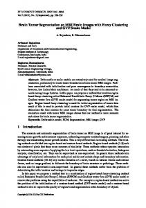
A medical image is often composed by a dark background and a light gray foreground. The presence of a strong edge between these two zones causes the arising of an over-illumination, called halo artifact, when a homomorphic filter is applied to the image. Guillemaud [8] proposed a homomorphic filtering scheme to compensate this over-illumination located on the boundaries using a binary image to identify the Region of Interest (ROI ) which bounds the foreground. This approach can be followed because there isn’t any useful information in the background: it contains only noise. In the left part of Fig.1 the filter scheme is shown. 12
10
8
6
4
2
entropy model 0
0
0.005
0.01
0.015
0.02
0.025
0.03
0.035
0.04
0.045
0.05
Butterworth cutoff frequency
Fig. 1. Left:Guillemaud scheme. Right: Bias local entropy curve and its estimated model. The point of the curve indicates the end of the transient phase whose abscissa corresponds to the desired cutoff frequency.
There is no trace in literature about the choice of the cutoff frequency, but this parameter is very important because it controls the filter. If it is too low no effect is visible on the restored image, while for high values a strong contrast loss can be noticed. A previous work from the authors [1] the E 2 D − HU M algorithm has been presented. The artifact is mainly located in the lower harmonics so a part of them has to be subtracted from the image spectrum to perform the restoration. These harmonics form the Bias image, which is a slow varying one: it doesn’t contain details and the image morphology isn’t involved. This harmonics transferring can be regarded as an information migration so that the information content in the Bias image grows according to an increment of the cutoff frequency. The information contained in the Bias image can be measured using the Shannon’s Entropy which is computed only on the foreground. Plotting the entropy amount vs. the Butterworth cutoff frequency diagram, a curve is obtained, which is an increasing monotonic function with an exponential profile. This behavior has been approximated by the capacitor charge mathematical
III
model and the cutoff frequency has been selected at the end of the transient phase, as depicted in the right part of Fig. 1. For more details we remaind to [1].
3
Preventing the Halo artifact using Fuzzy C-means
In section 2 we introduced the Guillemaud filter [8] to avoid the halo artifact between foreground and background but it is also produced by dark tissue in the foreground produces also the halo artifact on the surrounding tissues. Using the Guillemaud ROI shown in Fig.2g)g’), the halo arises as depicted in Fig.2d)d’). This is a particularly important problem in T1-weighted images, where the cerebrospinal fluid of the brain is located inside the ventricular system in the center position. As a consequence, a more careful image segmentation is required and we apply the same technique illustrated in [1]. The key idea consists in performing a fcm segmentation using 3 clusters (corresponding to dark, medium and light gray level) after the application of the filter with the Guillemaud ROI, so that strong shaded parts are restored. The darkest tissue, which corresponds to the cluster with the lowest centroid, is deleted from the Guillemaud ROI obtaining the masks shown in Figs.2h)h’). The filtering performed with this new ROI is shown in Figs.2f)f’), where the halos have disappeared. The experimentation has been performed on real images whose radiological parameters are shown in Table-2.
4
Image Segmentation
So far, fcm has been performed to avoid halos on the tissues produced by the filtering. Only gray levels in the ROI have been considered, because the background contains noise. Only the gray levels of the brain tissues are involved into the process and the algorithm speed is increased. The brain extraction can be performed using Brain Extraction Tool (BET) [5], already implemented in MRIcro [3]. A crucial task is the number of the clusters which. Usually the same number of clusters is chosen as the encephalic tissues: white matter, gray matter and cerebrospinal fluid. Using the proposed pre-processing, and a noise filter like the anisotropic diffusion filter [4] we obtain satisfactory results. Some results are shown in Fig. 3, which shows the slice n. 98 in a Brainweb simulated volume, and in Fig. 4 where a real slice is reported. The output of our implementation is the spatial distribution of the membership values of each cluster, whose values range in the interval ]0,1[. No limitation have been encountered as regards real data. Good segmentation has been obtained also from strongly corrupted slices (see Fig.4).
5
Results Evaluation and Measures
We performed E 2 D − HU M followed by fcm both on simulated T1-weighted images with 70% of RF-Inhomogeneity corruption provided by Brainweb [15][16] and real data. Once a slice has been extracted from the volume of Brainweb, it has been stored as raw data and converted into Analyze format using MRIcro [3] to be processed by SPM5 [17]. The segmentation using SPM5 fails both on real (Figs. 4-3rd row) and simulated data (Figs.3c)3d)3e)), while using our method the results are quite good (see Fig.3i)3j)3k) and Fig.4-2nd row). Brainweb provides also labelled tissues shown in Fig.3f3)g)3h) so that a visual comparison
IV
a)
c)
e)
g)
b)
d)
f)
h)
a’)
c’)
e’)
g’)
b’)
d’)
f’)
h’)
Fig. 2. Real T1-weighted image of the brain (up: dataset-1; down: dataset-2). For each group-Up:a)a’)original images and b)b’) their histogram equalization; c)c’) restored images and d)d’) their histogram equalization using Guillemaud ROI g)g’); e)e’) the restored ones and f)f’) their histogram equalization obtained using the masks h)h’). The dark boxes in figs d)d’) indicate the halo.
with a corrected segmentation can be performed. In order to provide an objective measure of the image segmentation, we introduce a segmentation index Si defined as follows: Ae − Ar Si = Ar where Ae is the area of the cluster spatial distribution; the same for Ar where it is computed on labelled tissues mentioned before. Si measures the amount in pixels of the relative difference between the areas of the ”true” segmented region (labelled in BrainWeb) and the one estimated by our algorithm. A positive value
V
a)
f)
b)
g)
c)
h)
i)
d)
j)
e)
k)
Fig. 3. Segmentation of slice 98 from T1-weighted Brainweb dataset with 70% of RFInhomogeneity. a) original corrupted image and b) its crisp representation after the application of Fuzzy C-Means. c)d)e) and i)j)k) are the spatial distributions of the clusters respectively for the Fuzzy C-Means after the application of E 2 D − HU M and SPM5 segmentation. f)g)h) are the labelled tissues which are also provided by Brainweb.
Fig. 4. Segmentation on real data. Fuzzy C-means segmentation on original image (first row) and after pre-processing (second row), the SPM5 results (third row). Here the spatial distribution of the each cluster is used instead of a crisp representation.
VI
of Si indicates an overestimation of the tissue, a negative value indicates an underestimation of the tissue and the zero value indicates a perfect identification of the cluster. Such a measure can be performed only on simulated data because we don’t posses a handmade segmentation of the slices.The results in the table of figure 3 show that our approach is better in tissues estimation with respect to SPM5. The estimation error is bounded under the 10%. Similar numerical results have been obtained for all the slices in the BrainWeb simulated data set.
6
Conclusions
SPM5 doesn’t use only raw image data in the file. It requires also spatial information: it is a strictly radiological tool, rather than an image processing method. SPM5 has an internal model so that a process consisting in a combination of registration, segmentation and RF-Inhomogeneity correction trying to find a best matching of the given volume with the internal atlas/model. This atlas is the result of a co-registration of statistical study performed on many subjects and provides a spatial estimation of the encephalic tissues. The presented segmentation scheme, which makes use of E 2 D − HU M and fcm doesn’t require such sophisticated input information and provides results that are comparable with the literature state of the art.
7
Acknowledgments
Particular thanks to Ernesto Basilico, eng. ;Mr. Filippo Longo; Maria Pia Pappalardo,Md. and all the staff of Palermo’s Ospedale Civico.
References 1. Ardizzone, E.; Pirrone ,R. and Gambino, O. :Exponential Entropy Driven HUM on Knee MR Images Proc. of IEEE XXVII Engineering in Medicine and Biology Conference - 4/7 September 2005 SHANGHAI (CHINA) 2. Nelder, J. A. and Mead, R. A Simplex Method for Function Minimization. Comput. J. 7, 308-313, 1965. 3. http://www.sph.sc.edu/comd/rorden/mricro.html 4. P. Perona and J. Malik. Scale-Space and Edge Detection Using Anisotropic Diffusion. IEEE Trans. on Pattern Analysis and Machine Intelligence, 12(7):629–639, Jul 1990. 5. S.M. Smith. Fast robust automated brain extraction. Human Brain Mapping, 17(3):143-155, November 2002. 6. B. Johnston, M. S. Atkins, B. Mackiewich, Member and M. Anderson. Segmentation of Multide Sclerosis Lesions in Intensity Corrected Multispectral MRI. IEEE Transaction On Medical Imaging, vol. 15, no. 2, April 1996 7. Mohamed N. Ahmed; Sameh M. Yamany; Nevin Mohamed: A Modified Fuzzy C-Means Algorithm for Bias Field Estimation and Segmentation of MRI Data. IEEE Transactions on Medical Imaging 21 (2002) 193–199 8. Guillemaud, R.: Uniformity Correction with Homomorphic filtering on Region of Interest. IEEE International Conference on Image Processing 2 (1998) 872–875 9. Axel L.; Costantini J.; Listerud J.: Intensity Correction in Surface Coil MR Imaging. American Journal on Roentgenology 148 (1987) 418–420 10. Lei Jiang,Wenhui Yang:A Modified Fuzzy C-Means Algorithm for Segmentation of Magnetic Resonance Images. Proc. VIIth Digital Image Computing: Techniques and Applications.Sun C., Talbot H., Ourselin S. and Adriaansen T. Editions. (2003) 225–231 11. Brinkmann B. H. , Manduca A. and Robb R. A.: Optimized Homomorphic Unsharp Masking for MR Greyscale Inhomogeneity Correction. IEEE Transactions on Medical Imaging. 17 (1998) 161–171 12. Likar B.; Viergever M.A.; Pernus F.: Retrospective Correction of MR Intensity Inhomogeneity by Information Minimization. IEEE Transactions on Medical Imaging 20 (2001) 1398–1410 13. Pham D.L.; Prince J.L.: Adaptive Fuzzy Segmentation of Magnetic Resonance Images. IEEE Transactions on Medical Imaging. 18(9), (1999) 737-752 14. Pham D.L.; Prince J.L.: An Adaptive Fuzzy C-Means Algorithm for Image Segmentation in the Presence of Intensity Inhomogeneities. Pattern Recognition Letters. 20(1), (1999) 57-68 15. Kwan R.K.S.; Evans A.C.; Pike G.B.: MRI simulation-based evaluation of image-processing and classification methods. IEEE Transactions on Medical Imaging. (1999) 18(11):1085–1097. 16. Kwan R.K.S.; Evans A.C.; Pike G.B.: An Extensible MRI Simulator for Post-Processing Evaluation. Visualization in Biomedical Computing (VBC’96). Lecture Notes in Computer Science, vol. 1131. Springer-Verlag, (1996) 135–140. 17. http://www.fil.ion.ucl.ac.uk/spm/software/spm5/