0.3456. 8 -0.0089. 0.2304. 0.8978. 0.4957. 0.3067. 0.3786. 9 0.3292. 0.2052. -0.2792. 0.4068. 0.0062. 0.3076. 10 0.2958. 0.2705. 0.9813. 0.8763. -0.3292.
Generating treatment bootstrap samples from random positioning uncertainty for IG-IMRT and IMRT treated patients Radiotherapy planning and image guidance Radiotherapy was planned in 1.8 Gy fractions to a total of 86.4 Gy using IMRT. The prostate, central part of the prostate (encompassing the urethra), rectum, bladder, bladder wall, were delineated. A planning target volume (PTV) margin of 1.0 cm except posteriorly where the margin was 0.6 cm was assigned to the clinical target volume (CTV), which encompassed the prostate plus seminal vesicles. Dose-volume limiting optimization objectives were assigned to the rectum and the bladder during the planning process. In addition, care was taken so that dose hot-spots did not overlap with the urethra. The PTV dose was within 95107% of the prescription dose if feasible, with the low dose tending to be located in the prostate/rectum interface. Patients treated with daily IG were positioned supine, and were initially set up to skin marks. Thereafter, orthogonal kilovoltage 2D radiographs (OBI, Varian Medical Systems, Palo Alto) were obtained, and the patients position were corrected using 3D translations if the deviation of fiducial marker positions exceeded 2 mm in any direction as compared to the position on the planning CT. A set of verification radiographs was afterwards taken to ensure that the new position was within 2 mm in all directions. The standard procedure was then to treat the patient without post-fraction imaging. Patients treated without daily IG were typically set up in prone position using skin marks, and the position was verified weekly using megavoltage images matched onto the bony anatomy of the planning CT.
Method for positioning used at MSKCC The IMRT group (treated without IGRT) were typically in prone position (64 out of 67). Patients were positioned towards bony structures once a week, and the correction vector from skin marks to bony structures was recorded and used throughout the rest of the week. This procedure was repeated for the course of 45-48 fractions. The patients treated with daily IGRT and IMRT (IG-IMRT) were first positioned using skin marks and this position was subsequently corrected using orthogonal x-rays daily and fiducial markers inside the prostate. The tolerance for the IGRT procedure was 2 mm (in each cardinal direction).
Method for sampling positioning variation The position (P) will be sampled in the following way: 𝑃!!"#
!
= 𝑆𝐸 !"#
!
+ 𝑅𝐸!!"#
!
Where x is sample number (1-200), and dir is direction (AP, CC, LR) and i is fraction number (typically 1-48). Note that SE is the systematic error and drawn once for each sample and RE is the random error. SE and RE are derived by 𝑆𝐸 !"#
!
= 𝑆𝐸 !"
!
+ 𝑆𝐸!!"#
!
Where, index IG is the inherent uncertainty introduced by the accuracy of the imaging system and drawn once for each sample; 0.75 mm (1SD) in each direction. For IG-IMRT patients, the term indexed P is zero, and for or IMRT patients, then this is picked randomly from the distribution (assumed normal distribution) and from the data below. 𝑅𝐸!!"# ! equals 1.4 mm (1SD) for patients treated with daily IGRT, stemming from the matching procedure (est. to be 1 mm) and the residual error tolerated of 2 mm (est. to equal to 2 SD). For IMRT patients the 𝑅𝐸!!"# ! varies with direction, and were estimated using the data previously presented by our group (Munck af Rosenschold et al, 2014).
We split patients into four risk groups in terms of positioning uncertainty (Group 1-4, below) using BV (bladder volume at CT simulation) and RCS (rectum cross section area at CT simulation). Then, we pick randomly a patient number (see below) within each patient group for each bootstrap sample (1-200). Obviously, we might then pick the same series of SE and RE for a subsequent bootstrap sample. However, given that the SE and the RE are used distributions used for subsequent sampling, the same series of positioning deviations will not be reproduced in multiple bootstrap samples. The advantage of the method is that it retains the internal correlation of uncertainty estimates in all directions (cf. Munck af Rosenschold et al, Radiother Onc 2014). SE and RE for the patient groups are found in tables below.
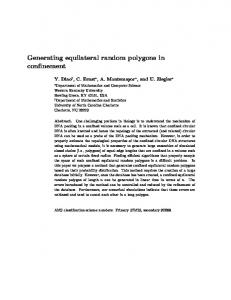
Definition of dorsolateral region For the purpose of this analysis, we defined 5 regions beyond the prostate gland. Each region concentrically were isotropically expanded out 0.25 cm from the dorsal half of the CTV (prostate). Thus, the regions encompass the posterolateral aspect of the gland including the neurovascular bundles. However, the regions were made such that they were not intersecting with the rectum (see schematic below).
Schematic of definition of dorsolateral regions for simulations (not to scale)
REGIONS 1-5
PROSTATE
CENTER OF GRAVITY
Re Re gion R gio 1 Re egion n 2 Re gion 3 gio 4 n5
RECTUM
Dosimetric calculations We attempted to quantify the radiation therapy dosimetry difference for the IMRT and IGIMRT cohorts by means of simulating the positioning uncertainty, following a similar procedure as Zhang et al (Phys Med Biol 2006). The patients’ planned dose distribution, CT
data set and contours were extracted from the in-house treatment planning software and imported into the CERR software (Deasy et al, Med Phys 2003). Using a programming script, the simulated isocenter position was sampled using a systematic and a random error. The sum of the dose is derived for each patient, each bootstrap sample and for each position in three-dimensional space: !
𝐷!"!
!
=
𝑑! !
!
Dtot is the sum dose matrix of a series of doses (di) from fractions (n), for bootstrap sample x (200 samples). From the Dtot dose matrix, we calculate the average and the standard deviation of the dose based on the fraction variations for the prostate. Finally, using the 200 bootstrap samples for each patient, the risk of receiving a dose reduction of at least 10 Gy lower than the prescription dose, in each of the Regions 1-5, is derived.
Positioning data Systematic deviation (SE) and random deviations (RE) (1 sd) in each direction given in cm below. Group 1 BV6.148 RE_CC 0.3476 0.2515 0.2465 0.3488 0.3079 0.3588
SE_AP -0.5729 0.9104 -0.5125 -0.2771 0.1867 -0.2021
RE_AP 0.3940 0.3191 0.2922 0.3771 0.4383 0.6809
SE_SD 0.4083 -0.0250 0.3187 -0.0125 -0.1933 -0.0667
RE_SD 0.3059 0.2654 0.2170 0.2247 0.2692 0.3205
7 8 9 10 11 12 13 14 15 16 17 18 19 20 21 22 23 24 25 26 27 28 29 30 31 32 33 34 35 36 37 38 39 40 41 42 43 44 45 46 47 48 49 50 51 52
0.2438 -0.1521 -0.0375 -0.0583 0.2489 0.1313 0.4729 -0.2652 -0.0178 -0.4688 0.2133 0.1109 0.1188 0.2333 0.3333 -0.1917 -0.7729 -0.6396 -0.2063 0.4604 0.1438 0.5938 0.3295 0.1604 0.1500 0.3417 0.3889 0.0271 -0.1458 0.5000 0.1625 -0.0400 0.2583 -0.1771 -0.0391 -0.1067 0.0644 0.3667 -0.1111 0.0851 0.0000 0.1396 -0.2979 0.0813 0.1479 0.3400
0.3798 0.2851 0.2376 0.2835 0.2096 0.9214 0.3752 0.3749 0.2026 0.2519 0.6642 0.4443 0.3272 0.4526 0.3097 0.3188 0.4186 0.2789 0.3111 0.2719 0.2287 0.5829 0.2775 0.2908 0.2917 0.3228 0.2773 0.1954 0.2432 0.3307 0.3133 0.3683 0.5910 0.2611 0.4404 0.3041 0.2248 0.3341 0.1898 0.2897 0.2895 0.3356 0.3864 0.4286 0.3268 0.2597
-0.2938 -1.4708 0.6167 0.3938 -0.0889 0.2083 -0.5292 1.5087 -0.1489 0.6604 -0.3356 -0.1696 0.8229 -0.3458 0.1133 -0.1188 0.7729 -0.7229 -0.3271 -0.2958 0.6896 -0.7917 -0.1750 -0.3271 -0.5312 -0.1917 0.6978 -0.3271 0.4313 -0.6609 1.1792 0.0756 -0.5062 0.3667 -0.0348 -0.8511 -0.7558 0.8792 0.4311 -0.1894 -0.7792 -0.0875 1.3043 -0.2375 -0.8208 0.0889
0.4479 0.3142 0.4478 0.2374 0.4458 0.4694 0.4594 0.5253 0.3195 0.3292 0.5068 0.4273 0.4926 0.3775 0.4832 0.5221 1.2576 0.4922 0.4685 0.4496 0.3544 0.7136 0.3404 0.3972 0.2918 0.5193 0.4309 0.3147 0.4043 0.8280 0.8508 0.3797 1.0022 0.3478 0.4463 0.4088 0.5624 0.4376 0.4461 0.5665 0.3038 0.4975 0.5793 0.5484 0.4749 0.3393
0.2125 0.5187 0.3521 0.1104 0.1089 -0.0333 0.3396 0.1500 -0.1200 0.0167 0.0267 -0.0239 0.3021 0.3833 0.1644 -0.2042 0.0396 -0.2063 0.2938 0.2354 -0.1333 -0.2188 -0.0045 -0.2146 0.4521 0.0021 0.3644 0.1354 -0.2125 0.0604 0.1187 0.0089 -0.2083 -0.0729 -0.4087 0.0489 -0.1602 0.2250 0.5000 0.2064 -0.0604 0.2542 0.1532 0.0687 -0.2667 0.1311
0.2170 0.1875 0.2010 0.2746 0.2485 0.2999 0.3344 0.2934 0.2616 0.5799 0.3236 0.3288 0.3949 0.2373 0.2656 0.3242 0.4471 0.2740 0.2629 0.1995 0.3110 0.2893 0.3510 0.2297 0.2903 0.2058 0.2846 0.2914 0.3577 0.2295 0.2871 0.3232 0.4292 0.2574 0.4386 0.2191 0.2985 0.2862 0.3148 0.2399 0.3133 0.3591 0.3966 0.3904 0.4334 0.2193
53 54 55 56 57 58 59 60 61 62 63 64 65 66 67
-0.0354 0.1146 -0.2104 0.0687 0.1354 -0.2833 0.2958 -0.3896 -0.7208 0.4596 -0.2178 0.0188 0.2708 0.0042 0.0917
0.3111 0.4736 0.2628 0.3163 0.2794 0.2596 0.2240 0.3137 0.4005 0.4277 0.2377 0.2498 0.3017 0.2657 0.3045
0.2813 0.1146 1.4729 -0.2333 -0.1375 0.1792 -0.7167 -0.4146 0.6604 0.5085 0.3422 -0.3313 0.2083 -0.4937 -0.2938
0.2929 0.3679 0.4671 0.4239 0.5193 0.4400 0.5158 0.5255 0.3852 0.4786 0.6514 0.3963 0.5603 0.4633 0.3303
0.3208 -0.1042 0.0563 0.1250 0.0396 0.3333 -0.0771 0.1687 0.2063 -0.1574 0.4311 0.2313 0.1208 -0.3833 0.1646
0.2526 0.2414 0.2492 0.2622 0.2796 0.3657 0.3484 0.2261 0.2226 0.2204 0.3759 0.3321 0.2729 0.3354 0.1695