other bacteria taxonomically similar (Brock 1995). They have formed a ...... Manual of Systematic Bacteriology (Claus and Berkeley 1986). ...... Microbiologia.
I. INTRODUCTION 1.1. Historical aspects The first record of bacteriocins dates back to 1925, when André Gratia demonstrated the activity of a highly specific inhibitory substance, produced by the bacterium Escherichia coli, with a bactericidal activity against other E. coli strains (de Lima and Filho 2005). It was observed that E. coli cells produced in liquid medium a substance stable to temperature oscillations and also possesses an inhibitory activity on the growth of other taxonomically related microorganisms. Since the discovery of the bacteriocins, most of the studies established in this field have been descriptive (Fredericq 1957 and 1963), and the basic methods of detections, assay and the bacteriocin typing of bacterial strains have been established. Genetic studies of colicinogeny and its transfer from one cell to another were also pioneered by Fredericq (1957 and 1963). Rogers (1928) reported that lactococci could also produce antibacterial substances. Whitehead (1933) then demonstrated that the lactococcal factor was proteinaceous. Mattick and Hirsch (1944) tested the concentrated factor against pathogenic streptococci, and Taylor et al. (1949) attempted to use the same inhibitory substance to treat bovine mastitis. Further studies on the molecular biology of the bacteriocins followed, with emphases on biosynthesis, liberation and the mode of action of these agents (Lakey et al. 1993). These data led to interesting use of the bacteriocins as a biochemical tool in cellular physiology (Becker et al. 1993). The occurrence of antagonistic interactions among bacteria was reported by Pasteur and Joubert at the end of the 19th century. These researchers observed that a bacterial isolate was able to interfere with the Bacillus antrachis growth (Jack et al. 1995). Similarly, inhibitory action exerted by a Staphylococcus spp. isolate when an interaction with Corynebacterium diphyteriae was also observed. This discovery led, at that time to the employment of Staphylococcal isolates as useful procedure to be applied in diphtheria treatment (Florey 1996). These activities were found to be produced by various species of enterobacteriaceae and for which the generic name (colicine) was proposed. Jacob et al. (1952) proposed that the general name (bacteriocine) should be used for highly specific ribosomally synthesized antibacterial proteins differentiated from the classical antibiotics, and produced by certain strains of bacteria and active mainly against strains of the same species. Bacteriocins have since gained new attentinon particularly in the epidemiology of nosocomial infections; they have been found to be very useful in typing organisms particularly those which are difficult to type by the usual methods (Lebek et al. 1993).
1.2. Microbial defense systems Microbes produce an extraordinary array of microbial defense systems to maintain their existence or ecological niche (Riley and Wertz 2002) against competitors or infections (Nissen-Meyer and Nes 1997). Microbial defense systems include broad-spectrum classical antibiotics so critical to human health concerns, metabolic by-products such as the lactic acids, lytic agents such as lysozymes found in many foods, and numerous types of protein exotoxins (James et al. 1991). The production of antimicrobial peptides is a first line of microbial defense, and also part of the innate immunity, found in a variety of species (Cleveland et al. 2001). Sometimes the peptides act against a specific group of competing organisms; sometimes their broad spectrum of activity serves as a more general defense
1
mechanism. Although such peptides may have a direct effect on the microbe, such as by damaging or destabilizing the bacterial, viral, or fungal membrane or acting on other targets, they appear to be broadly involved in the orchestration of the innate immune and inflammatory responses (Hancock and Diamond 2000a). Antimicrobial peptides are a universal feature of the defense systems of virtually all forms of life, with representatives found in organisms ranging from bacteria to plants and invertebrate and vertebrate species, including mammals. One family of microbial defense systems; antimicrobial peptides produced by bacteria; bacteriocins were among the first to be isolated and characterized (Mattick and Hirsch 1947). While they do not protect against infection in the classical sense, they contribute to survival of individual bacterial cells by killing other bacteria that might compete for nutrients in the same environment. Bacteriocins are thought to be produced by many or most bacteria (Klaenhammer 1988) and are generally extremely potent compared with most of their eukaryotic counterparts. Their activities may be either narrow or broad spectrum, capable of targeting bacteria within the same species or from different genera. Bacteriocins are found in almost every bacterial species examined to date, and within a species tens or even hundreds of different kinds of bacteriocins are produced (Riley and Gordon 1992).
1.3. Ecology On an evolutional basis, it appears that the ability to synthesize one or more bacteriocins has been a highly advantageous characteristic (Chen and Hoover 2003). A clear opportunity for survival and proliferation of an organism can be envisioned if it can eliminate a competing organism in its ecological niche, where competition can be quite intense given the diversity of species and rapid growth of bacteria (Dykes 1995). Bacteriocins play a fundamental role in bacterial population dynamics even though the degree of bacteriocin interactions is so complex at the ecological and evolutionary levels in mixed populations (such as biofilms) that much remains uncertain. Bacteriocins may serve as anti-competitors enabling the invasion of a strain into an established microbial community. They may also play a defensive role and act to prohibit the invasion of other strains or species into an occupied niche or limit the advance of neighboring cells. As noted by Riley (1998), examination of bacteriocins in natural settings, such as Lactobacillus plantarum in green olive fermentations, Escherichia coli in guinea pig conjunctivae, and Streptococcus mutans in the human oral cavity, have indicated that the competitive advantage is substantially increased for bacteriocin-producing cells against bacteriocin-sensitive bacteria in the same environments. In the L. plantarum example, a bacteriocin-producing strain was used to ferment Spanish-style green olives (Ruiz-Barba et al. 1994). Mathematical models have been devised to evaluate the interaction between bacteriocin producers and sensitive varieties. Most ecological modeling of bacteriocins has been with colicins (bacteriocins produced by E. coli and usually showing activity against other strains of E. coli and very closely related members of the Enterobacteriaceae). It is estimated that about 30% of the natural populations of E. coli produce bacteriocins (Riley 1998). Induction usually occurs under stressful conditions such as nutrient depletion or overcrowding (Riley and Gordon 1999).
2
1.4. Production of antimicrobial peptides 1.4.1. Definition of bacteriocins The definition of bacteriocins has changed rapidly since early this century. Antibiotics designate all substances originating from microorganisms, animals, plants or through the synthetic way that at low concentrations must have the following properties: i) lethal or inhibitory activity against microbial species; ii) ability to prevent the appearance of microbial resistance; iii) absence of undesirable effects to the host; and iv) chemical stability (Tavares 1984; Amato Neto et al. 1994; Abrahan et al. 1996). In general way, antibiotics can be defined as agents of great interference on the growth and/or microbial activities (Trabulsi et al. 2002). Substances recognized as antibiotics exert their activity by different ways that act on diverse targets in/on the microbial cell. It includes: i) damaging the cell wall; ii) destabilizing the cytoplasmic membrane; iii) changing the nucleic acid structure; iv) inhibiting the enzymatic action; v) antimetabolic action; and vi) affecting the acid nucleic synthesis (Pelczar et al. 1980). According to Tagg et al. (1976), bacteriocins are defined as proteinaceous compounds that kill closely related bacteria. This definition is true for the majority of bacteriocins investigated, but gradually it has become evident that certain bacteriocins may also elicit bactericidal activity against species that are more distantly related to the bacteriocin producer. Bacteriocins are one of the most abundant and diversified molecules with antibiotic activity produced by different bacterial genera including gram-positive and gram-negative species (Riley 1998). Bacteriocins have been designated as bacterial substances with capacity of inhibiting, even in low concentrations, the multiplication of other bacteria taxonomically similar (Brock 1995). They have formed a heterogeneous group about the producing-bacterial species, molecular size, antibacterial spectrum, stability, physical and chemical properties, and action mode (De Vuyst and Vandame 1994). Montville and Kaiser (1993) suggested that the definition of bacteriocins should be based on only two basic requisites, i.e. their protein nature and the absence of lethality to the producer cells. These authors confirmed that few antimicrobial proteins fit the classical definition proposed by Tagg et al. (1976). Bacteriocins have been designated as antibiotics by definition, and are differentiated from antibiotics on the basis of synthesis, mode of action, antimicrobial spectrum, toxicity, resistance mechanisms, and applications and clinical effects (Table 1) (Klement et al. 1990; Davis 1999). However, they were often confused in the literature with antibiotics (Hansen 1993). Antibiotic properties (Chen and Hoover 2003) should be safely and effectively used to control the growth of target pathogens in foods (Table 1).
1.4.2. Antimicrobial peptides from eukaryotes In plants, it is widely believed that antimicrobial peptides play an important and fundamental role in defense against infection by bacteria and fungi (Jensen et al. 2006). For instance, plant defensins display antibacterial and antifungal activities in vitro (Terras et al. 1992). Consistent with a defensive role, they are found in leaves, flowers, seeds, and tubers. Antimicrobial peptides have been also isolated from a wide range of invertebrate and vertebrate species, including fish, amphibians, and mammals, indicating that, even in
3
the presence of an adaptive immune response, these peptides have an important role in host defense. Amphibian skin glands have proven to be a rich source of antimicrobial peptides. Magainins (Zasloff 1987) are the prototypic amphibian antimicrobial peptides, with strong membrane-permeabilizing activity towards gram-positive and negative bacteria, fungi, yeasts, and viruses. Cathelicidins are a large and diverse group of vertebrate antimicrobial peptides. A second prominent group of mammalian antimicrobial peptides is the defensins (Ganz 2003). Histatin recovered from saliva from humans and primates and primarily directed against fungal pathogens, outstands for its distinctive mechanism of action which does not involve channel formation in the fungal cytoplasmic membrane but rather translocates efficiently into the cell and targets the mitochondrion (Tsai and Bobek 1998).
1.4.3. Antimicrobial peptides from bacteria Antimicrobial peptides have been produced and detected in all lineages of eubacteria and archaebacteria (Torreblanca et al. 1995). According to Klaenhammer (1988), 99% of all bacteria may make at least one antimicrobial peptide and the only reason that more have not been isolated is that very few researchers have looked for them. Microbes invest considerable energy into the production and elaboration of these antimicrobial mechanisms. Various environmental conditions, including pH, temperature, aeration, sugar concentration, buffering capacity of the medium and time of incubation, affect the production of bacteriocins (Lewus 1991) The physiological status of the sensitive strain has a great influence on its susceptibility to the lethal action of antimicrobial peptides, with metabolically active cells being more sensitive (Tag et al. 1976).
1.4.3.1. Bacteriocins of gram positive bacteria Bacteriocins are thought to be produced by many or most bacteria (Klaenhammer 1988) and are generally extremely potent compared with most of their eukaryotic counterparts. The most relevant active membrane peptides are those produced by gram positive bacteria (Oscáriz and Pisabarro, 2001). At one time it was an accurate statement to say that colicins were the most studied and most understood of the bacteriocins; however, it is now safe to say that the bacteriocins produced by gram-positive bacteria are the most investigated group of antibacterial peptides, given their potential for commercial applications in foods and other products (Schillinger et al. 1996). The activity of gram-positive bacteriocins may be either narrow or broad spectrum, capable of targeting bacteria within the same species or from different genera. Bacteriocins produced by gram-positive bacteria differ from those produced by gram-negative bacteria. These bacteriocins have a broader activity spectrum than those produced by gram-negative bacteria, and are often active against a wide range of gram-positive and sometimes against a few gram-negative bacteria. Specific immunity by a producing strain is less strong, most likely because the bacteriocins produced by gram-positive bacteria do not bind to a specific receptor protein. Instead, gram-positive bacteriocins are thought to interact directly with the phospholipid head groups of the membrane. The gram-positive bacteriocins are generally of the channel-forming type and not the nuclease type. Most of these bacteriocins are small (less than 5 KDa), heat stable, amphiphilic, membrane-permeabilizing peptides, cationic proteins that are structurally unlike the colicins. Most are synthesized as prepeptides in which the leader peptide is removed to form the biologically active bacteriocin. In this manner they resemble many of the antimicrobial peptides produced by eukaryotes (Jon Nissen and Ingolf 1997).
4
The most thoroughly studied bacteriocins are those produced by lactic acid bacteria (LAB), of which sakacins seem to be most unique (Jack et al. 1995), and the lantibiotics, which contain modified amino acid residues (Oscáriz and Pisabarro 2001). Bacteriocins of LAB are commonly classified into 3 groups that also include bacteriocins from other gram-positive bacteria (Klaenhammer 1993; Nes et al. 1996). Lantibiotics (from lanthionine-containing antibiotic) are small ( 10,000 Da) and the lower aliquot (< 10,000 Da) were collected and tested for antibacterial activity (Jiménez-Diaz et al. 1993).
2.2.12.2. SDS-PAGE technique The molecular weight of bacteriocins was estimated according to the method of Sambrook and Russell (2001) using discontinuous SDS-polyacrylamide gel electrophoresis (SDSPAGE) performed using a double slab electrophoresis cell (Cleaver scientific Ltd). The gel was 1.5 mm thick, with a 9 cm running gel of 15 % SDS-polyacrylamide resolving gel (5.0 ml of 30% (w/v). The acrylamide mix was composed of 29 grams of Acrylamide, 1 gram of N, N'-Methylene bisacrylamide in distilled water, stored at 4˚C in a dark bottle, 2.5 ml of 1.5 M Tris (pH 8.8), 0.1 ml 10 % Sodium dodecyl sulphate (SDS) 2.3 ml distilled H2O, 0.1 ml of 10 % (w/v) ammonium persulfate, and 0.004 ml of N,N,N',N'Tetramethylethylenediamine (TEMED). The mixture was overlaid with 5 ml solution of 5% SDS-polyacrylamide stacking gel ( 0.83 ml of 30 % acrylamide mix, 0.63 ml of 1.0 M Tris pH 6.8, 0.05 ml 10 % SDS, 3.4 ml distilled H2O, 0.05 ml of 10 % (w/ v) ammonium persulfate, and 0.005 ml of TEMED). 10 µl of the partially purified stationary-phase culture supernatant was mixed with 10 µl of 1x SDS gel-loading buffer (5ml of 1M Tris pH 6.8, 8 ml Glycerol, 16 ml 10% SDS, 5 ml distilled water, 1 ml β-Mercaptoethanol, 10 µg Bromophenol blue) and boiled at 100˚C for 5 minutes. 15 µl of the samples were loaded in a predetermined order onto the gel. The running buffer used consisted of: 1x Tris-glycine electrophoresis buffer (5x Tris-glycine buffer is composed of: 15.1 (g/l), Tris-
35
base, 94 glycine, and 50 ml of 10 % (w/v) SDS. Electrophoresis was carried out at (70 V/15 mA) in the stacking gel, increased to (100 V/25 mA) through the resolving gel until the tracking dye reached the bottom of the gel. The gel was stained overnight in 0.1% Comassie blue stain (0.25 g of Comassie brilliant blue R-250 in 90 ml of methanol: H2O (1:1, v/v) and 10ml glacial acetic acid). To diffuse excess dye from the gel, it was placed in a distaining solution (45 ml methanol, 10 ml glacial acetic acid and 45 ml H2O) for several hours. The apparent molecular masses of the samples were calculated by comparison with the mobility of standards.
36
III. RESULTS 3.1. Isolation of bacterial strains The first screening revealed about 30 isolates obtained from the selected sources (Table 5) that were collected from various locations. The largest number of isolates was obtained in LB medium. All strains isolated were gram-positive rods, aerobic or facultative; from which 18 isolates were spore-forming bacteria. Table 5: Origin of isolated bacilli strains used in the study Sources Milk and milk products Meat and meat products Soils Silage Fermented foods Mixture of fermented vegetable waste Mixture of fruit waste Pickles Cereals Spices Fermented olives Kitchen foods (raw and cooked) Compost Eggs Roots of beet and carrot Grass
Number of isolated Bacilli 2 3 6 1 1 2 1 2 1 3 1 1 1 1 3 1
3.2. Screening of isolated bacilli for bacteriocin production All isolated strains were screened for antibacterial activity spectrum against various grampositive and gram-negative bacteria using the AWD method. Table 6 shows the effect of crude bacteriocins produced by the isolated bacilli on the listed ten indicator strains including important pathogenic microorganisms. Among the 30 isolated strains of bacilli, 4 strains (Strain 9, 19, 22 and 27) inhibited the growth of representatives of at least two indicator strains whether gram positive or gram-negative strains. The average diameter of the inhibition zones obtained ranged from 0-4 mm in size. Few strains (strain 4, 10, 13, 18, and 25) showed insignificant inhibitory behaviour against the indicator strains (i.e. inhibited the growth of only one or two indicator strains). The other isolates had no capacity to inhibit the growth of any indicator strain. The majority of crude bacteriocin preparations inhibited the growth of E.coli. Alternatively, results were also confirmed by the growth kinetics method, where the inhibition of the CFS of strains19 and 22 was the greatest (data not shown). For these reason, these two strains were selected for strain identification and further antimicrobial activity characterization.
37
Table 6: Demonstration of the antimicrobial activity spectrum of the crude bacteriocins obtained from MRS cultures of bacilli isolates against gram-positive and negative indicator strains using the (AWD) method. Results were recorded after 24 hours of incubation at 30oC. Crude bacteriocin of Bacilli isolates 1
2 *
3 *
4 *
5 *
6
7 *
8 *
10 *
11
12
13 *
14 *
15 *
16 *
17 *
18 *
19 *
20
21
22 *
23
24
25 *
26 *
27 *
28
E. coli
-
-
-
-
-
-
-
-
+
+
-
-
+
-
-
-
-
+
+
-
-
+
-
-
+
-
+
-
Str. pyogenes β-hemolytic
-
-
-
-
-
-
-
-
-
-
-
-
-
-
-
-
-
-
-
-
-
-
-
-
-
-
+
-
P. aeruginosa
-
-
-
-
-
-
-
-
-
+
-
-
-
-
-
-
-
-
-
-
-
-
-
-
-
-
-
-
Proteus vulgaris
-
-
-
-
-
-
-
-
+
-
-
-
-
-
-
-
-
-
-
-
-
-
-
-
-
-
-
-
K.. pneumoniae
-
-
-
-
-
-
-
-
-
-
-
-
-
-
-
-
-
-
-
-
-
-
-
-
-
-
-
-
S. aureus
-
-
-
+
-
-
-
-
+
-
-
-
-
-
-
-
-
-
+
-
-
+
-
-
-
-
-
-
Str. α-hemolytic
-
-
-
-
-
-
-
-
-
-
-
-
-
-
-
-
-
-
-
-
-
-
-
-
-
-
-
-
S. typhimurium
-
-
-
-
-
-
-
-
-
-
-
-
-
-
-
-
-
-
+
-
-
+
-
-
-
-
-
-
S. para- typhimurium A
-
-
-
-
-
-
-
-
-
-
-
-
-
-
-
-
-
-
-
-
-
-
-
-
-
-
-
-
S. para- typhimurium B
-
-
-
-
-
-
-
-
-
-
-
-
-
-
-
-
-
+
-
-
-
-
-
-
+
-
+
-
Indicator strains
+ – *
9
: Inhibition zone of size (0–4 mm). : No inhibition zone. : Spore-forming bacilli. 38
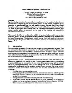
3.4. Identification and characterization of isolates with antimicrobial potential Two isolates (19 and 22) out of the bacilli strains were chosen for further experiments and characterization of their antimicrobial activity. Their selection was based on displaying high bacteriocin activity and their potential of inhibiting the growth of three indicator strains. The isolates were preliminarily identified by their morphological, cultural, physiological, and biochemical characteristics. Light microscopy showed that the two isolates were spore-forming, straight Gram-positive rods (Fig. 2). Biochemical patterns as tested using the API 20E system indicated that the isolates belonged to the genus Bacillus. The biochemical identification was confirmed by sequencing of the 16S rRNA gene from both isolates which was successful in identifying the isolates to the species level. The 16 rRNA gene sequence analysis of the two strains as determined by direct sequencing of PCR-amplified 16 rRNA was identical, having 100% sequence identity with Bacillus megaterium DSM 32. These two isolates were nominated as B. megaterium 19 (ID07-817) and B. megaterium 22 (ID07-818).
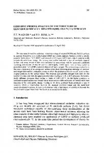
3.5. Bacterial growth curve In general, the two strains under investigation exhibited different growth paterns when cultivated under the same conditions. The lag phase for strain B. megaterium 19 was 3 hours shorter than that of strain B. megaterium 22 (which extended up to 6 hours of incubation), however, the growth of such strain in MRS broth produced a prolonged stationary phase (nearly up to 21 hours of incubation) compared to the latter strain when grown in the same medium. To determine the optimal growth phase for maximal bacteriocin production, the bacteriocin activity was monitored at 3 hours intervals up to 24 hours and in all the following experiments the decrease in bacteriocin activity was measured as % growth reduction. Bacteriocin activity was first observed after 1-2 hours of incubation of the two organisms (19 and 22, respectively). A low level of bacteriocin activity (almost 15% reduction) was noted during the lag phase of the producer strain 19. On the other hand, higher bacteriocin production of strain 22 was recorded during its lag phase (> 60 %). The maximum antibacterial activity of the bacteriocins against the indicator strains S. typhimurium and S. aureus was evaluated primarily during the stationary phase and peaks at its mid after 18 and 15 hours (for strains B. megaterium 19 and 22 respectively) of growth in MRS medium (Fig. 3 A and B), after which the growth (determined by culture turbidity) as well as the bacteriocin activity started to decline gradually within the 24 hours of experimentation.
39
A
B Fig. 2: Light micrographs of Gram-stained cells of isolates B. megaterium 19 (A), and B. megaterium 22 (B). Cells are gram-positive straight rods. Bar 12.5 = µm.
40
(A) 90
1.2
1.0
0.8 50 0.6 30
OD 600 nm
Reduction of growth (%)
70
0.4
10
0.2
-10
0.0 0
3
6
9
12
15
18
21
24
Time (h)
S. typhim urium
S.aureus
B. m egaterium 19
(B) 90
2.0 1.8 1.6 1.4
50
1.2 1.0
30
0.8
OD 600 nm
Reduction of growth (%)
70
0.6 10
0.4 0.2
-10
0.0 0
3
6
S. typhim urium
9
12 Time (h)
15
S.aureus
18
21
24
B. m egaterium 22
Fig. 3: Growth curve of B. megaterium 19 (A) and 22 (B) grown in MRS broth, incubated aerobically under shaking conditions at 30oC for 24 hours. O.D. values are averages of duplicate measurements (n=2). Activity of bacteriocin preparations against S. typhimurium ( ) and S. aureus ( ) was expressed as % reduction of growth. Error bars represent the standard deviation of mean values of activity % of bacteriocin preparations against each indicator strain.
41
3.6. Determination of the antimicrobial spectrum of bacteriocins The inhibitory spectrum of bacteriocin preparations of B. megaterium strains 19 and 22 was examined against a number of selected bacilli that responded variably. Additionally, the interactions between the two tests strains were also investigated (Table 7). It was observed that strains 22 and 7 were strongly inhibited by the CFS of strain 19, followed by strain 25 which was moderately inhibited by the same supernatant. The same pattern of inhibition was recorded for the CFS of strain 22. Strain 10 was the most resistant strain and was the least inhibited by the CFSs of both strains B. megaterium 19 and 22. Table 7: Effect of CFS of B. megaterium 19 and B. megaterium 22 on the growth of each other and the growth of related bacilli strains using the growth kinetics method. The optical density was measured at 600nm after 18 hours of incubation. B. megaterium strains Test strains Strain 19
Strain 22
7
41±4.9
57±0.7
10
27±3.5
44±2.1
19
⎯
71±5.7
22
49±5.7
⎯
25
39±4.9
63±6.4
Results are expressed as % of mean values of activity ± standard deviations (n=2). (%) growth reduction.
3.7. Bacteriocin production The effect of various physical treatments and chemical substances on the antimicrobial activity of the bacteriocin preparations of the two strains under study was tested.
3.7.1. Effect of growth media Five different media (M17, BHI, MRS, molasses, and whey) were first used to screen the antimicrobial activity. In general, the best results corresponding to the highest inhibitory effect on the pathogenic indicator strains were obtained in MRS broth for the two B. megaterium 19 and 22 strains after 12-18 hours of incubation at 30oC, followed by using M17 (after 24 hours of incubation), then finally by using whey (after 6-12 hours of incubation (Fig. 4 A, B, C, and D). Therefore, MRS broth was the medium selected for several further tests. However, very low levels of bacteriocin activity of both test strains against the indicator strains was recorded at early incubation (6 hours). The two test strains showed the least inhibition of the growth of the two indicator strains at 30oC when BHI medium was used. Good antimicrobial activity was recorded in the presence of 2 % (w/v)
42
molasses after 12-18 hours of incubation at 30oC, but no activity was detected after 24 hours of incubation.
3.7.2. Effect of incubation temperature Incubation temperature played a significant role on bacteriocins production. Bacteriocin preparations exhibited lower activity against the indicator strains upon increasing the incubation temperature when using MRS broth as the growth medium. This result was not recorded during the first 6 hours of incubation, but was observed after 12-24 hours at 37oC. Interestingly, CFS obtained when the test strains were grown in whey at 37 oC showed high inhibition activity to the indicator strains than those obtained at 30oC (Fig. 5 A, B, C, and D). The antimicrobial activity was observed after 12-18 hours of exposure to the bacteriocin preparations. On the other hand, increasing the incubation temperature had also an adverse effect on the activity of both bacteriocins against the indicator strains when molasses was used as the growth medium after 18 hours of exposure.
3.7.3. Effect of initial medium pH Different pH values ranging from 4.5-7.5 were used to test the production of bacteriocins produced by the two organisms under investigation. The results showed that the optimal bacteriocin production by B. megaterium 19 against S. typhimurium was recorded in MRS broth with an initial pH of 6.5, and 7.0 (Fig. 6 A and Fig. 6 B) after 12-18 hours of treatment, where the culture final pH (measured in the CFSs) reached pH of 4.5-4.6 respectively. Using the same above medium (pH 5.5, 6.5, and 7.0), the same bacteriocin showed a maximum activity of 95%, 93%, and 94% against S. aureus after 12, 18, and 24 hours of treatment respectively. The maximum values of bacteriocin activity (95%) were observed by preparations obtained from strain B. megaterium 22 grown in MRS broth at pH 6.5, against S. typhimurium and S. aureus (Fig. 6 C and Fig. 6 D) after 12 hours of incubation. In general, low levels of bacteriocin activity (45-65% of growth reduction) against the two indicator strains were recorded when both test strains were cultured in MRS broth with an initial pH of 4.5.
43
(A) B. megaterium 19 against S. typhimurium
(B) B. megaterium 19 against S. aureus 150
150
100
Reduction of growth (%)
Reduction of growth (%)
100 50
0 3
6
9
12
15
18
21
24
-50
-100
50
0 3
6
9
15
18
21
24
-100
-150
-150
-200
Time (h)
Time (h) M17
B HI
MRS
Molasses
M17
Whey
(C) B. megaterium 22 against S.. typhimurium
B HI
MRS
Molasses
Whey
(D) B. megaterium 22 against S. aureus 150
150
100
Reduction of growth (%)
100
Reduction of growth (%)
12
-50
50
0 3
6
9
12
15
18
21
24
-50
-100 -150
50 0 -50
3
6
9
12
15
18
21
24
-100 -150 -200 -250 -300
-200
Time (h) M17
B HI
MRS
Time (h) Molasses
M17
Whey
B HI
MRS
Molasses
Whey
Fig. 4: Effect of different growth media on the bacteriocin production of B. megaterium 19 and 22 (A, B) and 22 (C, D) against the indicator strains S. aureus and S. typhimurium when grown at 30°C, pH: 6.2 – 6.5. Results are expressed as % of mean values of activity (n=2) ± standard deviations. 44
(B) B. megaterium 19 against S. aureus 150
100
100
Reduction of growth (%)
Reduction of growth (%)
(A) B. megaterium 19 against S. typhimurium 150
50
0 3
6
9
12
15
18
21
24
-50
-100 -150
50 0 3
6
9
15
18
21
24
-100 -150 -200 -250
-200
Time (h)
Time (h) M17
B HI
MRS
Molasses
M17
Whey
(C) B. megaterium 22 against S.. typhimurium 150
100
100
50 0 3
6
9
12
15
18
B HI
MRS
Molasses
Whey
(D) B. megaterium 22 against S. aureus
150
Reduction of growth (%)
Reduction of growth (%)
12
-50
21
24
-50 -100 -150 -200
50 0 -50
3
6
9
12
15
18
21
24
-100 -150 -200 -250 -300
-250
Time (h)
Time (h) M17
B HI
MRS
Molasses
M17
Whey
B HI
MRS
Molasses
Whey
Fig. 5: Effect of different growth media on the bacteriocin production of B. megaterium 19 and 22 (A, B) and 22 (C, D) against the indicator strains S. aureus and S. typhimurium when grown at 37°C, pH: 6.2 – 6.5. Results are expressed as % of mean values of activity (n=2) ± standard deviations. 45
(A) 100
Reduction of growth (%)
90
80
70
60
50
40 3
6
9
12
15
18
21
24
Time (h) pH (4.5)
pH (5)
pH (5.5)
pH (6)
pH (6.5)
pH (7)
pH (7.5)
Fig. 6. A: Effect of the initial medium pH on bacteriocin production of B. megaterium 19 against S. typhimurium when grown in MRS at 30ºC. Results are expressed as % of mean values of activity (n=2) ± standard deviations. (B) 100
Reduction of growth (%)
90
80
70
60
50
40 3
6
9
12
15
18
21
24
Time (h) pH (4.5)
pH (5)
pH (5.5)
pH (6)
pH (6.5)
pH (7)
pH (7.5)
Fig. 6. B: Effect of the initial medium pH on bacteriocin production of B. megaterium 19 against S. aureus when grown in MRS at 30ºC. Results are expressed as % of mean values of activity (n=2) ± standard deviations.
46
(C) 100
Reduction of growth (%)
90
80
70
60
50
40 3
6
9
12
15
18
21
24
Time (h) pH (4.5)
pH (5)
pH (5.5)
pH (6)
pH (6.5)
pH (7)
pH (7.5)
Fig. 6. C: Effect of the initial medium pH on bacteriocin production of B. megaterium 22 against S. typhimurium when grown in MRS at 30ºC. Results are expressed as % of mean values of activity (n=2) ± standard deviations. (D) 100
Reduction of growth (%)
90
80
70
60
50
40 3
6
9
12
15
18
21
24
Time (h) pH (4.5)
pH (5)
pH (5.5)
pH (6)
pH (6.5)
pH (7)
pH (7.5)
Fig. 6. D: Effect of the initial medium pH on bacteriocin production of B. megaterium 22 against S. aureus when grown in MRS at 30ºC. Results are expressed as % of mean values of activity (n=2) ± standard deviations.
47
3.7.4. Effect of cultural conditions on bacteriocins production and antimicrobial activity of B. megaterium strains 19 and 22 3.7.4.1. Effect of carbon sources To optimize the role of MRS medium as the best medium for bacteriocin activity, the medium was supplemented with several carbon sources such as lactose, fructose, maltose, sucrose, and galactose which replaced glucose; and tryptone. The modified medium (mMRS) was used in the experiments to follow. Generally, the best results corresponding to the highest inhibitory effect on the indicator strains were obtained in mMRS broth containing sucrose, glucose, and maltose for the two B. megaterium 19 and 22 strains after 15-24 hours of incubation at 30oC, followed by fructose (after 18 hours of incubation), then finally when using lactose and galactose after 18-24 hours of incubation (Fig. 7 A, B, C, and D). However, very low activities of bacteriocins of both test strains grown in mMRS broth with sucrose concentrations in the range of 5.0 to 2.0 % against the indicator strains was recorded at early incubation (3 hours). Good antimicrobial activity was recorded in the presence of 5.0 % sucrose at 9-24 hours of incubation at 30oC for B. megaterium 19 and 2.0% sucrose after 9 to 21 hours of incubation for B. megaterium 22. (Fig. 8 A, B, C and D)
3.7.4.2. Effect of nitrogen sources In order to promote the bacteriocin production and antimicrobial activity, nine nitrogen sources were applied to the individually growth medium in concentrations equivalent to that in MRS broth medium. (Mentioned under 2.2.9.3.2) The results illustrated in figure 9 showed that the highest inhibitory effect on the indicator strains were obtained in MRS broth supplemented with beef extract (1.0%), tryptone (1.0%), and ammonium nitrate (0.4%) for B. megaterium 19 and 22 strains after 9-24 hours of incubation at 30oC, followed by using yeast extract (0.5%), ammonium sulphate (0.4 %), ammonium chloride (0.4 %), and ammonium acetate (0.4%) after 12 to 24 hours of incubation. Very low activities of bacteriocins were recorded using sodium nitrate 0.4 % and arginine 0.4 %. Concerning the activity level of the two bacteriocins produced by both test strains under investigation, it was strongly influenced by the nitrogen source combined with the MRS medium (Fig.10). MRS broth supplemented with yeast extract and beef extract are the favored organic nitrogen compounds, compared to the other nitrogen sources tested for bacteriocins production. Lowest production was obtained in MRS broth supplemented with wheat bran and CSL.
48
(A) B. megaterium 19 against S. typhimurium
(B) B. megaterium 19 against S. aureus
80
80 Reduction of growth (%)
100
Reduction of growth (%)
100
60
40
60
40
20
20
0
0 G lucose
Lactose
Fructose
15 h
18 h
Maltose
Sucrose
G lucose
G alactose
Fructose
15 h
(C) B. megaterium 22 against S.. typhimurium
18 h
Maltose
Sucrose
G alactose
24 h
(D) B. megaterium 22 against S. aureus 100
80
80 Reduction of growth (%)
100
Reduction of growth (%)
Lactose
24 h
60
40
20
60
40
20
0
0 G lucose
Lactose
Fructose
15 h
18 h
Maltose
Sucrose
G alactose
G lucose
24 h
Lactose
Fructose
15 h
18 h
Maltose
Sucrose
G alactose
24 h
Fig. 7: Effect of different carbon sources (g/l) on the bacteriocin production of B. megaterium 19 (A, B) and 22 (C, D) against the indicator strains S. aureus and S. typhimurium when grown at 30°C, pH: 6.5. Results are expressed as % of mean values of activity (n=2) ± standard deviations. 49
(B) B. megaterium 19 against S. aureus 150
100
100
50
50
Reduction of growth (%)
Reduction of growth (%)
(A) B. megaterium 19 against S. typhimurium 150
0 0
3
6
9
12
15
18
21
24
-50 -100 -150
0 0
3
6
9
12
18
21
24
-100 -150
-200
-200
-250
-250 Time (h)
10
20
30
Time (h)
50
70
10
100
(C) B. megaterium 22 against S. typhimurium 150
100
100
50
50
0 0
3
6
9
12
15
18
20
30
50
70
100
(D) B. megaterium 22 against S. aureus
150
Reduction of growth (%)
Reduction of growth (%)
15
-50
21
24
-50 -100
0 0
3
6
9
12
15
18
21
24
-50 -100 -150
-150
-200
-200
-250
-250
Time (h)
Time (h)
10
20
30
50
70
10
100
20
30
50
70
100
Fig. 8: Effect of different sucrose concentrations (g/l) on the bacteriocin production of B. megaterium 19 (A, B) and 22 (C, D) against the indicator strains S. aureus and S. typhimurium when grown at 30°C, pH: 6.5
50
(A) B. megaterium 19 against S. typhimurium
(B) B. megaterium 19 against S. aureus
Nitrogen sources (g/l) Yeast extract 5 g/l
Tryptone 10 g/l
Ammonium sulfate 4 g/l
ammonium chloride 4 g/l
Nitrogen sources (g/l) ammonium nitrate 4.0g/l
ammonium acetate 4.0g/l
sodium nitrate 4.0g/l
Argenine
Beef extract 10 g/l
4 g/l
100
100
50
50
Reduction of growth (%)
Reduction of growth (%)
Beef extract 10 g/l
0
-50
-100
-150
Yeast extract 5 g/l
Tryptone 10 g/l
Ammonium sulfate 4 g/l
ammonium nitrate 4.0g/l
ammonium acetate 4.0g/l
sodium nitrate 4.0g/l
Argenine 4 g/l
0
-50
-100
-150
15 h
-200
18 h
24 h
15 h
-200
(C) B. megaterium 22 against S. typhimurium Beef extract 10 g/l
Yeast extract 5 g/l
Tryptone 10 g/l
Ammonium sulfate 4 g/l
ammonium chloride 4 g/l
24 h
Nitrogen sources (g/l)
ammonium nitrate 4.0g/l
ammonium acetate 4.0g/l
sodium nitrate 4.0g/l
Argenine
Beef extract 10 g/l
4 g/l
100
50
50
Reduction of growth (%)
100
0
-50
-100
Yeast extract 5 g/l
Tryptone 10 g/l
Ammonium sulfate 4 g/l
ammonium chloride 4 g/l
ammonium nitrate 4.0g/l
ammonium acetate 4.0g/l
sodium nitrate 4.0g/l
Argenine 4 g/l
0
-50
-100
-150
-150
-200
18 h
(D) B. megaterium 22 against S. aureus
Nitrogen sources (g/l)
Reduction of growth (%)
ammonium chloride 4 g/l
15 h
18 h
-200
24 h
15 h
18 h
24 h
Fig. 9: Effect of different nitrogen sources on the bacteriocin production of B. megaterium 19 (A, B) and 22 (C, D) against the indicator strains S. aureus and S. typhimurium when grown at 30°C, pH: 6.5. 51
B. megaterium 19 against S. typhimurium
120
100
18 h
100
18 h
24 h
Reduction of growth (%)
Reduction of growth (%)
B. megaterium 22 against S. typhimurium
120
80
60
40
20
24 h 80
60
40
20
0
0 MRS + Beef extract
MRS+Beef extract+Yeast extract
MRS+Beef extract+Try ptone
MRS+Beef extract+Argenine
MRS +Beef extract+Ammonium chloride
MRS+Wheat bran
MRS+CSL
MRS + Beef extract
Nitrogen sources (g/l)
MRS+Beef extract+Yeast extract
MRS+Beef extract+Try ptone
MRS+Beef extract+Argenine
MRS +Beef extract+Ammonium chloride
MRS+Wheat bran
MRS+CSL
Nitrogen sources (g/l)
Fig. 10: Effect of combination of nitrogen sources on the bacteriocin production of B. megaterium 19 and 22 against the indicator strain S. typhimurium when grown in MRS broth pH: 6.5 and incubated at 30°C.
52
3.7.5. Effect of aeration conditions The effect of oxygen availability on bacteriocin activity was studied and was represented by the ratio of flask size to the different volume loads of the cultivation medium (MRS broth) inoculated with the test strains and incubated for 18 hours at 30ºC. Higher antimicrobial activities were recorded for the bacteriocin from B. megaterium 22 compared to those from B. megaterium 19 against the two indicator strains. The increase in the activity of the latter bacteriocin seemed to be correlated to the increase in the culture volume (decreasing the oxygen availability) from 20 ml up to 50 ml. On the other hand, the activity of the former bacteriocin reached the maximum value (91-92% growth reduction) in presence of 40 ml of the growth medium compared to the activity in presence of 20 ml, however, it slightly decreased with increasing the culture volume to 50 ml. The results obtained in Table 8 showed that B. megaterium 19 and B. megaterium 22 produced bacteriocins under limited or reduced aeration in the medium. The two bacteriocins activities where absent in presence of maximum level of aeration, but the detectable level of bacteriocin production was recorded as 43% and 50% with decrease of oxygen in the medium. Table 8: Effect of aeration on activity of bacteriocin from B. megaterium strain 19 and strain 22. Aeration is represented by the ratio of flask size to volume of the cultivation medium. Source of bacteriocin Strain 19 Strain 22
Indicator strain
Volume of medium (ml) 20
40
50
S. typhimurium
76±0.3
81±0.4
85±0.5
S. aureus
77±0.4
84±0.4
86±0.5
S. typhimurium
82±0.1
92±0.2
90±0.3
S. aureus
83±0.2
91±0.2
89±0.3
Results are expressed as % of mean values of activity (n=2) ± standard deviations. (%) growth reduction.
3. 8. Physical and biochemical characterization of bacteriocins 3.8.1. Heat resistance The thermal stability of bacteriocin preparations was assessed by exposing the CFSs to different temperatures ranging from 0°C to 121oC for 15 minutes before being tested for their antimicrobial activity. The effect of heat on bacteriocin activity was determined using S. typhimurium and S. aureus as indicator organisms. The inhibitory compound produced by the test isolates was considered to be heat stable as the activity remained constant after heating at 100ºC for 15 minutes and no bacteriocin activity was detected at 121ºC for 15 minutes exposure. (Fig. 11)
53
100
50
0
Reduction of growth (%)
0
30
40
50
60
70
80
90
100
121
Temperature (°C) -50
-100
-150
-200
B.megaterium megaterium 19against against S.typhimurium B.megaterium megaterium 19 S.S.typhimurium B. 19against against typhimurium. B. 19 S.typhimurium
B. megaterium 19 S. S. aureus B.megaterium megaterium 19against against S.aureus B. megaterium 19against against aureus. B. 19 S.aureus -250
B. megaterium 19 S. S. typhimurium B. megaterium 22against against typhimurium. B.megaterium megaterium 22against against S.typhimurium B. 22 S.typhimurium
B. megaterium 22against against aureus. B. megaterium 19 S. S. aureus B.megaterium megaterium 22against against S.aureus B. 22 S.aureus -300
Fig. 11: Effect of the heat treatment on the bacteriocins activity of B. megaterium 19 and 22 against S. typhimurium and S. aureus after 15 min exposure. Results are expressed as % of mean values of activity (n=2) ± standard deviations.
54
3.8.2. pH sensitivity The effect of pH on the activity of crude bacteriocins was tested by adjusting the CFSs to pH values from 2 to 12 and the samples were tested for antimicrobial activity by measuring the optical density. It was observed that bacteriocin produced by B. megaterium 19 was stable at pH 2 to 12 after 150 minutes. Concerning the results obtained with B. megaterium 22; it was found to be stable at pH 2 to 8 for the same period of time (Fig. 12 A, B). The two bacteriocins were found to be stable over a pH range between 2 and 7 after 24 hours of exposure.
3.8.3. Stability during storage The effect of time and temperature of storage on bacteriocins activity was also carried out. The bacteriocins produced by the test strains maintained a high stability and showed high values of reduction (ranging from 79-88%) to the growth of the indicator strains after 30 days of storage at +4ºC ((Fig. 14). Exposing the bacteriocin to longer periods of storage (45 days) resulted in the decrease in bacteriocin activity (at least by 3-10%); however the activity was almost retained up to 60 days of storage. Further influence of storage temperature below refrigerator conditions (freezer temperature -20ºC) on the bacteriocin activity was studied. The ability of the tested bacteriocins to inhibit the growth of the indicator strains declined significantly after 30 days of storage at -20oC to almost half the growth reduction values (Fig. 13). After 45 days, there was a faint (2-5% reduction of growth) or no detectable bacteriocin activity.
3.8.4. Effect of UV light Bacteriocin produced by B. megaterium 19 was stable after 15 minutes exposure to UV light. However, it was completely destroyed after 90 minutes. While the bacteriocin produced by B. megaterium 22 remained stable after the same period of exposure time. (Fig. 15 A, B)
55
B. megaterium 19 against S. typhimurium
B. megaterium 1919against aureus B. megaterium againstS.S.aureus
B. megaterium 19 against S.typhimurium
pH
pH 3
4
5
6
7
8
9
10
11
2
12
100
100
0
0
-100
-100
Reduction of growth (%)
Reduction of growth (%)
2
3
4
5
6
7
8
9
10
11
12
2:30 h 150 min
-200
-200
24hh 24
-300
-300
-400
-400
-500
-500
Fig. 12. A: Effect of pH on the bacteriocin activity of B. megaterium 19 against S. typhimurium and S. aureus. Results are expressed as % of mean values of activity (n=2) ± standard deviations.
56
2:30 h 24 h
B.B. megaterium typhimurium megaterium2222against againstS.S.typhimurium
B. megaterium 22 against aureus B. megaterium 22 S. against S.aureus
pH 3
4
5
6
7
8
9
10
11
12
2
500
500
0
0
-500
-500
-1000
-1500
-2000
Reduction of growth (%)
Reduction of growth (%)
2
pH 3
4
5
6
7
8
9
10
11
12
-1000
150 2:30min h -1500 24 24 hh
-2000
-2500
-2500
-3000
-3000
-3500
-3500
Fig. 12. B: Effect of pH on the bacteriocin activity of B. megaterium22 against S. typhimurium and S. aureus. Results are expressed as % of mean values of activity (n=2) ± standard deviations.
57
2:30 h 24 h
B. megaterium 19
100
B. megaterium 22
100
S. typhimurium S. aureus
60
40
20
S. typhimuriu
80
Reduction of growth (%)
Reduction of growth (%)
80
S. aureus
60
40
20
0
0
-20
-20 30
45
60
30
45
60
Time (Days)
Time (Days)
Fig. 13: Effect of long time storage at -20°C on the activity of bacteriocin produced by B. megaterium 19 and B. megaterium 22.
B. megaterium 19
100
S. typhimurium
90
S. aureus
70 60 50 40 30
S. aureus
70 60 50 40 30
20
20
10
10
0
S. typhimurium
90 80
Reduction of growth (%)
80
Reduction of growth (%)
B. megaterium 22
100
0 30
45
60
30
Time (Days)
45
60
Time (Days)
Fig. 14: Effect of long time storage at +4°C on the activity of bacteriocin produced by B. megaterium 19 and B. megaterium 22.
58
(A) B. megaterium 19
15
30
60
(B) B. megaterium 22
90
80
Time (min)
200
70
Reduction of growth (%)
0
Reduction of growth (%)
-200
-400
-600
60 50 40 30 20
-800
10 -1000
0 15
-1200
-1400
S. typhimurium
30
S. typhimurium
S. aureus
60
90
Time (min)
S. aureus
Fig. 15: Effect of exposure to UV light on the bacteriocins produced by B. megaterium 19 (A) and 22 (B) against the indicator strains S. aureus and S. typhimurium. Results are expressed as % of mean values of activity (n=2) ± standard deviations.
59
3.8.5. Sensitivity to proteolytic and other enzymes To test the effect of proteolytic and non-proteolytic enzymes on the activity of bacteriocin preparations, two concentrations were chosen; 1 and 2 mg ml-1 using the ODM method. Generally speaking, the bacteriocin from B. megaterium 19 was resistant to the tested enzymes and displayed a higher growth reduction potential against the two indicator strains compared to that of B. megaterium 22 even when the amounts of enzymes used were doubled (Tables 9 C and 9 D). In other words, there was a complete inactivation of the antimicrobial activity after treatment of the CFS of strain B. megaterium 22 with the different enzyme concentrations, where activity was totally lost except in case of treatment with 1 mg ml-1 of α-amylase enzyme (Tables 9 B). Bacteriocin of the former strain 19 was sensitive to amylase compared to other enzymes used in the study and showed an almost equal reduced activity towards S. aureus when treated with 1 and 2 mg ml-1 (18 and 17 % respectively) tables (9 A and C). However, the bacteriocin of strain 22 was sensitive to the rest of the selected enzymes (papain, pepsin, and trypsin) except for lipase when used at higher concentrations (2 mg ml-1) and tested against the same indicator strain. There was a significant effect of the activity of the enzyme-treated bacteriocin of B. megaterium 19 against S. typhimurium. Table 9 B shows an increase in the antimicrobial activity of bacteriocin 19 against S. typhimurium compared to that observed against S. aureus, where reduction of growth of the former strain by this bacteriocin was more obvious at lower enzyme concentrations (1 mg ml-1) with an exception of trypsin ( showed 88% reduction of growth). (Tables 9 C and 9 D) Table 9. A: Effect of elevated enzymes (0.1 mg/ml) on the activity of bacteriocin against S. aureus measured as % growth reduction. Activity of enzyme –treated bacteriocin against S. aureus Enzyme Bacteriocin from strain 19
Bacteriocin from strain 22
α-amylase
18±50
11±20
Trypsin
29±0.2
-154±17
Pepsin
49±40
-85±13
Papain
50±14
-212±17
Lipase
39±11
13±20
60
Table 9. B: Effect of different enzymes (0.1 mg/ml) on the activity of bacteriocin against S. typhimurium measured as % growth reduction. Enzyme
Activity of enzyme–treated bacteriocin against S. typhimurium Bacteriocin from strain 19
Bacteriocin from strain 22
α-amylase
38±4
34±11
Trypsin
46±5
-259±94
Pepsin
44±2
-28±11
Papain
64±9
-335±47
Lipase
19±0
-27±80
Table 9. C: Effect of elevated enzyme concentrations (0.2 mg/ml) on the activity of bacteriocin against S. aureus measured as % growth reduction. Enzyme
Activity of enzyme–treated bacteriocin against S. aureus Bacteriocin from strain 19
Bacteriocin from strain 22
α-amylase
17±20
-119±0.3
Trypsin
11±10
-120±40
Pepsin
10±0.5
-123±50
Papain
41±10
-115±90
Lipase
47±10
-125±80
Table 9. D: Effect of elevated enzyme concentrations (0.2 mg/ml) on the activity of bacteriocin against S. typhimurium measured as % growth reduction. Enzyme
Activity of enzyme–treated bacteriocin against S. typhimurium Bacteriocin from strain 19
Bacteriocin from strain 22
α-amylase
35±20
-118.1±0.7
Trypsin
88±0.9
-109.5±7.1
Pepsin
-6±0.3
-107.0±13.1
Papain
46±20
-101.3±19.9
Lipase
11±30
-122.8±7.6
3.8.6. Effect of organic solvents on bacteriocin activity It was observed that the best organic solvents for activity of bacteriocin produced by B. megaterium 19 ethanol followed by hexane, where the activity was (33%, 25%) respectively. Concerning B. megaterium 22 no activity was detected when applying the tested organic solvents. (Fig. 16 and Fig. 17)
61
Organic solvents (V) Hexane
Ethanol
Methanol
Chloroform
Acetone
100
-100
Reduction of growth (%)
-300
-500
-700
-900
-1100
-1300
S. typhimurium
S. aureus
-1500
Fig. 16: Effect of organic solvents on the activity of bacteriocin produced by B. megaterium 19.
Organic solvents (V) Hexane
Ethanol
Methanol
Chloroform
Acetone
100
-100
Reduction of growth (%)
-300
-500
-700
-900
-1100
-1300
-1500
S. typhimurium
S. aureus
Fig. 17: Effect of organic solvents on the activity of bacteriocin produced by B. megaterium 22.
62
3.8.7. Effect of inorganic salts on bacteriocin activity Different concentrations of NaCl, KCl and MnCl2 were selected to examine the effect of inorganic salts on the activity of the CFSs of B. megaterium 19 and 22 against the two indicator strains used after 2 hours of exposure. There was an increase in bacteriocin activity on the two indicator strains with the increase in the concentration of NaCl up to 5%. However the activity declined significantly in the presence of 5% NaCl for the former strain and only decreased by 3% for the latter strain when tested against S. typhimurium (Table 10 A). A more or less similar trend was observed upon treating the CFSs with different KCl concentrations. Bacteriocins of B. megaterium 19 and 22 displayed the highest activity in the presence of 1-3% KCl (95.4-95.9%, and 92.2-92.7% respectively) against the two indicator strains (Table 10 B). Activity of the latter strain was inhibited by more than 3% KCl against both indictor strains. Concentrations of MnCl2 above 0.25% inhibited the bacteriocin activity against the two indicator strains. (Table 10 C) Table 10. A: Effect of NaCl on the activity of crude bacteriocins obtained from B. megaterium 19 and 22. B. Indicator strain megaterium 19 22
S. typhimurium S. aureus S. typhimurium S. aureus
Concentration of NaCl (%) 0.5 1.0 3.0 83.5±0.3 85.4±1.2 87. 2±1.3 84.6±0.3 88.0±1.2 89.6±1.4 86.8±0.3 95.7±1.3 96.7±1.5 88.0±0.3 96.7±1.2 93.6±1.3
0.0 84.1±0.3 85.1±0.3 84.1±0.3 84.1±0.3
5.0 58.7±1.9 69.3±1.9 93.3±3.0 96.8±2.7
Table 10. B: Effect of KCl on the activity of crude bacteriocins obtained from B. megaterium 19 and 22. B. Indicator strain megaterium 19 22
S. typhimurium S. aureus S. typhimurium S. aureus
Concentration of KCl (%) 0.5 1.0 3.0
0.0
5.0
85.6±1.4
84.4±0.7
84.8±1.6
95.9±1.3
94.3±3.0
86.1±0.5
83.9±0.9
85.0±1.8
95.4±1.0
92.5±2.9
86.7±1.2
83.9±0.6
92.7±0.6
88.9±1.5
67.6±2.1
85.9±0.9
83.5±1.5
92.2±0.6
91.5±1.2
64.0±2.0
Table 10. C: Effect of MnCl2 on the activity of crude bacteriocins obtained from B. megaterium 19 and 22. B. Indicator strain megaterium S. typhimurium 19 S. aureus S. typhimurium 22 S. aureus
Concentration of MnCl2 (%) 0.25 0.5 1.0
0.0 82±0.5 85±0.5 85±0.5 86±0.6
67±0.8 69±0.9 79±1.0 76±0.9
63
5±0.3 13±0.6 15±0.8 16±0.7
6±0.4 8±0.5 6±0.4 3±0.2
2.0 24±1.4 3±0.2 21±1.2 10±0.6
3.8.8. Effect of spices used as food additives The spices used in this experiment w/v (curry, red pepper, black Pepper, and bastermy sheath) in concentrations ranging from 0.5-3% affected the bacteriocins activity differently. The addition of 0.5% curry solution to both bacteriocins resulted in a significant activity against the indicator strains (up to 94% reduction of growth) compared to the higher concentrations (1 or 3%) used, where a sharp decline in activity was detected (Table 11 A). In general, the indicator strains were strongly inhibited by the B. megaterium 19 bacteriocin more than that produced by strain 22 in presence of all the used curry concentrations. In contrast, increasing the concentration of red pepper solution seemed to stimulate the activity of B. megaterium 19 bacteriocin and had a lesser minor effect on that of strain 22 (Table 11 B). On the other hand, the indicator strains were strongly inhibited by the B. megaterium 22 bacteriocin more than that produced by strain 19 in the presence of all the red pepper concentrations used. Finally, the addition of 0.5% of black pepper solution minimized the bacteriocins activity compared to the effect of similar concentration of the previously mentioned spices (Table 11 C). Doubling the black pepper concentration (1% v/v) did not interfere with the bacteriocins activity against the indicator strains, where slightly reduced values were recorded. Upon increasing the concentration to 3%, the activity of B. megaterium 19 bacteriocin was halved, while that of B. megaterium 22 was slightly increased by a maximum of 10% against the indicator strains. Low values of growth reduction percentages to the indicator strains were recorded when adding 0.5% of bastermy sheath solution to either bacteriocin tested. Furthermore, hardly any antagonistic activity of bacteriocins against the indicator strains was detected when more than 0.5% of the bastermy sheath solution was added to either one (Table 11 D). Table 11. A: Effect of the addition of curry on the activity of bacteriocin obtained from B. megaterium 19 and 22. Bacteriocin activity (%) Source of Indicator strain Concentration of curry (g %) bacteriocin 0.5 1 3 S. typhimurium 93±0.2 64±0.2 53±0.2 Strain 19 S. aureus 94±0.2 72±0.2 49±0.1 S. typhimurium 70±0.2 53±0.1 46±0.1 Strain 22 S. aureus 71±0.2 54±0.1 46±0.1 Table 11. B: Effect of the addition of red pepper on the activity of bacteriocin obtained from B. megaterium 19 and 22. Bacteriocin activity (%) Source of Indicator strain Concentration of red pepper (g %) bacteriocin 0.5 1 3 S. typhimurium 60±0.3 64±0.4 66±0.3 Strain 19 S. aureus 60±0.3 64±0.3 67±0.3 S. typhimurium 84±0.4 82±0.4 84±0.4 Strain 22 S. aureus 83±0.4 82±0.4 84±0.4
64
Table 11. C: Effect of the addition of black pepper on the activity of bacteriocin obtained from B. megaterium 19 and 22. Source of bacteriocin Strain 19 Strain 22
Bacteriocin activity (%) Concentration of black pepper (g %) 0.5 1 3 53±0.1 60±0.1 33±0.1 54±0.1 62±0.1 32±0.1 64±0.1 69±0.2 79±0.2 63±0.2 69±0.2 79±0.
Indicator strain S. typhimurium S. aureus S. typhimurium S. aureus
Table 11. D: Effect of the addition of bastermy sheath on the activity of bacteriocin obtained from B. megaterium 19 and 22. Bacteriocin activity (%) Source of bacteriocin Strain 19 Strain 22
Indicator strain
Concentration of bastermy sheath solution (g%) 0.5
1
3
S. typhimurium
43±2.7
-81±5.5
-100±3.7
S. aureus
39±0.5
-98±5.6
-146±6.7
S. typhimurium
44±0.3
-103±8.4
-114±4.8
S. aureus 43±0.2 -92±5.3 -163±6.4 Results are expressed as % of mean values of activity ± standard deviations (n=2).
3. 9. Mode of action on target cells 3.9.1. Effect of bacteriocins on target cells in liquid medium The lytic effects of the bacteriocins under test on the target strain S. typhimurium were investigated. Figure 18 illustrates the effect of adding the crude bacteriocin to the indicator strain. The addition of the crude supernatant of B. megaterium 19 (Fig. 18 B) or strain 22 (Fig. 18. C) inhibited the growth of cells, where a significant reduction in the optical density of culture of rapidly dividing cells of S. typhimurium was observed compared to the turbid control culture that was free of either supernatant 105 CFU/ml. The direct plate count method supported this observation. In the case of the control tube (Fig. 18 A) inoculated with the indicator strain, but without either bacteriocin containing supernatants, the viable count increased to 2.5.106 CFU/ml (Fig. 18 a) after 24 hours of incubation at 37oC. Addition of bacteriocin preparation of B. megaterium 19 and 22 inhibited the growth of the cells, where the viable cell count of S. typhimurium in the early log phase cultures dropped to 1.7.105 CFU/ml (Fig. 18 b) and 2.4.10 5 CFU/ml (Fig. 18 c) respectively under the same previously mentioned conditions.
65
(a)
(b)
(c)
Fig. 18: Mode of action of a bacteriocin of B. megaterium 19 and bacteriocin of B. megaterium 22 against S. typhimurium. The indicator strain culture (A) was plated at 105 CFU/ml (a) without bacteriocin, mixed with bacteriocin of B. megaterium 19 (B, b), or mixed with bacteriocin of B. megaterium 22 (C, c).
66
3.9.2. Visualisation of bacteriocins mode of action using TEM Transmission electron microscopy of cells of S. typhimurium was further employed to confirm the mode of action of bacteriocins of strain B. megaterium 19 (Fig. 19). Figure 19 A shows intact cells and typical rods of S. typhimurium that were untreated with bacteriocin preparation of the test strain. On the other hand, (Fig. 19 B and C) illustrates the lysed cells of S. typhimurium (105 CFU/ml) after being treated with the bacteriocin preparation. Cells surfaces were damaged in addition to an alteration in the cell morphology. (A)
X 20.000
(B)
X 30.000
(C)
X 50.000
Fig. 19: Transmission electron micrographs (TEM) of S. typhimurium (105 CFU/ml). Control cells were left untreated (A) or were treated with bacteriocin preparation of B. megaterium 19 (B, C). Control cells remained as intact rod shape, while treated ones appeared as ghost cells with deformed morphology and distorted cell surface.
67
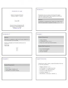
3. 10. Determination of bacteriocin molecular weight To determine the molecular weight of the two bacteriocins of the organisms under test, ammonium sulphate precipitation and ultrafiltration were applied for the partial purification of bacteriocins. Both methods resulted in the production of crude bacteriocin preparation. SDS-PAGE technique (Fig. 20) indicated that their antimicrobial peptides possessed a molecular weight ranging from 3.496 and 6.512 KDa.
(A)
St. Mw *
(B)
117 90 49
35 26.7 26 19
14.5 6.5 3.25 1.4
*Standard molecular weight (KDa) Fig. 20: SDS-PAGE electrophoresis gel of concentrated bacteriocins produced by strains B. megaterium 19 and B. megaterium 22 (A, B), (St. Mw) small and large standards prestained protein molecular weight markers.
68
IV. DISCUSSION Maintaining a safe food supply has become an ever-changing endeavor as new information on pathogenic bacteria is discovered. Consumer demands for minimally processed foods means that the food industry can no longer rely on traditional methods of thermal processing to create microbiologically safe foods. Minimally processed foods such as fresh fruits and vegetables have been shown to harbor pathogenic bacteria. Thermal processing of fresh fruits and vegetables is often not acceptable, so other methods of controlling pathogenic bacteria must be considered. Methods such as irradiation, highpressure processing, low-temperature storage, chemical preservatives, modification of atmosphere, and control of water activity, or combinations thereof may be considered. The use of antimicrobial compounds found within the foods themselves may be another way of controlling microorganisms. Bacteriocins are highly specific antibacterial proteins produced by strains of bacteria active mainly against some other strains of the same or related species (Gaur et al. 2004). The bacteriocins produced by LAB are potent biopreservative agents and the applications of these in food are currently the subject of extensive research. The search for new bacteriocins with wider spectrum of activity and compatibility with different food system is being studied by some investigators. Fruits and vegetable waste provide a good source for isolating LAB having antagonistic and probiotic properties. However, work on bacteriocinogenic LAB from agro-based waste has been limited (Lade et al. 2006). In this work, the search for bacteriocin production by some bacilli using agro-based waste was undertaken. Among references cited in this thesis only 6% were concerned with bacteriocin production using bacilli. (Ettayebi et al. 2000, Örnek et al. 2002, Ahern et al. 2003, Delmar et al. 2005) It is well documented that different Bacillus species are able to produce extracellular substances with antimicrobial activity against a wide variety of microorganisms. Many of them are indicated as preservative in food systems and beverages (Wang and Fung 1996; Zheng and Slavic 1999), as biological control agents against phytopathogens and as antibiotic producers (Zuber et al 1993). After the isolation of the strains from different sources, the cultures were tested for bacteriocin production using the well-diffusion assay, the deferred agar spot test, and the ODM at 600 nm wavelength. Of the 30 isolates, only nine bacilli were positive in the welldiffusion assay (0-4mm inhibition zone) and negative in the agar spot test. These bacteriocin-producing strains were gram-positive, rods and contain endospores. Results obtained by Schillinger and Lücke (1989) experimenting with L. saké were contradictory to our results. They reported that among the total of 19 strains, only six produced inhibition zones on agar in the well-diffusion assay. Lewus et al. (1991) found that only a few of the strains tested were positive when using the spot-on-the-lawn method, and also gave positive results in the well-diffusion assay. They considered that allowing some time for the bacteriocins to diffuse into the agar prior to incubation, or increasing the well size so that more sample could be applied, might increase the sensitivity of the assay. According to these authors, aggregation, non-diffusible bacteriocins, medium composition,
69
protease inactivation and concentration effects, can all lead to false negative results in the well-diffusion assay. Özlem and Feryal (2006) using L. casei and L. bulgaricus isolates showed weak antibacterial activity against E. coli, S. aureus, P. aeroginosa, B. subtilis, K. pneumonia, S. typhimurium, and Enterococcus cloacae. Abo-Amer (2007) reported that out of 73 isolated Lactobacillus strains; only four strains of L. plantarum (AA110, AA125, AA135 and AA140) demonstrated production of antagonistic activity against foodborne pathogens in LB medium at 37 oC by using AWD. Korenblum et al. (2005) isolated 40 strains of bacilli from drilling mud, core and water. 36 strains were able to inhibit at least one strain used as indicator and three of them were able to inhibit more than 65% of the strains tested in agar in the well-diffusion assay. In this study, our test strains exhibited a broad antimicrobial spectrum, capable of being active against gram positive and gram negative representatives. Also the maximum production and antibacterial activity of the bacteriocins against the indicator strains S. typhimurium and S. aureus was found to be during the stationary phase of both strains B. megaterium 19 and 22 respectively, after growth for 12-18 h in MRS broth at 30ºC and pH 6.2-6.5, also the bacteriocin activity started to decline gradually within 24 h of experimentation. This result indicates that the peptide is a primary metabolite. Similar results reported by NaClerio et al. (1993) on bacteriocin production and secretion by B. cereus was observed in the stationary phase, after 10 to 16 h of bacterial population growth in BHI broth at 30°C. While Cherif et al. (2001) reported that thuricin 7, produced by the very closely related species B. thuringiensis BMG1.7, and was expressed in the exponential growth phase. As it is well known, bacteriocins of LAB, particularly lantibiotics, are usually produced in the exponential phase (Hörner et al. 1990). Although some bacteriocins are mainly produced in the stationary growth phase (Biswas et al. 1991; Jiménez et al. 1993; Bárcena et al. 1998), generally bacteriocin production occurs only in the active growth phase. In contrast, Ten Brink et al. (1994) reported on acidocin B production by non growing L. acidophilus M46 cells. The bactericidal activity of cerein produced by B. cereus appeared at the beginning of stationary growth and declined 2 to 3 h after its appearance, probably as a consequence of its inactivation or degradation. These observations suggest that cerein synthesis, secretion, and action occur through a complex regulatory process, whose study will be facilated by the isolation of the cerein structural gene and its regulatory elements (Gino et al. 1993). Simonová and Lauková (2007) working on bacteriocin (PPB) EF2019 produced by a thermostable substance that is stable at pH 4.0, 7.0 and 9.0. Its production started at the early logarithmic growth phase and culminates at the late logarithmic growth phase of the EF2019 strain. Albano et al. (2007) reported that BacHA-6111-2 and BacHA-5692-3 produced by P. acidilactici were produced at their maximum level after 18 h of growth in MRS broth. While low levels of BacHA-6111-2 activity were recorded after 9 h of growth in MRS broth at 30ºC and 37ºC at pH 5.1. Similar results were reported for BacHA-5692-3 at pH 5.05. After many experiments using the ODM, strains B. megaterium 19 and B. megaterium 22 were found to produce and secrete bacteriocins, which were strongly effective against gram negative bacteria E. coli and Str. pyogenes β-hemolytic and gram positive bacteria S. typhimurium, S. aureus and against other bacilli strains. Two species namely S. typhimurium and S. aureus were chosen as the important indicator strains because they are the cause of a potential public health hazard, since nine of them produce
70
enterotoxins that cause food poisoning if ingested and deteriorates the organoleptic characterization of foods. De Kwaadsteniet et al. (2005) demonstrated that bacteriocin ST15 produced by E. mundtii ST15 was active against gram positive and gram negative bacteria such as Acinetobacter baumanii, B. cereus, Clostridium tyrobutyricum, E. feacalis, K. pneumoniae, L. saké, S. aureus and Str. pneumoniae. Screening for the bacteriocin production of strains of LAB from various meat and meat products resulted in the detection of a bacteriocin-producing L. lactis subsp. cremoris CTC 204, isolated from chicken (Bromberg et al. 2005). The bacteriocin inhibited not only closely related LAB (L. helveticus), but also pathogenic microorganisms (S. aureus, L. monocytogenes, B. cereus and C. perfringens). Furthermore, Abo-Amer (2007) found that the antimicrobial agent excreted by L. plantarum AA135 was the most active against a wide range of grampositive and gram-negative pathogens. Earlier reports (Tagg et al. 1976; Daeschel et al. 1990; Sanni et al. 1999) have shown that some bacteriocins produced by gram-positive bacteria have a broad spectrum of activity. However, it was generally observed that bacteriocin from the producer organism had no inhibitory effect on the organism producing it. JoshiI et al. (2006) reported that the antimicrobial activity of partially purified bacteriocin produced during natural lactic acid fermentation of carrot, radish and cucumber was assessed and characterized. Out of ten strains, the isolated strain of Lactobacillus genus from carrot fermentation produced bacteriocin with maximum antimicrobial activity against E. coli, S. aureus and B. cereus, though it was more effective against E. coli than others on growth in the MRS broth at 32 ºC for 24-48 h. In another part of this investigation, different experiments were designed to attain the best available cultural conditions which could help in the production of bacteriocins, with the least economical costs possible and with the simplest methods available. As large amounts of bacteriocin are necessary to test their preservative efficiency in natural environments, establishment of the factors and levels influencing maximal production would lead to a more effective recovery of these antimicrobial compounds from a defined laboratory culture medium. Specific environmental conditions, including those found in food, have been studied by Leroy and De Vuyst (2003) and Mota and Bradelli (2003) to determine their effect on the production of bacteriocins. According to Leal Sánchez et al. (2002) bacteriocin production changes dramatically upon altering of environmental conditions and optimum production may require a specific combination of environmental parameters. High bacteriocin production in this study was obtained with MRS broth within 12 to 18 h and M17 broth after 24 h at 30ºC followed by 37 ºC. The low production levels recorded in BHI broth, molasses, and whey, may suggest that specific nutrients are required for bacteriocin production. This result was consistent with that found by De Kwaadsteniet et al. (2005) where the highest activity of bacteriocin ST15 produced by E. mundtii ST15 was recorded after 14 hours of growth in MRS broth at 30ºC. A low level of activity was recorded in M17 broth, BHI broth, soymilk and molasses. Gino et al. (1993) reported that cerein synthesis and/or secretion was observed when B. cereus strain GN105 was grown in glucose-supplemented BHI broth at 30ºC, while no antimicrobial activity was observed for growth in various other minimal or rich media. Also no bacteriocin was detected when strain GN105 was grown in BHI broth at 25 ºC, while only a reduced amount of cerein was found at 37 ºC. Similar results for L. plantarum strains ST23LD and ST341LD that were grown in BHI or M17 broth adjusted to pH 6.5 were described by Todorov and Dicks (2006). Growth of the latter two strains in 10%
71
(w/v) soy milk or 10% (w/v) molasses yielded low levels of bacteriocin production. The low activity levels recorded in M17 broth, BHI broth, soy milk and molasses, despite relatively good growth, suggest that specific nutrients are required for the production of bacteriocin ST23LD and ST341LD. Production of these bacteriocins was much higher at 30 ºC than at 37ºC, which suggests that growth temperature also plays an important role. Some of the results obtained in this study were supported by the observation of Bizani and Brandelli (2002) on bacteriocin production from B. cereus 8A, which was higher at 30 °C than at 25 °C. An important decrease in bacteriocin activity was observed at 37 °C. They deduced that the relationship between growth and specific production rates, as a function of the temperature, showed different kinetics of production. Also Guerra and Pastrana (2002) found that the nisin and pediocin produced by L. lactis and P. acidilactici obtained on whey were lower than those obtained on MRS broth. The same observation was reported by Liao et al. (1993) experimenting with L. plantarum UG1 when grown in whey permeates producing plantaricin UG1. Few publications also reported the feasibility of the production of both nisin and pediocin AcH bacteriocins in whey permeate. The maximum values of bacteriocin activity (95%) was observed by preparations obtained from strain B. megaterium 22 grown in MRS broth at pH 6.5, against S. typhimurium and S. aureus after 12 hours of incubation. The final pHs of medium dropped to 5.56 respectively. In general, low levels of bacteriocin activity against the two indicator strains were recorded when both test strains were cultured in MRS broth with an initial pH of 4.5 (45-65%). Todorov et al. (2006) showed that L. lactis subsp. Lactis HV219 strain did not grow in MRS with an initial pH of 4.5. Low levels of bacHV219 were recorded in MRS broth adjusted to pH of 5.0. In the same medium adjusted to pH of 5.5 and 6.5, bacHV219 was recorded. The highest level was recorded after 24 h in MRS medium adjusted to an initial pH of 6.0. The final pH values of the cultures were more or less the same (pH 4.5), irrespective of the initial pH. Todorov and Dicks (2007) reported that the bacteriocin ST712BZ (14.0 KDa in size) inhibits the growth of L. casei, E. coli, P. aeruginosa, E. faecalis, K. pneumoniae and L. curvatus. The growth of this strain in BHI, M17, soy milk and molasses was similar to the growth in MRS (pH.6.0), with optimal bacteriocin production recorded in MRS after 24 h. The same level of bacteriocin production was recorded in MRS broth with an initial pH of 6.5, 6.0 and 5.5. Low levels of bacteriocin activity were recorded in MRS broth with an initial pH of 5.0 and 4.5, which is consistent with our results. In order to select the best carbon and nitrogen sources for maximum bacteriocin production by B. megaterium 19 and B .megaterium 22, the production medium previousely mentioned in section (2.2.10.3), was used. The best results reflecting the highest inhibitory effect on the indicator strains (S. typhimurium and S. aureus) were obtained in MRS broth supplemented with the following carbon sources (2%) in a descending order: sucrose, glucose, maltose, fructose, galactose and finally lactose, for both B. megaterium 19 and B. megaterium 22 after 15 to 24 h of incubation at 30ºC. Since sucrose showed best productivity, different concentrations were prepared (1-10%). The results showed that 5% and 2% sucrose were best for B. megaterium 19 and 22 productivities respectively, after 9 to 21h incubation at 30ºC. The significance of this finding lies in the low cost of sucrose compared to glucose, an aim achieved in this research. On the other hand, Todorov et al. (2006) showed that L. lactis subsp. Lactis HV219 strain grown in the MRS broth with glucose concentrations in the range of 5.0 to 20.0 g/l yielded the same bacHV219 activity.
72
In the presence of higher concentrations of glucose, reduction of bacteriocin HV219 was observed. In case of fructose, concentrations of 10.0 to 30.0 g/l have a stimulating affect to bacHV219 production. Similar results were recorded for sakacin P (Aasen et al. 2000) and nisin (De Vuyst and Vandamme 1992). The present work demonstrated that the activity level of the two bacteriocins produced by both strains under test was strogly influenced by the nitrogen sources added one at a time to MRS medium as follows: beef extract, yeast extract, tryptone, ammonium sulphate, ammonium nitrate, ammonium chloride, ammonium acetate, sodium nitrate and argenine. Improved productivity was recorded with beef extract, yeast extract and ammonium nitrate for B. megaterium 19, and beef extract, ammonium nitrate and tryptone for B. megaterium 22. Combinations of beef extract (1.5%) and yeast extract (0.5%) provided the highest bacteriocins production upon cultivating the cells in mMRS broth for 9 to 24 h at 30ºC. Lowest production was obtained in MRS broth supplemented with WB and CSL. The suitability of mMRS as an efficient medium that promotes bacteriocin production was studied by many workers (Jurgen et al. 2003, Adenike et al. 2007). Todorov et al. (2006) reported that the activity level of bacteriocin HV291 produced by L. lactis subsp. Lactis was strongly influenced by the nitrogen source added to MRS medium. They also found that the yeast extract is the favored organic nitrogen compound, compared to the others tested for bacHV219 production. While Verellen et al. (1998) published that in the case of plantaricin 423, highest production was obtained in MRS broth supplemented with bacteriological peptone, followed by amino acids; tryptone and meat extract. On the other hand, B. megaterium 19 and B. megaterium 22 produced bacteriocins under limited or reduced oxygen in the medium and/or stress conditions (86% and 89%). The two bacteriocins activities (Table 8) was not detected in maximum level of aeration, whereas the detectable levels of bacteriocin production was recorded (43% and 50%) with decreased oxygen in the medium. This finding was supported by the results of Jurgen et al. (2003) who reported that oxidative stress caused a higher level of bacteriocin production from L. curvatus LTH 1174. In an attempt to characterize the physical and biochemical properties of the bacteriocins produced by the two strains B. megaterium 19 and B. megaterium 22 presented in this study, different factors such as heat, pH, storage, UV light, enzymes, inorganic salts, organic solvents and spices were determined using S. typhimurium and S. aureus as indicator organisms. The antimicrobial agents produced by B. megaterium 19 and B. megaterium 22 were heat-stable retaining their activities after heating at 100 oC for 15 minutes. The total loss of activity was observed at 121 ºC for 15 min. This is an interesting feature in view of its potential use as a food additive, in process like pasteurization, drying, tundalization, refrigeration and freezing; the resistance of bacteriocins in these range of temperatures make them effective against psychrophile, thermophile and mesophile food borne pathogens, and also to different temperatures during production and storage. Tagg et al. (1973) found that the crude bacteriocin produced by Str. pyogenes strain FF-22, was particularly stable to heating, with no apparent decrease in activity on heating for 60 min at 96 ºC. The results are in accordance with those stated in this thesis. Luca et al. (2002) indicated that the bacillocin 490 produced by a thermophilic strain of B. licheniformis showed high stability at 100°C with 46.4% residual activity after 1 hour of
73
exposure at this temperature. It also showed remarkable stability at 4°C (no loss of activity after 12-month storage was observed). On the other hand, Karmen and Bogovič (2003) comparing some bacteriocins produced by LAB with those of B. cereus reported that the latter bacteriocins were not extremely heat stable. Bromberg et al. (2005) showed that the bacteriocin-producing L. lactis subsp. cremoris CTC 204 was heat stable even at autoclaving temperature (121°C for 10 min) and was produced during refrigerated storage. Vinod et al. (2006) described that the bacteriocin produced by Lactobacillus CA44 was stable at up 100°C for 60 minutes but its activity declined compared to that at 68°C and was completely lost at 121°C for 15 minutes. Contradictory results were obtained by Valdes and Scherer (1994), where they found that the bacteriocin linocin M18 produced by Brevibacterium linens was heat sensitive. The activity of bacteriocins produced by the test organisms in this work was pH dependent. The bacteriocins retained more than 60% of its stability after exposure to pH values ranging from (2-12) after 90 minutes of incubation at 30ºC and 30% from (pH 2-7) after 24 hours of incubation. These results were in accordance with those obtained by Ogunbanwo et al. (2003a), when using bacteriocin produced by L. brevis OG1. The greater stability of bacteriocin in a wide ranges of pH, proposes a beneficial application and a great advantage for the food industry. This property makes them very effective against molds, yeast, acetobacter lactic acid bacteria and other deteriorized food microorganisms in neutral, alkaline and acidic foods (dairy products, meat and poultry, some fish products), as the pH of many foods varies from very acidic to basic. In this connection it is interesting to notice that nisin, the only bacteriocin so far employed commercially as food additive against gram-positive bacteria, acts in acidic conditions, as its maximum stability is at pH 2.0, and it is radically not soluble at pH 8.0. According to De Vuyst and Vandamme (1994) L. plantarum J-51 antimicrobial activity was stable in the pH range of 1–9, being inactive at higher pHs, as most bacteriocins and bacteriocin-like substances produced by LAB, such as nisin, pediocins, sakacin A. Brevicin B37, caseicin 80 (Rammelsberg and Radler 1990) and plantaricin D (Franz et al. 1998), which show activity in a wide range of pHs. L. plantarum J-53, J-65 and J-75 strains produced antimicrobial activities which were stable only in the pH range of 1–4, becoming inactive at higher pH. A similar behaviour has been reported for plantaricin A (Daeschel et al. 1990), plantaricins S and T (Jiménez-Diaz et al. 1993), plantaricin C (Gonzalez et al. 1994), plantaricin F (Fricourt et al. 1994) and other bacteriocins (De Vuyst and Vandamme 1994), which are active only in the acid range. Tagg et al. (1973) indicated that the crude bacteriocin produced by Str. pyogenes strain FF22 was stable at pH 2 did not loose activity, but was completely inactivated at pH 11. Özlem et al. (1998) reported that the purified pediocin F, a bacteriocin produced by P. acidilactici F was found to be stable over a wide pH range between 3 and 9. Activity was lost at pH 10 and above after 24 hr at 25 °C and heat treatment. The loss of activity at higher pH could be due to degradation of the molecule. This is similar to the properties reported for bacteriocins produced by other lactic acid bacteria such as pediocin AcH, Pediocin A, pediocin PA-1, lactacin, lactacin 27, acidolin, nisin and diplococcin. Navarro et al. (1999) found that the bacteriocin produced by LAB isolated from Rioja red wines was found to be stable between pH 3 and 9 and under strong heating conditions (100 °C for 60 min).
74
Rodriguez et al. (2002) described that pediocin PA-1 produced by P. acidilactici PA-1was perfectly stable after 21 days of storage at 15 °C at pH 4 to 6; however, half of the activity was lost at pH 7. Larsen et al. (1993) found that bavaricin A produced by L. saké MI401 was very stable at pH 2.0 to 9.7, but storage of bavaricin A at pH 12.5 for 4 h resulted in the complete loss of activity. Karmen and Bogovič (2003) on the other hand, observed that the bacillocin 490 produced from B. licheniformis, showed great stability in a wide pH range, since it had optimum activity at neutral and moderately basic pH and maintained some 60% of activity after incubation at pH 3.5 and 11, with 95–100% of activity detected rather uniformly from pH 4.5 to 9.0. Todorov and Dicks (2006) published that the BacHV219 remained stable after incubation for 2h at pH values between 2.0 and 10.0. The effect of period and temperature of storage on bacteriocin activity was also carried out. It was observed that the bacteriocins produced by the test strains in this investigation maintained high stability ranging from 79-88% after 30 days of storage at +4ºC. Exposing the bacteriocin to longer periods of storage (45 days) resulted in the decrease in the bacteriocin activity (at least by 3-10%). However the activity remained consistent up to 60 days of storage. The ability of the tested bacteriocins decreased the growth of the indicator strains significantly after 30 days of storage at -20oC to almost half. After 45 days, there was a faint (2-5% reduction of growth) or no detectable bacteriocin activity. These results indicate that cold temperatures may be the most appropriate preservation technique. According to Navarro et al. (1999) L. plantarum J-51 bacteriocin remained stable at storage temperatures, such as 4 °C, –20°C and room temperature, and under strong heating conditions (100 °C for 60 min). Nevertheless, thermostability was lower after a previous storage at – 20°C. Zeliha and Metin (2001) found that buchnericin LB produced by L. buchneri LB was resistant to high temperatures at 4ºC, -20ºC or -70ºC and long storage periods for 3 months. Ogunbanwo et al. (2003a) showed that bacteriocins produced by L. plantarum F1 and L. brevis OG1were fully stable after storage for 60 days at -20°C, but declined or became non-detectable after storage for 80 to 120 days at 37°C. The influence of UV light on bacteriocin activity was studied on both organisms. It was observed that bacteriocin produced by B. megaterium 19 was stable after 15 minutes exposure to UV light. However, it was completely destroyed after 90 minutes, while the bacteriocin produced by B. megaterium 22 maintained stable after 90 min. Results from UV light were not surprising and confirmed the protein status of bacteriocins. This might be due to the effect of ultraviolet light on the protein nature causing modification and/or change in the ring structure of peptides, or affecting the protein function. The results of B. megaterium 19 are consistent with those described for bacteriocins produced by L. plantarum F1 and L. brevis OG1 after exposure to UV light from 0 to 5 minutes (Ogunbanwo et al. 2003a). The effect of different enzymes on the activity of bacteriocin produced by the test organisms, proteolytic and non proteolytic enzymes were used (trypsin, papain, pepsin, lipase and α-amylase) have been the subject of several studies. The present results reflected a complete inactivation of the antimicrobial activity after treatment of the CFS of strain B. megaterium 22 with the different enzymes and their selected concentrations, where activity was totally lost except in case of treatment with 1
75
mg/ml α-amylase enzyme. Bacteriocin of B. megaterium 19, on the other hand, was sensitive to amylase treatment compared to other enzymes used in this study and showed an almost equal reduced activity (18 and 17%) respectively towards S. aureus when treated with 1 and 2 mg/ml. However, the bacteriocin was sensitive to the rest of the selected enzymes (papain, pepsin, and trypsin) except for lipase when used at higher concentration (2 mg ml-1). The sensitivity of the antibacterial substance produced by B. megaterium 22 to trypsin, pepsin, papain and lipase and their resistance to α-amylase was attributed to their proteinaceous nature and suggests that the bacteriocin is not glycosylated and proves the presence of a lipid moiety in the bacteriocin. However, the activity of the antimicrobial substance produced from B. megaterium 19 was resistant to papain, trypsin and pepsin, reflecting the modified form of their amino acid residues. The resistance to proteolytic enzymes can be explained on the basis of the presence of unusual amino acids in their bacteriocin structures, presence of N-and / or C- terminally blocked peptides. Which cyclic peptides can be resistant to hydrolysis by proteases due to their cyclic structure renders them relatively inflexible. This may make cleavage sites inaccessible because of steric hindrance (Eckart 1994). This work mentioning that the compounds produced by our strains were inactivated by an array of proteolytic enzymes, including those of pancreatic origin like trypsin and of gastric origin, pepsin. These results suggest that bacteriocins produced by the organisms under investigation could be possibly added to dairy and meat packaging materials. The results of several authors are in accordance to those stated in this research. However, most of their researchers results were focused on the relation between enzymes and bacteriocins isolated from organisms other than bacilli. Valdes and Scherer (1994) reported that the bacteriocin linocin M18 produced by Brevibacterium linens grown at 30°C in tryptose agar medium was sensitive against a number of proteases, which reveals its proteinaceous nature. No inhibition of bacteriocin activity by nucleases, lipase, catalase, α-amylase, and the proteases ficin and papain was observed. Contreras et al. (1996) reported that L. amylovorus LMG P-13139, isolated from corn steep liquor, produces two bactericidal peptides sensitives to trypsin, but insensitive to α-amylase, and lipase. Zeliha and Metin (2001) indicated that the commercially fermented vegetable product produced a bacteriocin-designated buchnericin LB produced by L. buchneri LB was hydrolyzed by trypsin, papain and pepsin. However, it was observed that it was resistant to lipase, α-amylase. Guinan et al. (2005) reported that the lactococcal bacteriocin from L. lactis (lacticin 3147) would easily be broken down by digestive enzymes which does not pose a threat to humans, making it a safe and economically viable alternative to chemical preservatives presently used in the food industry. Sathish et al. (2005) found a novel bacteriocin-like substance (BLIS) from a pathogenic strain of Vibrio harveyi. The crude material (CFS) was inactivated by proteolytic enzymes. Of interest was the inactivation by lipase, which suggests the presence of a lipid moiety in the BLIS. In contrast, α-amylase did not have any effect on the inhibitory activity. Bromberg et al. (2005) showed that bacteriocin-producing L. lactis subsp. cremoris CTC 204 was inactivated by papain but not by lipase or pepsin.
76
Also De Kwaadsteniet et al. (2005) reported that the complete inactivation of antimicrobial activity was observed after treatment of the CFS of bacteriocin ST15 produced by E. mundtii ST15 with pepsin. No change in activity was recorded with α-amylase. De Kwaadsteniet et al. (2006) found that bacteriocin T8 produced by E. faecium T8, isolated from vaginal secretions of children infected with human immunodeficiency virus was sensitive to proteinase K, pepsin, and pronase. Treatment with amylase had no effect on bacteriocin activity. Albano et al. (2007) found that the complete inactivation or significant reduction in antimicrobial activity was observed after treatment of the cell-free supernatant of BacHA-6111-2 and BacHA-5692-3 produced by P. acidilactici with trypsin. Treatment with papain at 0.1 mg/ml decreased the activity of BacHA-6111-2 to 50 %. Treatment with pepsin (1 mg/ml) reduced the activity levels of the bacteriocins by more than 75 %. Treatment with α-amylase did not change the antimicrobial activity. The effect of some organic solvents on bacteriocin activity of the two bacilli under test was also examined using hexane, ethanol, methanol, chloroform and acetone. Ethanol and hexane did not affect the activity of bacteriocin from B. megaterium 19. On the other hand, all organic solvents applied completely destroyed the bacteriocin produced from B. megaterium 22. These results suggest that ethanol and hexane could be used in the extraction procedure to remove the bacteriocin from the aqueous phase and recover from the organic phase. Another explanation could prove the presence of lipid moiety in bacteriocin. Gino et al. (1993) found that the antimicrobial activity of cerein produced by B. cereus was resistant to acetonitrile, chloroform, acetone, methyl alcohol, and ethyl alcohol. Özlem et al. (1998) indicated that the antimicrobial activity of pediocin F produced by P. acidilactici F overcome the treatment with the organic solvents, acetonitrile, chloroform, acetone, formaldehyde, ethyl alcohol, hexane and isopropanol. Some of results are in accordance to these stated in this thesis for B. megaterium 19. Zeliha and Metin (2001) showed that the buchnericin LB produced by L. buchneri LB completely lost its biological activity after treatment with organic solvents as formaldehyde, chloroform, acetone, 2-propanol, ethyl alcohol, hexane, iso-butanol and ethyl ether at a concentration of 10 mg/ml. Some of these results are similar to our results obtained for B. megaterium 22. Also Korenblum et al. (2005) found that the treatment of bacteriocin produced by B. licheniformis T6-5 with acetone, ethanol and methanol led to their total or partial inactivation. In order to show the effect of the addition of different ingredients on the bacteriocin efficacy, NaCl, KCl and MnCl2 were used. There was an increase in bacteriocin activity of B. megaterium 19 and B. megaterium 22 against the two indicators strains (S. typhimurium and S. aureus) on increasing the concentration of NaCl up to 5%, however the activity declined significantly in the presence of 5% NaCl for B. megaterium 19 and only decreased by 3% for B. megaterium 22. A more or less similar trend was observed upon treating the CFS with different KCl concentrations. Bacteriocins of B. megaterium 19 and 22 displayed the highest activity in the presence of 3% KCl (95.4-95.9%, and 92.2-92.7%, respectively) against the two indicator strains. Activity of B. megaterium 22 was inhibited by more than 3% KCl against both indicator strains. Concentrations of MnCl2 above 0.25% inhibited the bacteriocin activity against the two test strains. These data suggest that the inorganic salts have a synergistic effect on bacteriocin efficacy when added with specific concentrations in foods. Monovalent cations could mediate the adsorption of bacteriocin to the indicator strain; however, divalent ions could mask such effect.
77
Gänzle et al. (1999) and Gänzle (1999) reported that a synergistic effect has already been shown for curvacin A and sodium chloride, rendering the gram-negative pathogens E. coli and S. enterica susceptible, while at low pH these pathogens also show increased sensitivity to the bacteriocins tested. Hugas et al. (2002) indicated that some common ingredients have a synergistic effect on bacteriocin efficacy or production, while some ingredients can reduce antimicrobial activity. NaCl at the concentration found in a number of processed meat products, including fermented meats can reduce the growth of protective cultures and thereby reduce the production of bacteriocins, such as sakacin K. Leroy and De Vuyst (1999) reported that sodium chloride and sodium nitrite can inhibit the bacteriocinogenic effect of bacteriocin-producing cultures in a meat system. This problem was overcome with a preinoculation enrichment procedure, whereby bacteriocin DPC4275 produced by L. lactis subsp. lactis DPC3147 isolated from an Irish kefir grain (Ryan et al. 1996) was treated with 2.5 ppm manganese and 250 ppm magnesium before the fermentation process (Scannell et al. 2001). Manganese and magnesium can promote the metabolic activities of lactic acid bacteria, thereby possibly favoring bacteriocin production, which is not the case in this study. Spices and herbs are used for imparting desirable sensory properties to food products. Different mixtures of spices are added to many kinds of fermented sausages, all of which have their own specific organoleptic properties. The starter culture that is added to ensure rapid acidification and a safe end product is also influenced by the spices added, and hence it is important to know the effects that different food ingredients will have on bacteriocin activity. All of the spices tested in this study in concentrations ranging from 0.5-3% affected bacteriocins activity differently. The addition of 0.5% curry solution to both bacteriocins produced by B. megaterium 19 and 22 resulted in a significant activity against the indicator strains (up to 94% reduction of growth). In contrast, increasing the concentration of red pepper solution seemed to stimulate the activity of bacteriocin of B. megaterium 19 and had a lesser minor effect on that of strain 22. Finally, the addition of 0.5% black pepper solution minimized the bacteriocins activity compared to the effect of similar concentration of the previously mentioned spices. Lower values of growth reduction percentages to the indicator strains were recorded when adding 0.5% of bastermy sheath solution to either bacteriocin tested. Above 1% of bastermy sheath solution inactivated completely the bacteriocins used. This indicates that small amounts of additives should be used in some foods to promote the activity of the bacteriocins applied, especially when more than one spice is used. Many spices display different antimicrobial activities. The antiseptic potential of spices resides in the essential oils (Zaika 1988). One of the most potent spices is garlic, in which several antimicrobial components are present where the principal active substance was identified as allicin (Ankri and Mirelman 1999; Harris et al. 2001). Extensive studies have been performed to determine the inhibitory properties of garlic, and many food-borne pathogens, both gram-positive and gram-negative bacteria, have been shown to be inhibited by it (Kumar and Berwal 1998; Ross et al. 2001; Leuschner and Zamparini 2002). Singh et al. (2001) indicated that bacteriocins or bacteriocinogenic starter cultures can be applied in various food products and may provide a synergistic
78
effect using nisin and garlic extract. In both, such a positive interaction between nisin and garlic extract has been shown towards strains of L. monocytogenes. Ettayebi et al. (2000) reported that the concentration of nisin necessary for effective control of L. monocytogenes and B. subtilis could be lowered when it was used in combination with thymol. Ettayebi et al. (2000) and Singh et al. (2001) described that the combination of spices together with bacteriocins that, albeit in a lower amount, can be produced in situ by the bacteriocinogenic starter culture may lead to a synergistic effect, rendering pathogens susceptible to L.monocytogenes. Such a synergistic inhibition has been shown between nisin on the one hand and garlic extract or thymol on the other. Lemon (2001) reported that in the sausage environment, the bacteriocin that is produced in situ may be aided in its bactericidal action by the lactic acid that is produced and hence the drop in pH, the presence of relative high amounts of salt, and the antimicrobial activity of some of the spices present. He also found that garlic, black walnut leaves, cloves, goldenseal root, barberry root and fenugreek help maintain and promote healthy intestinal ecology. Bestermy sheath contains more than one spice with different amounts, according to different producers (garlic, salt, chili, feunogreek and pepper….etc). Therefore the results concerning the use of bastermy in this work might be misleading. This might be due to the presence of an unidentified compound that may have led to the inhibition of bacteriocin activity. Moreover, higher concentrations of NaCl or NaNo3 which may be added to bastermy sheath could result in that reduction on activity. To test the mode of action of bacteriocins produced by the two bacilli (B. megaterium 19 and 22) on the target cells of S. typhimurium, CFU and TEM were used. Based on the results obtained, it appeared that the two bacteriocins have a bactericidal effect on the indicator strain, since the decrease in viable cell numbers was observed, with obvious cell lysis as observed by the TEM. According to the results reported by Ahern et al. (2003), it appeared that thuricin 439 produced by B. thuringiensis strain B439 has a bactericidal effect on the indicator L. innocua and with obvious cell lyses occurring after 5.5 hours. This is in accordance with our results. This suggest that the bacteriocin may cause death of the indicators strains cells and/or induce leakage of K+, amino acids, uncouple mitochondrial respiration and depletion of the intracellular ATP, or cell wall lyses (Chikindas et al. 1993). Chumchalová et al. (2004) reported that the partial purification and characterization of heat-stable acidocin CH5, a bacteriocin produced by L. acidophilus CH5, showed that the active compound caused fast decrease in viable cell count of the indicator strain L. delbrueckii subsp. Lactis LTI30 which was in accordance to results obtained in this study. Bactericidal mode of action is typical for bacteriocins produced by lactic acid bacteria (Davey et al. 1981), such as lactacin B (Barefoot and Klaenhammer 1983), sakacin P (formerly bavaricin A) (Larsen et al. 1993) and gassericin KT7. These observations are parallel with the results described in this work for both bacilli strains.
79
Also De Kwaadsteniet et al. (2006) reported that the addition of bacteriocin T8 (produced by E. faecium T8) to actively growing cells of E. faecalis resulted in a rapid decrease in cell density indicating that the mode of action is bactericidal, similar to that recorded for enterocin P and the results of bacteriocins of B. megaterium strains 19 and 22. The molecular weight of bacteriocins was estimated according to the method of Sambrook and Russell (2001), using discontinuous SDS-polyacrylamide gel electrophoresis (SDSPAGE). The determined molecular mass of two bacteriocins of B. megaterium 19 and B. megaterium 22 ranged from 3.496 to 6.512 KDa, which is in agreement with the results obtained from the SDS-PAGE assay of some other bacteriocins like BacHA-6111-2 and BacHA-5692-3 produced by P. acidilactici where molecular weights were estimated to range from 3.5 to 6.5 KDa (Albano et al. 2007).The molecular mass of bacteriocin ST15 is 3944 KDa (De Kwaadsteniet et al. 2005) and is close to that reported for enterocin CRL35 (Wachsman et al. 2003) and enterocin 012 (Jennes et al. 2000), but smaller than the 4.287 KDa reported for mundticin ATO6 and KS (Bennik et al. 1998; Kawamoto et al. 2002). According to the chemical structure and antimicrobial activities of the resulting bacteriocins, they may be contained in Class II group IIa which includes the small (< 10 KDa), heat stable, non lanthionine containing peptides (Cleveland et al. 2001). Which might be proved by the mode of action, where bacteriocin produced by B. megaterium strain 19 functions by lysing cells as illustrated by the TEM micrographs. Future research The potential of the two bacteriocins produced by the two bacilli under investigation to inhibit foodborne pathogenic bacteria, such as S. aureus, Bacillus spp, S. typhimurium and E. coli, is of a crucial interest. Some of these bacteria can produce toxins resulting in human illness. In addition to the broad inhibition spectrum, its technological properties and especially their heat and storage stability indicate that both bacteriocins have potential for application as a biopreservative to control pathogens in processed foods. Future work require more pratical studies that provide detailed information on the synthesis, regulatory processes, purification and amino acid sequencing for the full characterization of that resulting bacteriocins. Based on the fact that the two bacilli strains are bacteriocin producers, their desirable technological properties may be further investigated to determine their potential as candidates employed as biopreservation in food products. Also, the fact that the type of medium and medium components can affect the bacteriocin activity can be taken into consideration when developing food products and modifiying their consumption acceptability properties including texture and taste perception.
80
V. SUMMARY Bacteriocins are proteinaceous toxins produced by bacteria to inhibit the growth of similar or closely related bacterial strain(s). They are phenomenologically analogous to yeast and paramecium killing factors, and are structurally, functionally, and ecologically diverse. Over the last 2 decades, a variety of bacteriocins, produced by bacteria that kill or inhibit the growth of other bacteria, have been identified and characterized biochemically and genetically. Bacteriocins can be exploited in food in a variety of imaginative and commercially significant applications in bio-preservation, shelf-life extension, industrial fermentation, and in agriculture. However, in order to fully realize this potential, this thesis focused on screening of bacteriocinogenic bacilli isolated from native food products and wastes in Egypt. The cultures were tested for the potential antimicrobial activity of their supernatant against some selected pathogenic and spoilage indicator organisms. The mode of action and influence of several physiological factors on bacteriocins activity were investigated. The results of this study are summarized as follows: 1. Thirty bacilli isolates were obtained from different sources; milk and milk products, meat and meat products, soils, silage, pickles, cereals, spices, compost, eggs, roots of beet and carrot, grass, fermented foods, vegetables, and olives, edible and non edible foods, and food wastes. The isolates were screened for bacteriocin production using different assays against the indicator strains S.typhimurium and S. aureus. The selection was based on the fact that they are the causative agents of food poisoning, and hence present potential health hazards. 2. Two bacilli strains (19 and 22) were selected for their high antibacterial activites. The strains were identified by API 20E tests, partial 16S rRNA gene sequences conducted at the German culture collection of microorganisms and cell cultures (DSMZ) as B. megaterium (ID 07-817) and B. megaterium (ID 07-818). The bacteriocins of the two bacilli were maximally produced in the early stationary phase as estimated from their growth curves. 3. The inhibitory spectrum of the bacteriocin preparations from B. megaterium strains 19 and 22 was examined against S. typhimurium, S. aureus, E. coli, Str. pyogenes βhemolytic and a number of the other bacilli that responded variably. A result which demonstrated the wide antimicrobial spectrum of the bacteriocins produced. 4. Five different media (M17, BHI, MRS, molasses, and whey) were first used to screen the antimicrobial activity. In general, the best results corresponding to the highest inhibitory effect on the indicator strains were obtained in MRS broth at 30ºC and pH: 6.5 for the both B. megaterium test strains, followed by using M17, then finally by using whey. Satisfactory antimicrobial activity was recorded in the presence of 2% (w/v) molasses after 12-18 hours of incubation; however no activity was detected on molasses after 24 hours of incubation. 5. The effect of different concentrations of medium ingredients in the bacteriocin production was evaluated using MRS broth. The optimum bacteriocin production was obtained in the presence of 2 and 5% sucrose (for B. megaterium 19 and 22 respectively), and in the presence of a combination of beef extract (1.5%) and yeast extract (0.5%) as nitrogen sources. The results of these experiments proposed an
81
ingredient combination for an optimized medium for bacteriocin production. On the other hand, B. megaterium 19 and B. megaterium 22 produced bacteriocins under limited or reduced oxygen in the medium and/or stress conditions. 6. Variation of physiological conditions on bacteriocins activities showed that the two bacteriocins were thermotolerant (0ºC to 100ºC) for 15 min, and significantly stable over a wide range of pH (2-12) after 150 min and pH (2-7) after 24 h of incubation at 30ºC. 7. The effect of time and temperature of storage on bacteriocins activity was studied. Exposing the bacteriocins of both organisms to longer periods of storage (30-45 days) resulted in the increase in the bacteriocin activity at 4ºC and decreased after 30 days of storage at -20ºC. 8. The bacteriocin produced by B. megaterium 19 retained their antimicrobial activity upon exposure to UV light for 15 minutes, while bacteriocin produced by B. megaterium 22 retained its activity after exposured for 90 minutes. 9. The bacteriocins produced responded variably to proteolytic and non-proteoloytic enzymes. Bacteriocin produced by B. megaterium 19 was significantly resistant to trypsin, α-amylase, pepsin, papain, and lipase, while that of B. megaterium 22 was only resistant to α-amylase but was sensitive by pepsin, papain, and lipase. 10. The treatment of bacteriocins with all organic solvents used with an exception of ethanol and hexane resulted in the complete removal of the bacteriocins produced at the aqueous phase to the organic phase. 11. There was an increase in bacteriocin activity of the two bacilli strains B. megaterium 19 and 22 against the two indicator strain with the increase in the concentration of NaCl up to 5%. Bacteriocins of B. megaterium 19 and 22 displayed the highest activity in the presence of 3% KCl. Concentrations of MnCl2 above 0.25% inhibited the bacteriocin activity against the two test strains. 12. All spices used in this study affected the bacteriocins activity differently. The indicator strains were strongly inhibited by the B. megaterium 19 bacteriocin more than that produced by strain 22 in presence of all the added curry concentrations. In contrast, increasing the concentration of red pepper solution seemed to stimulate the activity of bacteriocin for B. megaterium 19 and had a lesser minor effect on that of strain 22. Hardly any antagonistic activity of bacteriocins against the indicator strains was detected when more the 0.5% of the bastermy sheath solution was added to either one. 13. To test the mode of action and influence of bacteriocins produced by the two bacilli (B. megaterium 19 and 22) on the target cells of S. typhimurium, CFU and TEM were used. Based on the results obtained, it appeared that the two bacteriocins have a bactericidal effect on the indicator strain, since the decrease in viable cell numbers was observed, with obvious cell lysis occurred as seen from TEM. 14. SDS-PAGE analysis of bacteriocins produced by B. megaterium 19 and B. megaterium 22 revealed that they prossessed an average molecular weight ranging from 3.496 to 6.512 KDa.
82
VI. REFERENCES Aasen I M, Moreto T, Katla T, Axelsson L and storo I 2000. Influence of complex nutrients, temperature and pH on bacteriocins production by Lactobacillus saké CCUG 42687. Appl. Microbiol. Biotechnol. 53: 159-166. Abee T, Krockel L and Hill C 1995. Bacteriocins: modes of action and potentials in food preservation and control of food poisoning. Int. J. Food Microbiol. 28: 169–185. Abo-Amer 2007. Characterization of a bacteriocin-like inhibitory substance produced by Lactobacillus plantarum isolated from Egyptian home-made yogurt. Microbiol. 33: 313-319. Abrahan A M L, Gomez A L B and Andrew C F 1996. Aislamento de dermatófitos a partir de niños sin signos clínicos de dermatofitoses. Rev.Medic. Tropica. Adenike A O, Mopelola A O and Adeleye A J 2007. In vitro antimicrobial characteristics of bacteriocin producing Lactobacillus strains from Nigerian indigenous fermented foods. Appl. Microbiol. J. Biotechnol. 6: 445-453. Ahern M, Verschueren S and van Sinderen D 2003. Isolation and characterization of a novel bacteriocin produced by Bacillus thuringiensis strain B439. FEMS. Microbiol. Lett. 220: 127-131. Alatossva T 1994. Analogies between superinfection exclusion and bacteriocin immunity. Trends Microbiol. 2: 215-216. Albano H, Todorov S D, Van reenen C A, Hogg T, Dicks L M and Teixeira P 2007. Characterization of two bacteriocins produced by Pediococcus acidilactici isolated from Alheira, a fermented sausage traditionally produced in Portugal. J. Food Microbiol. 116: 239-247. Allgaier H, Jung G, Werner G G, Schneider U and Zahner H 1986. Epider minisequencing of a heterotetracyclic 21-peptide amide antibiotic. Eur. J. Biochem. 160: 9-22. Allison G, Fremaux C A and Klaenhammer T R 1994. Expansion of the bacteriocin activity and host range upon complementation of two peptides encoded within the lactacin F operon. J. Bacteriol. 176: 2235–2241. Altena K, Guder A, Cramer C and Bierbaum G 2000. Biosynthesis of the lantibiotic mersacidin: organization of a type B lantibiotic gene cluster. Appl. Environ. Microbiol. 66: 2565– 2571.
83
Amato Neto V, Levi G C and Lopes H V 1994. Antibióticos na Prática Médica. São Paulo : Roca. Ankri S and Mirelman D 1999. Antimicrobial properties of allicin from garlic. Microbes Infect. 2: 125–129. Appendini P and Hotchkiss J H 2002. Review of antimicrobial food packaging. Innov .Food Sci. Emerg .Technol. 3: 113-126. Ariyapitipun T, Mustapha A and Clarke A D 1999. Microbial shelf life determination of vacuum-packaged fresh beef treated with polylactic acid, lactic acid, and nisin solutions. J. Food Prot. 62: 913–920. Ariyapitipun T, Mustapha A and Clarke A D 2000. Survival of Listeria monocytogenes Scott A on vacuum-packaged raw beef treated with polylactic acid, lactic acid, and nisin. J. Food Prot. 63: 131–136. Banerjee S and Hansen J N 1988. Structure and expression of a gene encoding the precursor of subtilin, a small protein antibiotic. J. Biol. Chem. 263: 9508-9514. Bárcena J M, Siñeriz F, González de Llano D, Rodríguez A and Suárez J E 1998. Chemostat production of plantaricin C by Lactobacillus plantarum LL441. Appl. Environ. Microbiol. 64: 3512–3514. Barefoot S F and Klaenhammer TK 1983. Detection and activity of lactacin B a bacteriocin produced by Lactobacillus acidophilus. Appl. Environ. Microbiol. 45: 1808-1815. Barefoot S F and Klaenhammer T R 1984. Purification and characterization of the Lactobacillus acidophilus bacteriocin lactacin B. Antimicrob. Agents Chemother. 26: 328-334. Becker R J, Cooper A J and Starzyka M J 1993. Evidence for association of bacteriocinogenic activity with membrane vesicles of thermus rubens. Microbios. 73: 123-133. Bennik M, Vanloo B, Brasseur R, Gorris M and Smid J 1998. A novel bacteriocin with an YGNGV motif from vegetable associated Enterococcus mundii: full characterization and interaction with target organisms. Biochim. Biophys. Acta. 1373: 47-58. Bergey`s manual of determinative bacteriology 1994. Holt J G, Kreg N R, Sneath P H A and Williams S T (eds). Ninth edition, p. 559. Wilkins, Baltimove, London, Munich, and Tokyo.
84
Bhunia A K, Johnson M C, Ray B and Belden E L 1990. Antigenic property of pediocin AcH produced by Pediococcus acidilactici H. J. Appl. Bacteriol. 69: 211–215. Biswas S, Ray P, Johnson C and Ray B 1991. Influence of growth conditions on the production of bacteriocin, pediocin AcH by Pediococcus acidilctici H. Appl. Environ. Microbiol. 57: 1265-1267. Bizani D and Brandelli A 2002. Characterization of a bacteriocin produced by a newly isolated Bacillus sp. Strain 8 A. J. Appl. Microbiol. 93: 512–519. Blackburn P, Polak J, Gusik S and Rubino S 1998. Nisin compositions for use as enhanced broad range bactericides. Int. Patent. Appl. WO. 8911-2399. Boemare N E, Boyer Giglio M H, Thaler J O, Akhust R J and Behelin M 1992. Lysogeny and bacteriogeny in Xenorhabdus nematophilus and other Xenorhabdus spp. Appl. Environ Microbiol. 58: 3032-3037. Boulnois G J, Paton J C, Mitchell T J and Andrew P W 1991. Structure and function of pneumolysin, the multifunctional, thiol-activated toxin of Str. pneumoniae. Mol. Microbiol. 5:2611–2616. Bowdish D M, Davidson D J, Lau Y E, Lee K, Scott M G and Hancock R E 2005. Impact of LL-37 on anti-infective immunity. J. Leukoc. Biol. 77:451–459. Bradley D E 1966. The structure of pyocin particles released from Pseudomonas aeruginosa by mitomycin C. Int. Congr. Elect. Microsc. 6: 115-116. Bradely D 1967. Ultrastructure of bacteriophage abd bacteriocins. Bacteriol. Rev. 31: 230-314. Brashears M, Reilly S and Guilland S E 1998. Antagonistic action of cells of Lactococcus lactis toward Escherichia coli O157:H7 on refrigerated raw chicken meat. J. Food Prot. 61: 166-179. Braun V, Pilsl H and Groß P 1994. Colicins: structures, modes of actions, transfer through membranes, and evolution. Arch. Microbiol. 161: 199–206. Brock T D 1995. Biology of microorganisms. New Jersey : Prentice Hall.
85
Bromberg R, Moreno I, Delboni R, Cintra H and Oliveira P 2005. Characteristics of the bacteriocin produced by Lactococcus lactis subsp. cremoris CTC 204 and the effect of this compound on the mesophilic bacteria associated with raw beef. J. Microbiol. Biotechnol. 21: 351-358. Chatterjee S, Chatterjee D K, Jani R H, Blumbach J, Ganguli B N, Klesel N, Limbert M and Seibert G 1992a. Mersacidin, a new antibiotic from Bacillus. In vitro and in vivo antibacterial activity. J. Antibiot. 45 (39): 839–845. Chatterjee S, Chatterjee S, Lad S J, Phansalkar M S, Rupp R H, Ganguli B N, Fehlhaber H W and Kogler H 1992b. Mersacidin, a new antibiotic from Bacillus. fermentation, isolation, purification and chemical characterization. J. Antibiot. 45: 832–838. Chen H and Hoover D J 2003. Bacteriocins and their food applications. Comp. Rev. Food. Sci. Food Saf. 2 (3): 82–100. Cherif A, Ouzari H, Daffonchio D, Cherif H, Ben Slama K, Hassen A, Jaoua S and Boudabous A 2001. Appl. Microbiol Lett. 32: 243–247. Cheung J, Danna K, O’Connor E, Price L and Shand R 1997. Isolation, sequence, and expression of the gene encoding halocin H4, a bacteriocin from the halophilic archaeon Haloferax mediterranei R4. J. Bacteriol. 179: 548–551. Chikindas M L and Montville T J 2002. Perspectives for application of bacteriocins as food preservatives. In: Control of food borne microorganisms. Juneja, VK, Sofos, JN, (eds). New York: Marcel Dekker. Inc. 303321. Chikindas M L, Garcia M, Drissen A, Ledeboer A, Nissen mayer, Nes I, Abee T, Konings W and Venema 1993. Pediocin PA-1, a bacteriocin from Pediococcus acidilactici PAC1.0, forms hydrophilic pores in the cytoplasmic membrane of targent cells .Appl. Environ. Microbiol. 59: 35773584. Chumchalova´ J, Stiles J, Josephsen J and Plockova´ M 2004. Characterization and purification of acidocin CH5, a bacteriocin produced by Lactobacillus acidophilus CH5. J. Appl. Microbiol. 96: 1082–1089. Cintas L M, Casaus P, Holo H, Hernandez P E, Nes I F and Harvastein L S 1998. Enterocins L50A and L50B, two novels bacteriocins from Enterococcus feacium L50, are related to staphylococcal hemolysins. J. Bacteriol. 180: 1988-1994.
86
Claus D and Berkely R 1986. Genus Bacillus Cohn 1972, 174 Al . In: Bergey`s manual of Systematic Bacteriology. P H A Sneath, N S Mair, M E S and Harpe and J G Holt (eds). Vol 2, pp 1105-1110. Wilkins, Balimore, London, Los Angles. Sydney. Claypool L, Heinemann B, Voris L and Stumbo C R 1966. Residence time of nisin in the oral cavity following consumption of chocolate milk containing nisin. J. Sci. 49: 314–316. Cleveland J, Mantiville T J, Ness I. F and Chiknids M L 2001. Bacteriocins: safe antimicrobials for food preservation. Int. J. Food Microbiol. 71:1-20. Contreras B G, DE Vuyst L, Devreese B, Busanyova K, Raymaeckers J, Bosman F, Sablon E and Vandamme E J 1996. Isolation, purification, and amino acid sequence of lactobin A, one of the two bacteriocins produced by Lactobacillus amylovorus LMG P-13139. Appl. Environ. Microbial. 63: 13– 20. Cruickshank R, Duguid J P, Marmion B P and Swain R H A 1975. Medical. Microbiol. 12th edn. Churchill Livingstone. NY. Daeschel M A and klaenhamner T R 1985. Association of a 13.6 megadalton plasmid in Pediococcus pentosaceus with bacteriocin acidity. Appl. Environ. Microbiol. 50: 1538 – 1541. Daeschel M A, McKenney M C and McDonald L C 1990. Bacteriocidal activity of Lactobacillus plantarum C-11. Food Microbiol. 7: 91–98. DSM catalogue of strains 1993. DSMZ: Deutch Sammlung von Mikroorganismen und Zelkulturen GmbH (German culture collection of microorganisms and cell cultures). Davey G P, Richardson P N and Nebo B C 1981. Purification and some properties of diplococcin from Streptococcus cremoris 346. Appl. Environ. Microbiol. 41: 84–89. Davis I 1999. What are antibiotics? Archaic functions for modern activities. Molec. Microbiol. 4: 12271232. Daw M A 1989. Ph.D thesis. Dublin University. Trinity College Dublin. Ireland. Daw M A and Falkiner F R 1993. The ultrastructure of cloacin C5 of Enterobacter cloacae. Proc. Roy. Microscp. Soc. 28(1): 24.
87
Daw M A and Fredrick R F 1996. Bacteriocins: nature, function and structure. Micron. 27(6): 467–479. De Clercq E 2004. Antiviral drugs in current clinical use. J. Clin. Virol. 30: 115–133. De Kwaadsteniet M, Todorov S D, Knoetze H and Diks L M T 2005. Characterization of a 3944 Da bacteriocin produced by Enterococcus mundtii ST15, with activity against gram positive and gram negative bacteria. Int. J. Food Microbiol. 105: 433444. De Kwaadsteniet M, Todorov S D, Knoetze H and Diks L M T 2006. Bacteriocin T8, a novel class IIa sec-dependent bacteriocin produced by Enterococcus faecium T8, isolated from vaginal secretions of children infected with human immunodeficiency virus. Appl. Environ. Microbiol. 72: 4761–4766. de Lima E T and Filho R L 2005. Bacteriocins: nomenclature, detection, mechanism of action and potential use in poultry production. J. Food. Agric. Environ. 3 (2): 62-66. de Lima, Vinderola C G, Mocchiutti P and Reinheimer J A 2002. Interactions among lactic acid starter and probiotic bacteria used for fermented dairy products. J. Sci. 85: 721-729. De Man J C, Rogoza M and Sharpe M E 1960. A medium for the cultivation of Lactobacilli. J. Appl. Bacteriol. 23: 130-135. de Souza E L, Clemilson A and de Sousa1 C P 2005. Bacteriocins: Molecules of fundamental impact on the microbial ecology and potential food biopreservatives. Braz. Arch. Biol.Technol. 48(4): 559-566. De Vuyst and Vandamme E J 1992. Influence of carbon source on nisin production in Lactococcus lactis subsp. Lactis batch fermentation. J. Gen. Microbiol. 138: 571-578. De Vuyst L and Vandamme E J 1994. Antimicrobial potential of lactic acid bacteria. In: Bacteriocins of lactic acid bacteria. De Vuyst L and Vandamme E J, (eds) pp. 91–142. Glasgow: Blackie Academic and Professional. Delmar B, Amanda S, Motta1 J, Morrissy1 R, Terra2 A, Souto2 A and Brandelli1 2005. Antibacterial activity of cerein 8A, a bacteriocin- like peptide produced by Bacillus cereus. Microbiol. 8: 125-131. Diep D B, Havarstein L S and Nes I F 1996. Characterization of the locus responsible for the bacteriocin production in Lactobacillus plantarum C11. J. Bacteriol. 178: 4472-4483.
88
Diep D B, Havarstein L S, Nissen-Meyer J and Nes I F 1994. The gene encoding plantaricin A, a bacteriocin from Lactobacillus plantarum C11, is located on the same transcription unit as an agr-like regulatory system. Appl. Environ. Microbiol. 60: 160-166. Dykes G A 1995. Bacteriocins: ecological and evolutionary significance. Trends. Ecol. Evol. 10(5): 186-189. Eckart K 1994. Mass spectrometry of cyclic peptides. Mass Spectrometry Rew. 13: 23-55. Ehrman M A, Remiger A, Eijsink V G and Vogel R F 2000. A gene cluster encoding plantaricin 1.25 beta and other bacteriocin like peptides in Lactobacillus plantarum TMW 1.25. Biochem. Biophys. Act. 1490: 355-361. Einarsson H and Lauzon H L 1995. Biopreservation of brined shrimp (pandalus-borealis) by bacteriocins from lactic-acid bacteria. Appl. Environ. Microbiol. 61: 669-676. Engelke G, Gutowski-Eckel Z, Hammelmann M and Entian K D 1992. Biosynthesis of the lantibiotic nisin: genomic organization and membrane localization of the NisB protein. Appl. Environ. Microbiol. 58: 3730–3743. Engelke G, Gutowskieckel Z, Kiesau P, Siegers K, Hammelman M and Entian K D 1994. Regulation of nisin biosynthesis and immunity in Lactococcus lactis 6F3. Appl. Environ. Microbiol. 60: 814–825. Ennahar S, Sashihara T, Sonomoto K and Ishizaki A 2000. Class IIa bacteriocins: biosynthesis structure and activity. FEMS. Microbiol. Rev. 24: 85106. Ettayebi K, El Yamani J and Rossi-Hassani B D 2000. Synergistic effects of nisin and thymol on antimicrobial activities in Listeria monocytogenes and Bacillus subtilis. FEMS. Microbiol. Lett. 183: 191–195. Ferrer S, Viejo M B, Guasch J F, Enfedaque J and Regue M 1996. Genetic evidence for an activator required for induction of colicin-like bacteriocin 28b production in Serratia marcescens by DNA-damaging agents. Am. Soc. Microbiol. 178: 951– 960. Filatova N A, Anikin V B, Platonov V G and Bulet P 2002. Antiviral and antitumor peptides from insects. Proc. Nat. Acad. Sci. USA. 99(20): 1262812632.
89
Fleming D W, Cochi S L, MacDonald K L, Brondum J, Hayes P S, Plikaytis B D, Holmes M B, Audurier A, Broome C V and Reingold A L 1985. Pasteurized milk as a vehicle of infection in an outbreak of listeriosis. New England J. Med. 312: 404-407. Florey H W 1996. The use of microorganisms for therapeutic e structure. Micron. 27: 467-479. Franz M P, Du Toit M, Olasupo N A, Schillinger U and Holzapfel W H 1998. Plantaricin D, a bacteriocin produced by Lactobacillus plantarum BFE 905 from ready-toeat salad. Appl. Microbiol. Lett. 26: 231–235. Fredericq P 1957. Colicins. Ann. Rev. Microbiol. I1: 7-22. Fredericq P 1963. On the nature of colicinogenic factors: a review. J. Theor. Biol. 4: 159- 161. Fricourt B V, Barefoot S F, Testin R F and Hayasaka S S 1994. Detection and activity of plantaricin F, an antibacterial substance from Lactobacillus plantarum BF001 isolated from processed chant catfish. J. Food Prot. 57: 698–702. Fritze D J, Flossdorf and Claus D 1990. Taxonomy of alkaliphilic Bacillus strains. Int. J. Syst. Bacteriol. 40: 92-97. Ganz T 2003. Defensins: antimicrobial peptides of innate immunity. Nat. Rev. Immunol. 3:710–720. Gänzle M 1999. Effect of ecological factors on the inhibitory spectrum and activity of bacteriocins. J. Food Microbiol. 46: 207–217. Gänzle M G, Hertel C and Hammes W P 1999. Resistance of Escherichia coli and Salmonella against nisin and curvacin A. Int. J. Food Microbiol. 48: 37-50. Gaur Y, Narayan K, Chauhan S and Ali A 2004. Bacteriocinogeny: concept, nomenclature, prevalence and application. Indian. J. Microbiol. 44(1): 1-30. Ge Y, MacDonald D L, Holroyd K J, Thornsberry C, Wexler H and Zasloff M 1999. In vitro antibacterial properties of pexiganan, an analog of magainin. Antimicrob. Agents Chemother. 43: 782–788. Gino N, Ezio R, Margherita S and Mauriljo D F 1993. Antimicrobial activity of a newly identified bacteriocin of Bacillus cereus. Microbiol. American. Soc. 4313.
90
Appl.
Gonzalez J E, San Millan J L, Moreno F 1994. The smallest known gene. Nature. 369: 281. Gratia A 1925. C R Seanc. Soc. Biol. 93: 1040-1041. In: Mayr-Harting and others. (1972). Guder A, Wiedemann I and Sahl H G 2000. Post-translationally modified bacteriocins-the lantibiotics. Biopolymers 55: 62-73. Guerra N P and Pastrana L 2002. Modelling the influence of pH on the kinetics of both nisin and pediocin production and characterization of their functional properties. Proces. Biochem. 37: 1005–1015. Guinan C M, Cotter P D, Hill C and Ross R P 2005. Microbial solutions to microbial problems; lactococcal bacteriocins for the control of undesirable biota in food. J. Appl. Microbiol. 98: 1316–1325. Hagas M, Pagés F, Garriga M and Monfort J M 1998. Application of the bacteriocinogenic Lactobacillus saké CTC494 to prevent growth of Listeria in fresh and cooked meat products packed with different atmospheres. Food Microbiol. 15: 639-650. Hagas M, Garriga M, Pascual M, Aymerich M T, and Monfort J M 2002. Enhancement of sakacin K activity against Listeria monocytogenes in fermented sausages with pepper or manganese as ingredients. Food Microbiol. 19: 519-528. Hancock R E and Chapple D S 1999. Peptides antibiotics antimicrobial. Agents in Chemotherapy. 43: 1317-1323. Hancock R E and Diamond W 2000a. Cationic antimicrobial peptides: towards clinical applications. Expert opinion on investigational drugs. 9: 1723-1729. Hancock R E and Diamond G 2000b. The role of cationic antimicrobial peptides in innate host defences. Trends. Microbiol. 8: 402–410. Hancock R E and Rozek A 2002. Role of membranes in the activities of antimicrobial cationic peptides. FEMS. Microbiol. Lett. 206: 143–149. Hansen J N 1993. Antibiotics synthesized by post translational modification. Annu. Rev. Microbiol. 47:535– 564. Hardy K G 1975. Colicinogeny and related phenomena. Bacteriol. Rev. 39: 464-515.
91
Hardy K G and Meynell G C 1972a. Colicin factors and mitomycin. J. Gen. Microbiol. 73: 547-549. Hardy K G and Meynell G C 1972b. Induction of colicin factor E2-P9 by mitomycin. J. Bacteriol. 112: 1007- 1009. Harris J C, Cottrell S L, Plummer S and Lloyd D 2001. Antimicrobial properties of allium sativum (garlic). Appl. Microbiol. Biotechnol. 57: 282– 286. Håvard J, Pamela H and Robert E W H 2006. Peptide Antimicrobial Agents. Clin. Microbiol. Rev. 19(3): 491–511. Havarstein L S, Holo H and Nes I F 1994. The leader peptide of colicin V shares consenus sequence with leader peptides that are common among peptide bacteriocin produced by gram positive bacteria. Microbiol. 140: 2383-2389. Henning S, Metz R and Hammes W P 1986. New aspects for the application of nisin to foods based on its mode of action. Int J. Food Microbiol. 3: 135-142. Holt J G, Krieg N R, Sneath P H A, Staley J T and Williams S T 1994. Editors. In: Bergey’s Manual of Determinative Bacteriology. 9th ed. Baltimore: Williams and Wilkins, p. 559. Hörner T, Ungermann V, Zahner H, Fiedleer H P, Utz R, Kellneer R, Jung G 1990. Appl. Microbiol. Biotechnol. 167: 439–446. Howards P, Cavard D and Lazdunski C 1989. Amino acid sequence and length requirements for assembly and function of the colicin A lysis protein. J. Bacteriol. 171: 410-418. Hurst A 1978. Nisin: its preservative effect and function in the growth cycle of the producer organism. Soc. Appl. Bacteriol. Symp. Ser. 7: 297-314. Hurst A and Hoover D G 1993. Nisin. In: Antimicrobials in foods. Davidson P M, Branen A L (eds). pp 369-340. Issacson R E and konisky J 1974. Studies on the regulation of the colicin Ib synthesis: replication of the Co1Ib-P9 plasmid during colicin induction. Antimicrob. Agents Chemother. 6: 848-852. Ito S and Kageyama M 1970. Relationship between pyocins and a bacteriophage in pair. J. Gen. Appl. Microbiol. 16: 231-240.
92
Ito S, Kageyama M and Egami F 1970. Isolation and characterization of pyocins from several strains of Pseudomonas aeruginosa. J. Gen. Appl. Microbiol. 16: 205-214. Jack R W, Tagg J R and Ray B 1995. Bacteriocins of gram positive bacteria. Microbiol. 59(2): 171-200. Jacob F, Siminovitch L and Wollman E 1952. Sur la biosynthese d'une colicine et sur son mode d'action. Ann. Inst. Pasteur. 83: 295-315. James S M, Fannin S L, Agee B A, Gall B, Parker E, Vogt J, Run G, Williams J, Lieb L, Prendergast T, Werner S B and Chin J 1985. Listeriosis outbreak associated with Mexican-style cheese- California. Morbidity. Mortality Weekly Report. 34: 357. James R, Lazdunski C and Pattus F 1991. Bacteriocins, microcins and lantibiotics. 65: 519. James R, Kleanthous C and Moore G R 1996. The biology of E -colicins: paradigms and paradoxes. Microbiol.142:1569–1580. Jay J M 1996. Modern food microbiology. Inc. 635. Jensen H, Hamill P and Hancock R E 2006. Peptides antimicrobial agents. Clin. Microbiol. Rev. 19 (3): 491-51. Jennes W, Dicks L M and Verwoerd D J 2000. Enterocin 012, a bacteriocin produced by Enterococcus gallinarum isolated from the intestinal tract of ostrich.J. Appl. Environ. Microbiol. 88: 349–357. Joerger M C and Klaenhammer T R 1986. Characterization and purification of helveticin J and evidence for a chromosomally determined bacteriocin produced by Lactobacillus helveticus 481. J. Bacteriol. 167: 439446. Jiménez- Diaz R, Rios-Sánchez R M, Desmazeaud M, Ruiz-Barba J L and Piard J C 1993. Plantaricins S and T, two new bacteriocins produced by Lactobacillus plantarum LPCO10 isolated from a green olive fermentation. Appl. Environ. Microbiol. 59: 1416–1424. Jon Nissen M and Ingolf F N 1997. Ribosomally synthesized antimicrobial peptides: their function, structure, biogenesis, and mechanism of action. Arch. Microbiol. 167: 67–77. Joshi1 V K, Sharma1 S and Neerja S R 2006. Production, purification, stability and efficacy of bacteriocin from isolates of natural lactic acid fermentation of vegetables. Food Technol. Biotechnol. 44 (3): 435–439.
93
Jung G 1991. Lantibiotics: ribosomally synthesized biologically active polypeptides containing sulfide bridges and a, b didehydroamino acids. Angew. Chem. Int. Ed. Engl. 30: 1051-1068. Jung G and Sahl H G 1991. Lantibiotics: a survey. In: Nisin and Novel Lantibiotics, pp. 1–34. Leiden. The Netherlands. Jurgen V, Winy M and Luc De Vuyst 2003. The curing agent sodium nitrite, used in the production of fermented sausages, is less inhibiting to the bacteriocin-producing meat starter culture Lactobacillus curvatus LTH 1174 under anaerobic conditions. Appl. Environ. Microbiol. 69: 3833–3839. Kaletta C, Entian K D and Jung G 1991. Prepeptide sequence of cinnamycin (Ro 09-0198): the first structural gene of a duramucin – type lantibiotic. Eur. J. Biochem. 199: 411-415. Karaoğlu S A, Aydin F, Kilic S S and Kilic A O 2003. Antimicrobial activity and characteristics of bacteriocins produced by vaginal Lactobacilli. Turk. J. Med. Sci. 33: 7-13. Karmen G T and Bogovič B M 2003. Partial characterization of bacteriocins produced by Bacillus cereus isolates from milk and milk products. Food. Technol. Biotechnol. 41 (2): 121-129. Katla T, Møretrø T, Aasen I M, Holck A, Axelsson L and Naterstad K. 2001. Inhibition of L. monocytogenes in cold smoked salmon by addition of sakacin P and/or live Lactobacillus saké cultures. Food Microbiol. 18: 431-9. Kavanagh K and Dowd S 2004. Histatins: antimicrobial peptides with therapeutic potential. J. Pharm. Pharmacol. 56:285– 289. Kawamoto S, Shima J, Sato R, Eguchi T, Ohmomo S, Shibato J, Horikoshi N, Takeshita K and Sameshima T 2002. Biochemical and genetic characterization of mundticin KS, an antilisteral peptide produced by Enterococcus mundtii NFRI 7393. Appl. Environ. Microbiol. 68: 3830-3840. Kingsbury D 1966. Bacteriocin production by strains of Neisseria meningitidis. J. Bacteriol. 91: 1696-1699. Klaenhammer T R 1988. Bacteriocins of lactic acid bacteria. Bioch. 70: 337–349. Klaenhammer T R 1993. Genetics of bacteriocins produced by lactic acid bacteria. FEMS. Microbiol. Rev. 12: 3986.
94
Klein C and Entian K-D 1994. Genes involved in self-protection against the lantibiotic subtilin produced by Bacillus subtilis ATCC6633. Appl. Environ. Microbiol. 60: 2793-2801. Klein C, Kaletta C and Entian K-D 1993. Biosynthesis of the lantibiotic subtilin is regulated by a histidine kinase/response regulator system. Appl. Environ. Microbiol. 59: 296-303. Klein C, Kaletta C, Schnell N and Entian K-D 1992. Analysis of genes involved in biosynthesis of the lantibiotic subtilin. Appl. Environ. Microbiol. 58: 132-142. Klement Z, Rodolph K and Sands D C 1990. Methods in phytobacteriology. Bacteriocins, p. 245–249. Akademiai Kiado, Budapest, Hungary. Koebink R and Braun V 1993. Insertion derivatives containing segments of up to 16 amino acids identify surface and periplasm exposed regions of the outer memberane receptor of Escherichia coli K-12. J. Bacteriol. 3: 826-839. Konisky R and Richard S F 1970. Characterisation of colicin Ia and colicin Ib. J. Biol. Chem. 245: 2972-2978. Korenblum E, von der Weid I, Santos A L S, Rosado 1 A S, Sebastian G V, Coutinho C M, Magalhães F C, de Paiva M M and Seldin L 2005. Production of antimicrobial substances by Bacillus subtilis LFE-1, B. firmus H2O-1 and B. licheniformis T6-5 isolated from an oil reservoir in Brazil. J. Appl. Microbiol. 98: 667–675 Kozak W, Bardowski J, Dobrzanski WT 1977. Lacstrepcin – a bacteriocin produced by Streptococcus lactis. Bull. Acad. Pol. Sci. [Biol]. 25: 217-221. Kozar W, Bardowski J E and Dobrzanski W T 1978. Lactostrepcins-acid bacteriocins produced by lactic Streptococci. J. Res. 45: 247-257. Kruszewska D, Sahl H G, Bierbaum G, Pag U, Hynes S O and Ljungh A 2004. Mersacidin eradicates methicillin-resistant Staphylococcus aureus (MRSA) in a mouse rhinitis model. J. Antimicrob. Chemother. 54: 648–653. Kumar M and Berwal J S 1998. Sensitivity of food pathogens to garlic (Allium sativum). J. Appl. Microbiol. 84: 213–215. Kupke T and Gotz F 1996. Post-translational modifications of lantibiotics. Antonie van Leeuwenhoek. 69: 139–150.
95
Lade H S, Chitanand M P, Gyananath G and Kadam T A 2006. Studies of some properties of bacteriocins produced by Lactobacillus species isolated from agro-based waste. Int. J. Microbiol. 2(1):1-5. Lakey J H, Duche D, Gonzalez-Manas J M, Baty D and Pattus F 1993. Flouresence energy transfer distance measurements the hydrophopic helical hairpin of colicin A in the membrane bound state. J. Mol. Biol. 230: 1055-1067. Larsen A, Vogensen G and Josephsen J 1993. Antimicrobial activity of lactic acid bacteria isolated from sour doughs: Purification and characterization of bavaricin A, a bacteriocin produced by Lactobacillus bavaricus MI401.J. Appl. Bacteriol.75: 113-122. Lauková A, Czikková S, Laczková S and Turek P 1999. Use of enterocin CCM 4231 to control Listeria monocytogenes in experimentally contaminated dry fermented Hornád salami. Int. J. Food Microbiol. 52: 115-119. Leal-Sánchez M V, Jiménez-Diaz R, Maldonado-Barragán A, Garrido-Fernandez A and Ruiz-Baeba J L 2002. Optimization of bacteriocin production by batch fermentation of Lactobacillus plantarum LPCO10. Appl. Environ. Microbiol., 68:4465-4471. Lebek G, Teysseire P and Baugartner A 1993. A method of typing Listeria monocytogenes strains by classification of listeriocin and phage receptors. Int. J. Microbiol. Virol. Parasitol. Infect. Dis. 278: 58-65. Leistner L 2000. Basic aspects of food preservation by hurdle technology. Int. J. Food Microbiol. 55: 181– 186. Lemon R J 2001. Office of special nutritionals (HFS-450). Food and drug administration. Center for food safety and applied nutrition. Leroy F and De Vuyst L 1999. The presence of salt and curing agent reduces bacteriocin production by Lactobacillus saké CTC 494, a potential starter for fermented sausage. Appl. Environ. Microbiol. 65: 5350– 5356. Leroy F and De Vuyst L 2003. A combined model to predict the functionality of the bacteriocin-producing Lactobacillus saké strain CTC494, Appl. Environ. Microbiol., 69:1093-1099. Leuschner R G and Zamparini J 2002. Effects of spices on growth and survival of Escherichia coli O157 and Salmonella enterica serovar Enteriditis in broth model systems and mayonnaise. Food. Control. 13: 399–404.
96
Lewus C B 1991. Characterization of bacteriocins produced by lactic acid bacteria isolated from meat. Appl. Environ. Microbiol. 153. Lewus C B, Sun S and Montville T J 1992. Production of an amylase sensitive bacteriocin by an paramesenteroides strain. Appl. Environ. Microbiol. 58:143–149.
atypical
Leuconostoc
Lewus C B, Kaiser A and Montville T J 1991. Inhibition of food-borne pathogens by bacteriocins from lactic acid bacteria isolated from meat. Appl. Environ. Microbiol. 57(6): 1683-1688. Liao CC, Yousif AE and Chism GW. 1993. Pediococcus acidilactici PO2 bacteriocin production in whey permeate and inhibition of Listeria monocytogenes in foods. J. Food Sc. 58: 430-434. Liggette W R and Koffler A 1948. Corn steep in microbiology. Bactriol. Rev. 12: 297-312. Luca M, Mario V, Gino NC2 and Maurilio De F 2002. Purification and partial characterization of bacillocin 490, a novel bacteriocin produced by a thermophilic strain of Bacillus licheniformis. Microbial. Cell Factories. 1: 1. Maidak B, Cole L, Parker J R, Jr CT, Garitty G M, Larsen N, Li B, Lilburn T G, McCaughey M J, Olsen G j, Overbeek R, Pramanik S, Schmidt T M, Tiedjie J M and Woese C R 1999. A new version of the RDP (Ribosomal database project). Nucl. Acids Res. 27: 171-173. Marugg J D, Gonzalez C F, Kunka B S, Ledeboer A M, Pucci M J, Toonen M Y, Walker S A, Zoetmulder L C and Vandenbergh P A 1992. Cloning, expression, and nucleotide sequence of genes involved in production of pediocin A-1, and bacteriocin from Pediococcus acidilactici PAC1.0. Appl. Environ. Microbiol. 8: 2360-7. Mattick A T R and Hirsch A 1947. Further observations on an inhibitory substance (nisin) from lactic Streptococci. Lancet. 11: 5–7. Mattick A T R and Hirsch A 1944. A powerful inhibitory substance produced by group N streptococci. Nature. 154: 551. McAuliffe O, Ross R P and Hill C 2001. Lantibiotics: structure, biosynthesis and mode of action. FEMS. Microbial. Rev. 25: 285308.
97
McCormick E M and Savage D C 1983. Characterization of Lactobacillus sp. strain 100-37 from murine gastrointestinal tract. Ecology, plimid content, and antagonistic activity toward Clostridium raumrim H1. Appl. Environ. Microbiol. 46: 103. Montville T J and Kaiser A 1993. Antimicrobial proteins: Classification nomenclature, diversity and relationship to bacteriocins. In: Bacteriocins of lactic acid bacteria. Hoover D G and Steenson L R (eds). Academic. Press, pp 1-22. Montville T J and Winkowski K 1997. Biologically base preservation systems and probiotic bacteria. In: Food Microbiol. Doyle M P, Beuchat L R and Montville T J (eds). ASM Press. 57-77. Montville T J, Winkowski K and Lusdescher R D 1995. Models and mechanisms for bacteriocin action and application. Int. J. 5: 797-814. Montville T J, Chung H J, Chikindas M I and Chen Y 1999. Nisin A depletes intracellular ATP and acts in a bactericidal manner against Mycobacterium smegmatis. Lett. Appl. Microbiol. 28: 189-193. Moreno I, Learyer A L S and Leitão M F 2000. Detection and characterization of bacteriocins producing of Lactococcus lactis strains. Brazilian J. Microbiol. 31: 184-192. Morris S L, Walsh R C and Hansen J N 1984. Identification and characterization of some bacterial membrane sulphydryl groups which are targets of bacteriostatic and antibiotic action. J. Biol. Chem. 259: 13590– 13594. Motta A S, Cladera-Olivers F and Brandelli A 2004. Screening for antimicrobial activity among bacteria isolated from the Amazon basin. Braz. J. Microbiol. 35: 307-310. Motta A Sand Brandelli A. 2003. Influence of growth conditions on bacteriocin production by Bacillus linens. Appl. Mirobiol. Biotechnol., 62: 163-167. Murray M and Richard J A 1997. Comparative study of the antilisterial activity of nisin A and pediocin AcH in fresh ground pork stored aerobically at 5°C. J. Food Prot. 60: 1534-1540. NaClerio G, Ricca E, Sacco M and de Felice M 1993. Antimicrobial activity of a newly identified bacteriocin of Bacillus cereus. Appl. Environ. Microbiol., 59(12): 4313- 4316. Navarro L, Zarazaga M, Sa´enz J, Ruiz-Larrea F and Torres C 1999. Bacteriocin production by lactic acid bacteria isolated from Rioja red wines. Agricult. Aliment. 3: 7135-7199.
98
Nes I F and Tagg J R 1996. Novel lantibiotics and their prepeptides. Antonie van Leeuwenhoek. 69: 89-97. Nes I F, Diep D B, Havarstein L S, Brurberg M B, Eijsink V and Holo H 1996. Biosynthesis of bacteriocins in lactic acid bacteria. Antonie van Leeuwenhoek. 70: 113128. Nissen-Meyer J and Nes I F 1997. Ribosomally synthesized antimicrobial peptides: their function, structure, biogenesis, and mechanism of action. Arch. Microbiol. 167: 67–77. Nissen-Meyer J, Holo H, Håvarstein L S, Sletten K and Nes I F 1992. A novel lactococcal bacteriocin whose activity depends on the complementary action of 2 peptides. J. Bacteriol. 174: 5686-5692. Nielsen J W, Dickson J S and Crouse J D 1990. Use of a bacteriocin produced by Pediococcus acidilactici to inhibit Listeria monocytogenes associated with fresh meat. Appl. Environ. Microbiol. 56: 2142–2145. Nieto-Lozano J C, Meyer J N, Sletten K, Pleaz C and Nes I F 1992. Purification and amino acid sequence of a bacteriocin produced by Pediococcus acidilactici. J. Gen. Microbiol. 138: 1985-1990. Nilsson L, Huss H H and Gram L 1997. Inhibition of Listeria monocytogenes on cold-smoked salmon by nisin and carbon dioxide atmosphere. Int. J. Food Microbiol. 38: 217-2797. Nomura M 1967. Colicins and related bacteriocins. Ann. Rev. Microbiol. 12: 257-284. Ogunbanwo S T, Sanni A I and Onilude A A 2003a. Characterization of bacteriocin produced by Lactobacillus plantarum F1 and Lactobacillus brevis OG1. Afr. J. Biotechnol. 2 (8): 219-227. Ogunbanwo S T, Sanni A I and Onilude A A 2003b. Influence of cultural conditions on the production of bacteriocin by Lactobacillus brevis OG1. Afr. J. Biotechnol. 2 (7): 179–184. Örnek D, Jayaraman A, Syrett B C , Hsu C H, Mansfeld F B and Wood T K 2002. Pitting corrosion inhibitin of aluminium 2024 by Bacillus biofilms secreting polyaspartate or γ-polyglutamate . Appl. Microbiol. Biotechnol. 58:651-657. Oscáriz and Pisabarro 2001. Classification and mode of action of membrane-active bacteriocins produced by grampositive bacteria. Int. Microbiol. 4(1): 13-19.
99
Özlem E and Feryal E 2006. Isolation and characterization of Lactobacillus bulgaricus and Lactobacillus casei from various foods. Food Engineer. 30: 39-44. Özlem OS, Ufuk GÜ, Yavuz BE and Cumhur ÇÖ 1997. Purification and characterization of pediocin F, bacteriocin produced by Pediococcus acidilactici F. Tr. J. of Biology. 22: 217-228. Padgett T, Han I and Dawson P 1998. Incorporation of food-grade antimicrobial compounds into biodegradable packaging films. J. Food Prot. 61: 1330-1335. Patrzykat A, Friedrich C L, hang L Z, Mendoza V and Hancock R E W 2002. Sublethal concentrations of pleurocidin-derived antimicrobial peptides inhibit macromolecular synthesis in Escherichia coli. Antimicrob. Agents Chemother. 46: 605– 614. Pelczar M, Reid R and Chan E C S 1980. Microbiology. International edition. Tata McGraw Hill Publishing Company Ltd., New Delhi. 4 ed, Microbiology. pp 936. Price L and Shand R 2000. Halocin S8: a 36- amino acid microhalocin from the haloarchaeal strain S8a. J. Bacteriol. 182: 4951– 4958. Pugsley A P 1984a. Genetic analysis of ColN plasmid determinants for colicin production, release, and immunity. J. Bacteriol. 158: 523-529. Pugsley A P and Oudega B 1987. Methods for studying colicins and their plasmids. In: Plasmids, a practical approach. Hardy K G. (ed) 105–61. Qiao M, Ye S, Koponen O, Ra R, Usabiaga M, Immonen T and Saris P E 1996. Regulation of the nisin operons in Lactococcus lactis N8. J. Appl. Bacteriol. 80: 626-634. Rainey F A, Ward-Rainey, Kroppenstedt N and Stackebrandt E 1996. The genus Nocardiopsis represents a phylogenetically coherent taxon and a distinct actinomycete lineage; proposal of nocardiopsaceae fam. Nov. Int. J. Syst. Bacteriol. 46: 1088-1092. Ramme lsberg, M and Radler F 1990. Antibacterial polypeptides of Lactobacillus species. J. Appl. Bacteriol. 69: 177–184. Rauch P J G and de Vos W M 1992. Characterization of the novel nisin-sucrose conjugative transposon Tn5276 and its insertion in Lactococcus lactis. J. Bacteriol. 174: 1280-1287.
100
Ray B 1996. Fundamental food microbiology. Washington : CRC Press. Reeves P 1965. The bacteriocins. Bacteriol. Rev. 29: 2445. Reeves P 1972. The bacteriocins, p. 87-111. In: Molecular biology, biochemistry and biophysics, vol. 11. A Kleinzeller, G F Springer and H G Wittmann (ed). Springer-Verlag, New York. Reis M, Eschbach-Bludau M, Iglesias-Wind M I, Kupke T and Sahl H G 1994. Producer immunity towards the lantibiotic Pep5: Identification of the immunity gene pepI and localization and functional analysis of its gene product. Appl. Environ. Microbiol. 60: 2876-2883. Riley M A 1998. Molecular mechanisms of bacteriocins evolution. Ann. Rev. Gen. 32: 255-278. Riley M A and Gordon D M 1992. A survey of col plasmids in natural isolates of Escherichia coli and an investigation into the stability of col-plasmid lineages. J. Microbiol. 138: 1345–5132. Riley M A and Gordon D M 1999. The ecological role of bacteriocins in bacterial competition. Trends. Ecol. Evol. 7(3): 129133. Riley M A and Wertz J E 2002. Bacteriocins: evolution, ecology, and application. Annu. Rev. Microbiol. 56: 117-137. Rodriguez J M, Martínez M I and Kok J 2002. Pediocin PA-1, a wide-spectrum bacteriocin from lactic acid bacteria. Crit. Rev. Food Sci. Nutr. 42: 91-121. Rogers L A 1928. The inhibitory effect of Streptococcus lactis on Lactobacillus bulgaricus. J. Bacteriol. 16: 321–325. Ross Z M, O’Gara E A, Hill D J, Sleightholme H V and Maslin D J 2001. Antimicrobial properties of garlic oil against human enteric bacteria: evaluation of methodologies and comparisons with garlic oil sulfides and garlic powder. Appl. Environ. Microbiol. 67: 475–480. Ruiz-Barba J L, Cathcart D P, Warner P J and Jimenez-Diaz R 1994. Use of Lactobacillus plantarum LPC010, a bacteriocin producer, as a starter culture in spanish-style green olive fermentations. Appl. Environ. Microbiol. 60: 2059-2064. Ryan M P, Rea M C, Hill C and Ross R P 1996. An application in Cheddar cheese manufacture for a strain of Lactococcus lactis producing a novel broad-spectrum bacteriocin, lacticin 3147. Appl. Environ. Microbiol. 62: 612–619.
101
Ryser E T and Marth E H 1999. Listeria, listeriosis and food safety. 2nd ed. New York: Marcel Dekker, Inc. 738 p. Sahl H G 1994. Staphylococcin 1580 is identical to the lantibiotic epidermin. Implications for the nature of bacteriocins from gram-positive bacteria. Appl. Environ. Microbiol. 60:752–755. Sambrook J, Fritsch E F and Maniatis T 1989. Molecular cloning. A laboratory manual. Cold Spring Harbor Laboratory. Cold Spring Harbor. NY. 1659. Sambrook J and Russell D W 2001. Molecular cloning: A Laboratory Manual. 3rd ed. Cold Spring Harbor Laboratory Press, Cold Spring Harbor, New York. 144 p. Sanni A I, Onilude A A, Ogunbanwo S T and Smith S I 1999. Antagonistic activity of bacteriocin produced by Lactobacillus species from Ogi, an indigenous fermented food. J. Basic Microbiol. 39: 189 – 195. Sano Y, Kobayashi M and Kageyama M 1993. Functional domains of S-type pyocins deduced from chimeric molecules. J. Bacteriol. 175: 6179–6185. Sathish P, Peter C M, Rasmus H, Philip G M and Brian A 2005. A novel bacteriocin-like substance (BLIS) from a pathogenic strain of Vibrio harveyi. Microbiol. 151: 3051–3058. Scannell A G, Schwarz G, Hill C, Ross R P and Arendt E K 2001. Pre-inoculation enrichment procedure enhances the performance of bacteriocinogenic Lactococcus lactis meat starter culture. Int. J. Food Microbiol. 64: 151–159. Schillinger U and Lüke F 1989. Antimicrobial activity of Lactobacillus saké from meat. Appl. Environ. Microbiol. 55: 1901-1906. Schillinger U, Geisen R and Holzapeel W H 1996. Potential of antagonistic microorganisms and bacteriocins for the biological preservation of food. Trends. Food Sci. Technol. 7(5): 158-164. Shand R, Price L and O’Connor E M 1998. Halocins: protein antibiotics from hypersaline environments. In Microbiology and Biogeochemistry of Hypersaline Environments (ed). A Oren, Chapter 24, pp. 295–306. Boca Raton, FL: CRC. Shtenberg A I 1973. Toxicity of nisin. Food Cosmet.
102
Shtenberg A J and Ignatev A D 1970. Toxicological evaluation of some combinations of food preservatives. Food Cosmet. Toxicol. 8: 369-380. Siezen R J, Kuipers O P and de Vos W M 1996. Comparison of lantibiotic gene clusters and encoded proteins. Antonie van Leeuwenhoek. 69: 171–184. Simonová M |and Lauková A 2007. Bacteriocin activity of Enterococci from rabbits. Veter. Res. Com. 31(10): 143-152. Singh B, Falahee M B and Adams M R 2001. Synergistic inhibition of Listeria monocytogenes by nisin and garlic extract. Food Microbiol. 18: 133– 139. Steinberg D A, Hurst M A, Fujii C A, Kung A H, Ho J F, Cheng F C, Loury D J and Fiddes J C 1997. Protegrin-1: a broad-spectrum, rapidly microbicidal peptide with in vivo activity. Antimicrob. Agents Chemother. 41:1738–1742. Stevens K A, Sheldon B W, Klapes N A and Klaenhammer T R 1991. Nisin treatment for inactivation of Salmonella species and other gram-negative bacteria. Environ. Microbiol. 57: 3612-3615. Tadashi B and Schnneewind O 1998. Instruments of microbial warfare: bacteriocin synthesis, toxicity and immunity. Trends. Microbiol. 6: 66-71. Tagg, J R 1992. Bacteriocins of gram-positive bacteria; an opinion regarding their nature, nomenclature, and numbers. In: Bacterioeins, microcins and lantibiotics, James R, Lazdunski C and Pattus F (eds). 33-36. Tagg J R, Readi S D and McGiven2 A R 1973. Bacteriocin of a group a Streptococcus: Partial purification and properties. Antimicrobial agents and chemotherapy. 214-221. Tagg J R, Dajani A S and Wannamaker L W 1976. Bacteriocins of gram-positive bacteria. Bacteriol. Rev. 40: 722- 756. Tanaka K 2001. P-I3 - kinase p85 is a target molecule of proline-rich antimicrobial peptide to suppress proliferation of ras - transformed cells. J. Cancer Res. 92(9): 959-967. Tavares W 1984. Manual de antibióticos. São Paulo : Atheneu.
103
Taylor J I, Hirsch I and Mattick A T R 1949. The treatment of bovine streptococcal and staphylococcal mastitis with nisin. Vet. Rec. 61: 197–198. 38: 147-150. Ten Brink B, Minekus M, Vander Vossen J M B M, Leer R J and Huis J H J 1994. Antimicrobial activity of lactobacilli: preliminary characterization and optimization of production of acidocin B., a novel bacteriocin produced by Lactobacillus acidophilus M46. J. Appl. Bacteriol. 77: 140-148. Terebiznik M R, Jagus R J, Cerrutti P, de Huergo M S and Pilosof A M 2000. Combined effect of nisin and pulsed electric fields on the inactivation of Escherichia coli. J. Food Prot. 63: 741–746. Terras F R, Schoofs H M, De Bolle M F, Van Leuven F, Rees S B, Vanderleyden J, Cammue B P and Broekaert W F 1992. Analysis of two novel classes of plant antifungal proteins from radish (Raphanus sativus L.) seeds. J. Biol. Chem. 267: 15301–15309. Todorov S and Dicks M 2006. Effect of medium components on bacteriocin production by Lactobacillus plantarum strains ST23LD and ST341LD, isolated from spoiled olive brine. Process. Biochem. 41: 11-19. Todorov S D and Dicks L M 2007. Bacteriocin production by Lactobacillus pentosus ST712BZ isolated from boza. Braz. J. Microbiol. 38: 166- 172. Todorov S D, Danova S T, van Reenen C A, Meincken M, Dinkova G, Ivanova I V and Dicks L M 2006. Characterization of bacteriocin HV219, produced by Lactococcus lactis subsp. Lactis HV219 isolated from human vaginal secretions. J. Basic Microbiol. 46 (3): 226-238. Torreblanca M, Meseguer I and Ventosa A 1995. Production of halocin is a practically universal feature of archael halophilic rods. Lett. Appl. Microbiol. 19: 201–5. Tossi A and Sandri L 2002. Molecular diversity in gene encoded, cationic antimicrobials polypeptides. 8(9): 743-761. Trabulsi L R, Alterthum F, Gompertz O F and Candeias J A N 2002. Microbiologia. Atheneu. São Paulo. Trotti A, Garden A, Warde P, Symonds P, Langer C, Redman R, Pajak T F, Fleming T R, Henke M, Bourhis J, Rosenthal D I, Junor E, Cmelak A, Sheehan F, Pulliam J, Devitt-Risse P, Fuchs H, Chambers M, O’Sullivan B and Ang K K 2004. A multinational, randomized phase III trial of iseganan HCl oral solution for reducing the severity of oral mucositis in patients receiving radiotherapy for head-and-neck malignancy. Int. J. Radiat. Oncol. Biol. Phys. 58: 674–681.
104
Tsai H and Bobek L A 1998. Human salivary histatins: promising anti-fungal therapeutic agents. Cri. Rev. in Oral Biol. Medic. 9(4): 480- 497. Upreti G C and Hinsdill R D 1975. Production and mode of action of lactocin 27: Bacteriocin from homofermentative Lactobacillus. Antimicrob Agents Chemother. 7: 139-45. Valdes N and Scherer S 1994. Isolation and characterization of linocin M18, a bacteriocin Produced by Brevibacterium linens. Appl. Environ. Microbiol. 3809-3814. van Belkum M J, Hayema B J, Geis A, Kok J and Venema G 1991. Organization and nucleotide sequences of 2 lactococcal bacteriocin operons. Appl. Environ. Microbiol. 57: 492-498. Vaughan E E, Daly C and Fitzgerald G F 1992. Identification and characterization of helveticin V-1829, a bacteriocin produced by Lactobacillus helveticus 1829. J. Appl. Bacteriol. 73: 299–308. Venema K, Abee T, Haandrikman A J, Leenhouts K J, Kok J, Konings W N and Venema G 1993. Mode of action of lactococcin B, a thiolactivated bacteriocin from Lactococcus lactis. Appl. Environ. Microbiol. 59: 1041–1048. Verellen T L, Bruggeman G, Van Reenen C A, Dicks L M and Vandamme E J 1998. Fermentation optimization of plantericin 423, a bacteriocin produced by Lactobacillus plantarum 423. J. Ferment. Bioenerg. 86: 174-179. Verluyten V, Leroy F and de Vuyst L 2004. Effects of different spices used in production of fermented sausages on growth of and curvacin A production by Lactobacillus curvatus LTH 1174. Appl. Environ. Microbiol. 70 (8): 4807–4813. Vinderola C G, Costa G A, Regenhardt S and Reinheimer J A 2002. Influence of compounds associated with fermented dairy products on the growth of lactic acid starter and probiotic bacteria. Int. Dairy J. 12: 579-589. Vinod K J, Somesh S and Neerj R 2006. Production, purification, stability and efficacy of bacteriocin from isolates of natural lactic acid fermentation of vegetables. Food. Technol. Biotechnol. 44: 435-439. Vizioli J and Salzet M 2003. Antimicrobial peptides: new weapons to control parasitic infections. Trends. Parasit. 19. In press.
105
Wachsman M B, Castilla V, de Ruiz Holdago A P, De Torres R A, Sesma F and Coto C E 2003. Enterocin CRL35 inhibits late stage of HSV-1 and HSV-2 replication in vitro. Antivir. Res. 58: 17-24. Wanda J and Bonita A 1991. Partial purification and characterization of a bacteriocin produced by Propionibacterium thoenii. Appl. Environ. Microbiol. 57: 701-706. Wang J and Fung D Y C 1996. Alkaline fermented foods: a review with emphasis on pidan fermentation. Crit. Rev. Microbiol. 22: 101-138. Wessels S, Jelle B and Nes I 1998. Bacteriocins of lactic acid bacteria. Toxicol. Denmark. Whitehead H R 1933. A substance inhibiting bacterial growth, produced by certain strains of lactic Sstreptococci. Biochem. J. 27: 1793–1800. Zaika L L 1988. Spices and herbs: their antimicrobial activity and its determination. J. Food Safety. 9: 97– 118. Zasloff M 1987. Magainins, a class of antimicrobial peptides from Xenopus skin: isolation, characterization of two active forms, and partial cDNA sequence of a precursor. Proc. Natl. Acad. Sci. USA 84: 5449–5453. Zasloff M 2002. Antimicrobial peptides of multicellular organisms. Nat. 415(6870): 389-395. Zeliha Y and Metin Y 2001. Characterization of buchnericin LB produced by Lactobacillus buchneri LB. Food Engineer. Agric. 25: 73-82. Zhang S and Mustapha A 1999. Reduction of Listeria monocytogenes and Escherichia coli O157:H7 numbers on vacuum packaged fresh beef treated with nisin or nisin combined with EDTA. J. Food Prot. 62: 1123–1127. Zhang L, Rozek A and Hancock R E 2001. Interaction of cationic antimicrobial peptides with model membranes. J. Biol. Chem. 276: 35714– 35722. Zhang L, Parente J, Harris S M, Woods D E, Hancock R E W and Falla T J 2005. Antimicrobial peptide therapeutics for cystic fibrosis. Antimicrob. Agents Chemother. 49:2921–2927.
106
Zheng G and Slavik M F 1999. Isolation, partial purification and characterization of a bacteriocin produced by a newly isolated Bacillus subtilis strain. Appl. Microbiol. Lett. 28: 363-367. Zottola E A, Yezzi T L, Ajao D B and Roberts R F 1994. Utilization of cheddar cheese containing nisin as an antimicrobial agent in other foods. Int. J. Food Microbiol. 24: 227-238. Zuber P, Nakano M M and Marahiel M A 1993. Peptide antibiotics. In: Bacillus subtilis and other gram positive bacteria ed. Sonenshein A L, Hoch J A and Losick R. (eds). Washeington D C: Amer. Soc. Microbiol, pp 897-916.
107
TABLE OF CONTENT Content
Page
ACKNOWLEDGEMENT ..................................................................................................... i TABLE OF CONTENT .....................................................................................................iii LIST OF TABLES ..............................................................................................................vii LIST OF FIGURES ...........................................................................................................viii LIST OF ABBREVIATIONS ............................................................................................... x PREFACE AND OBJECTIVES .........................................................................................xii I. INTRODUCTION .............................................................................................................. 1 1.1. Historical aspects ........................................................................................................ 1 1.2. Microbial defense systems .......................................................................................... 1 1.3. Ecology ....................................................................................................................... 2 1.4. Production of antimicrobial peptides .......................................................................... 3 1.4.1. Definition of bacteriocins .................................................................................3 1.4.2. Antimicrobial peptides from eukaryotes...........................................................3 1.4.3. Antimicrobial peptides from bacteria ...............................................................4 1.4.3.1. Bacteriocins of gram positive bacteria...........................................................4 1.4.3.2. Bacteriocins of gram negative bacteria ..........................................................5 1.4.4. Antimicrobial peptides from archaebacteria .....................................................6 1.5. Nomenclature .............................................................................................................. 7 1.6. Classification of bacteriocins ...................................................................................... 8 1.6.1. Class I bacteriocins .........................................................................................10 1.6.2. Class II bacteriocins ........................................................................................10 1.6.3. Class III bacteriocins ......................................................................................10 1.6.4. Class IV bacteriocins ......................................................................................11 1.7. Synthesis and transport of bacteriocins .................................................................... 11 1.8. Characteristics of bacteriocins .................................................................................. 12 1.8.1. Protein structure of bacteriocins .....................................................................12 1.8.2. Bacteriocinogeny and lysogeny ......................................................................13 1.9. Mode of action .......................................................................................................... 14 1.10. Toxicity and safety considerations ......................................................................... 16 1.11. Applications of bacteriocins ................................................................................... 16 1.11.1. In clinical purposes .......................................................................................16 1.11.2. In human health ............................................................................................17 1.11.3. Bacteriocins in food systems ........................................................................18 1.11.3.1. Biopreservation of dairy products..............................................................18 1.11.3.2. Biopreservation of meat products ..............................................................20 1.11.3.3. Biopreservation of seafood products .........................................................20
i
1.11.3.4. Bacteriocins in packaging film ..................................................................20 1.11.4. Use of bacteriocins in hurdle technology .....................................................21 1.12. Future of bacteriocins ............................................................................................. 21 II. MATERIALS AND METHODS .................................................................................... 23 2.1. Materials ................................................................................................................... 23 2.1.1. Chemicals, reagents and solutions ......................................................................... 23 2.1.1.1. Alcohols .......................................................................................................23 2.1.1.2. Salts, solvents and hydrocarbons .................................................................23 2.1.1.3. Stains ............................................................................................................23 2.1.1.4. Buffers and solutions ...................................................................................23 2.1.1.4.1. PBS (Phosphate buffer saline) ..................................................................23 2.1.1.5. SDS-PAGE reagents ....................................................................................23 2.1.2. Enzymes ................................................................................................................. 24 2.1.3. Media ..................................................................................................................... 24 2.1.3.1. Medium for preliminary screening of bacilli strains ...................................24 2.1.3.2. Media utilized for bacteriocin production ...................................................24 2.1.3.2.1. De Man Rogosa Sharpe (MRS) medium ..................................................24 2.1.3.2.2. Brain Heart Infusion (BHI) broth .............................................................24 2.1.3.2.3. M17 medium .............................................................................................25 2.1.3.2.4. Sugar-cane molasses .................................................................................25 2.1.3.2.5. Whey .........................................................................................................25 2.1.3.3. Medium used for propagation of pathogenic strains ...................................25 2.1.3.3.1. MacConkey agar .......................................................................................25 2.1.3.3.2. Nutrient agar (NA) ....................................................................................25 2.1.4. Carbohydrates sources ........................................................................................... 25 2.1.5. Nitrogen sources .................................................................................................... 26 2.1.6. Pathogenic strains .................................................................................................. 26 2.1.7. Spices ..................................................................................................................... 26 2.2. Methods .................................................................................................................... 26 2.2.1. Sample collection ................................................................................................... 26 2.2.2. Isolation, enumeration, and purification of bacteria .............................................. 27 2.2.3. Maintenance of bacteriocinogenic test strains ....................................................... 27 2.2.4. Inoculum preparation and culture conditions ........................................................ 27 2.2.5. Screening of isolated bacilli for bacteriocin production ........................................ 28 2.2.5.1. Preparation of cell free supernatant (CFS) ..................................................28 2.2.5.2. Bacteriocin assays ........................................................................................28 2.2.5.2.1. Detection of bacteriocin activity using the agar well diffusion assay (AWD) ......................................................................................................................28 2.2.5.2.2. Detection of bacteriocin activity using the deferred agar spot test (DAS) ........................................................................................................................28
ii
2.2.5.2.3. Detection of bacteriocin activity by the growth kinetics method (the optical density measurements; ODM) ......................................................................28 2.2.6. Identification and characterization of bacteriocin producer isolates ..................... 29 2.2.7. Bacterial growth curve ........................................................................................... 30 2.2.8. Determining the antimicrobial spectrum of bacteriocins ....................................... 30 2.2.9. Factors affecting bacteriocin production ............................................................... 30 2.2.9.1. Effect of different growth media and incubation temperature .....................31 2.2.9.2. Effect of growth medium pH .......................................................................31 2.2.9.3. Effect of cultural conditions on bacteriocins production and antimicrobial activity of B. megaterium strains 19 and 22 .......................................31 2.2.9.3.1. Effect of sugar sources ..............................................................................31 2.2.9.3.2. Effect of nitrogen sources .........................................................................32 2.2.9.4. Effect of aeration conditions ........................................................................32 2.2.10. Physical and biochemical characterization of bacteriocins ................................. 32 2.2.10.1. Heat resistance ...........................................................................................32 2.2.10.2. pH sensitivity .............................................................................................32 2.2.10.3. Stability during storage ..............................................................................33 2.2.10.4. Effect of UV light ......................................................................................33 2.2.10.5. Sensitivity to proteolytic and other enzymes .............................................33 2.2.10.6. Effect of organic solvents on bacteriocin activity .....................................33 2.2.10.7. Effect of inorganic salts on bacteriocin activity ........................................34 2.2.10.8. Effect of spices used as food additives on bacteriocin activity .................34 2.2.11. Mode of action on target cells .............................................................................. 34 2.2.11.1. Mode of action on target cells in liquid medium .......................................34 2.2.11.2. Visualization of bacteriocins mode of action using TEM .........................34 2.2.12. Determination of bacteriocin molecular weight .................................................. 35 2.2.12.1. Partial purification of bacteriocins .............................................................35 i. Ammonium sulfate precipitation ...........................................................................35 ii. Ultrafiltration ........................................................................................................35 III. RESULTS ...................................................................................................................... 37 3.1. Isolation of bacterial strains ...................................................................................... 37 3.2. Screening of isolated bacilli for bacteriocin production ........................................... 37 3.4. Identification and characterization of isolates with antimicrobial potential ............. 39 3.5. Bacterial growth curve .............................................................................................. 39 3.6. Determination of the antimicrobial spectrum of bacteriocins................................... 42 3.7. Bacteriocin production .............................................................................................. 42 3.7.1. Effect of growth media ...................................................................................42 3.7.2. Effect of incubation temperature ....................................................................43 3.7.3. Effect of initial medium pH ............................................................................43
iii
3.7.4. Effect of cultural conditions on bacteriocins production and antimicrobial activity of B. megaterium strains 19 and 22 .......................................48 3.7.4.1. Effect of carbon sources ..............................................................................48 3.7.4.2. Effect of nitrogen sources ............................................................................48 3.7.5. Effect of aeration conditions ...........................................................................53 3. 8. Physical and biochemical characterization of bacteriocins ..................................... 53 3.8.1. Heat resistance ................................................................................................53 3.8.2. pH sensitivity ..................................................................................................55 3.8.3. Stability during storage ...................................................................................55 3.8.4. Effect of UV light ...........................................................................................55 3.8.5. Sensitivity to proteolytic and other enzymes ..................................................60 3.8.6. Effect of organic solvents on bacteriocin activity ..........................................61 3.8.7. Effect of inorganic salts on bacteriocin activity .............................................63 3.8.8. Effect of spices used as food additives ...........................................................64 3. 9. Mode of action on target cells.................................................................................. 65 3.9.1. Effect of bacteriocins on target cells in liquid medium ..................................65 3.9.2. Visualisation of bacteriocins mode of action using TEM ...............................67 3. 10. Determination of bacteriocin molecular weight .................................................... 68 IV. DISCUSSION ................................................................................................................ 69 V. SUMMARY .................................................................................................................... 81 VI. REFERENCES .............................................................................................................. 83 VII. ARABIC SUMMARY ............................................................................................. 107
iv
LIST OF TABLES Table no
Page
Table 1: Bacteriocin vs. antibiotics........................................................................................ 6 Table 2: Antibiotic peptides classified................................................................................... 9 Table 3: Examples of world wide use of nisin..................................................................... 19 Table 4: Increased activity of bacteriocins used as a part of hurdle technology. ................ 22 Table 5: Origin of isolated bacilli strains used in the study................................................. 37 Table 6: Demonstration of the antimicrobial activity spectrum of the crude bacteriocins obtained from MRS cultures of bacilli isolates against gram-positive and negative indicator strains using the (AWD) method.. ................................................... 38 Table 7: Effect of CFS of B. megaterium 19 and B. megaterium 22 on the growth of each other and the growth of related bacilli strains using the growth kinetics method.. ............................................................................................................................... 42 Table 8: Effect of aeration on activity of bacteriocin from B. megaterium strain 19 and strain 22.. ....................................................................................................................... 53 Table 9. A: Effect of elevated enzymes (0.1mg/ml) on the activity of bacteriocin against S. aureus. ................................................................................................................. 60 Table 9. B: Effect of different enzymes (0.1mg/ml) on the activity of bacteriocin against S. typhimurium ......................................................................................................... 61 Table 9. C: Effect of elevated enzyme concentrations (0.2 mg/ml) on the activity of bacteriocin against S. typhimurium.................................................................................. 61 Table 9. D: Effect of elevated enzyme concentrations (0.2 mg/ml) on the activity of bacteriocin against S. typhimurium.................................................................................. 61 Table 10. A: Effect of NaCl on the activity of crude bacteriocins obtained from B. megaterium 19 and 22.......................................................................................................... 63 Table 10. B: Effect of KCl on the activity of crude bacteriocins obtained from B. megaterium 19 and 22.......................................................................................................... 63 Table 10. C: Effect of MnCl2 on the activity of crude bacteriocins obtained from B. megaterium 19 and 22. .................................................................................................... 63 Table 11. A: Effect of the addition of curry on the activity of bacteriocin obtained from B. megaterium 19 and 22. ........................................................................................... 64 Table 11. B: Effect of the addition of red pepper on the activity of bacteriocin obtained from B. megaterium 19 and 22.............................................................................. 64 Table 11. C: Effect of the addition of black pepper on the activity of bacteriocin obtained from B. megaterium 19 and 22.............................................................................. 65 Table 11. D: Effect of the addition of bastermy sheath on the activity of bacteriocin obtained from B. megaterium 19 and 22. .......................................................... 65
v
LIST OF FIGURES Figure no
Page
Fig. 1: Morphological comparison of (a) phage PS3 and (b) R-type pyocin of P. aeruginosa. .......................................................................................................................... 13 Fig. 2: Light micrographs of Gram-stained cells of isolates B. megaterium 19 (A), and B. megaterium 22 (B).. .................................................................................................. 40 Fig. 3: Growth curve of B. megaterium 19 (A) and 22 (B) grown in MRS broth.h.. .......... 41 Fig. 4: Effect of different growth media on the bacteriocin activity of B. megaterium 19 and 22 (A, B) and 22 (C, D) against the indicator strains S. aureus and S. typhimurium when grown at 30°C, pH: 6.2 – 6.5.. .................................................. 44 Fig. 5: Effect of different growth media on the bacteriocin activity of B. megaterium 19 and 22 (A, B) and 22 (C, D) against the indicator strains S. aureus and S. typhimurium when grown at 37°C, pH: 6.2 – 6.5.. .................................................. 45 Fig. 6. A: Effect of the initial medium pH on bacteriocin activity and production of B. megaterium 19 against S. typhimurium when grown in MRS at 30ºC........................ 46 Fig. 6. B: Effect of the initial medium pH on bacteriocin activity and production of B. megaterium 19 against S. aureus when grown in MRS at 30ºC.. ............................... 46 Fig. 6. C: Effect of the initial medium pH on bacteriocin activity and production of B. megaterium 22 against S. typhimurium when grown in MRS at 30ºC....................... 47 Fig. 6. D: Effect of the initial medium pH on bacteriocin activity and production of B. megaterium 22 against S. aureus when grown in MRS at 30ºC.. .............................. 47 Fig. 7: Effect of different carbon sources (g/l) on the bacteriocin activity of B. megaterium 19 (A, B) and 22 (C, D) against the indicator strains S. aureus and S. typhimurium when grown at 30°C, pH: 6.5.. ....................................................................... 49 Fig. 8: Effect of different sucrose concentrations (g/l) on the bacteriocin activity and production of B. megaterium 19 (A, B) and 22 (C, D) against the indicator strains S. aureus and S. typhimurium when grown at 30°C, pH: 6.5 .................................. 50 Fig. 9: Effect of different nitrogen sources on the bacteriocin activity and production of B. megaterium 19 (A, B) and 22 (C, D) against the indicator strains S. aureus and S. typhimurium when grown at 30°C, pH: 6.5. ............................................. 51 Fig. 10: Effect of combination of nitrogen sources on the bacteriocin activity and production of B. megaterium 19 and 22 against the indicator strain S. typhimurium when grown in MRS broth pH: 6.5 and incubated at 30°C. .......................... 52 Fig. 11: Effect of the heat treatment on the bacteriocins activity of B. megaterium 19 and 22 against S. typhimurium and S. aureus after 15 min exposure.. ........................... 54 Fig. 12. A: Effect of pH on the bacteriocin activity of B. megaterium 19 against S. typhimurium and S. aureus.. ............................................................................................ 56 Fig. 12. B: Effect of pH on the bacteriocin activity of B. megaterium22 against S. typhimurium and S. aureus.. ................................................................................................ 57
vi
Fig. 13: Effect of long time storage at -20°C on the activity of bacteriocin produced by B. megaterium 19 and B. megaterium 22. ....................................................... 58 Fig. 14: Effect of long time storage at +4°C on the activity of bacteriocin produced by B. megaterium 19 and B. megaterium 22. ...................................................... 58 Fig. 15: Effect of exposure to UV light on the bacteriocins produced by B. megaterium 19 (A) and 22 (B) against the indicator strains S. aureus and S. typhimurium.. ....................................................................................................................... 59 Fig. 16: Effect of organic solvents on the activity of bacteriocin produced by B. megaterium 19. .................................................................................................................... 62 Fig. 17: Effect of organic solvents on the activity of bacteriocin produced by B. megaterium 22. .................................................................................................................... 62 Fig. 18: Mode of action of a bacteriocin of B. megaterium 19 and bacteriocin of B. megaterium 22 against S. typhimurium................................................................................ 66 Fig. 19: Transmission electron micrographs (TEM) of S. typhimurium.. ............................ 67 Fig. 20: SDS-PAGE electrophoresis gel of concentrated bacteriocins produced by strains B. megaterium 19 and B. megaterium 22.. ............................................................... 68
vii