The aorto-enteric fistula was associated with persistent inflammatory aortitis, stent graft kinking, and infection. Five cases of secondary aorto-enteric fistulas ...
Isolated amyloid deposition has been reported in almost every organ systems including the genitourinary tract (1). Localized amyloidosis in the seminal vesicle ...
Schwannomas are benign peripheral nerve sheath tumors arising from ... 0.3%â5% of all retroperitoneal tumors are giant paraspinal schwannomas (1).
Revascularisation by axillobifemoral bypass has been challenged by the possibility of aortic stump blowout, a risk that also remains during long-term follow up.
Myung-geun Noh,1 Sang Soo Shin,2 Yoo Duk Choi,1 Taek Won Kang3,4* ... *Correspondence: Department of Urology, Chonnam National University Hospital and Medical School, 42 Jebong-ro, Dong- .... Park U, Han KC, Chang HK, Huh MH.
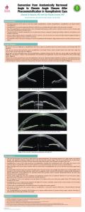
Ahmed Al Habash, MD and Earl Randy Craven, MD. Conversion from Anatomically Narrowed ... Burgoyne C, Tello C, Katz LJ. Nanophthalmia and chronic angle ...
Arrhythmias e.g. sinus tachycardia, AF, broad complex arrhythmias, VT and VF. ⢠Catecholamine induced cardiomyopathy (Takotsubo. Cardiomyopathy) due to ...
Shawn E. Cowper, MD; James V. Fiorica, MD; Edward M. Haller;. Santo V. Nicosia, MD; .... Randall ME, Andersen WA, Mills SE, et al. Papillary squamous cell ...
usually presents with dyspnea (platypnea and orthodexia). [2]. Diagnostic criteria are: Chronic liver disease with portal hypertension arterial oxygen tension less ...
Key words: Adenocarcinoma, rectal neoplasms, vaginal neoplasms. How to cite this article: ... vulva, cervix, uterus and bilateral adnexal structures are normal.
Bisphosphonates for treatment of osteoporosis: Expected benefits, potential harms, and drug holidays. Canadian Family Physician. 2014;60(4):324-333. 5.
Vallecular cyst : A rare cause of Respiratory Distress and Stridor in. New Born. Saurabh Patel M.D.. 1. , Ankita Shukla, M.D.. 1. , Shardha Polam, M.D.. 1,2.
Carhill AA, Cabanillas ME, Jimenez C, Waguespack SG, Habra MA, Hu M, ... Brose MS, Elisei R, Dutcus CE, de las Heras B, Zhu J, Habra MA, Newbold K, Shah.
Case Report. We present a case of a 30 year old African American Male with. Morquio syndrome and widespread metastatic gastric adenocarcinoma.
This is a case of a 22-year-old Filipino male, morbidly obese, not known to have diabetes mellitus (DM) who presented with diabetic ketoacidosis on initial ...
Apr 13, 2015 - Pericardial effusion is a rare complication of Graves' disease. A pregnant Filipino .... pericardial effusion in tamponade.17 We also regarded the.
Mir Mohsin, MBBS, MS, MCh;3 Peerzada Umar Farooq Baba, MBBS, MS, MCh;3 ... reporting a 65-year-old man with a giant lipoma involving his left buttock and.
gastrointestinal (GI) tract.1 We present a single case of a 9-year-old girl ... 1. Chest X-ray of 9-year-old Sotho girl. ... Cohen NP, Booth IW, Parashar K, Corkery JJ.
This syndrome shows no predilection for sex and has no ... the timing of treatment but orthodontic treatment should start as soon as ... (Amniotic band syndrome).
Jun 10, 2012 ... 1PG Scholar Dept of Shalya Tantra, N.K.J. Ayurvedic medical college, Bidar,
Karnataka, India. 2Assistant professor, of Shalya Tantra, N.K.J. ...
Fig. 3 Lower labial appliances bonded to complete space closure and correct rotation of lower molars. ***InVu Aesthetic Brackets, registered trademark of TP ...
changes of UC (cryptitis, crypt abscess, crypt distortion, crypt branching and inflammatory infiltrate) and no protozoa or parasite. With a diagnosis of acute severe.
Aug 4, 2014 - ABSTRACT: Ceruminous adenoma is a benign glandular neoplasm of ceruminous glands that arises solely from the external auditory canal.
Roseman JM, Wyche D True aneurysm of the profunda femoris artery. Literature review, differential diagnosis, management. J Cardiovasc Surg 1987;28:701â5.
Endovascular repair of a bilateral deep femoral artery aneurysm Lucas Van Houtven, Patrick Lauwers, Frank De Belder (*), Steven Laga (**), Jeroen Hendriks and Paul Van Schil Departments of Thoracic and Vascular Surgery; Radiology (*); and Cardiac Surgery (**)
Introduction:
Ø Deep femoral artery aneurysms (DFAA) are very rare Ø Only 0,5% of all peripheral artery aneurysms ; 1-6% of all femoral artery aneurysms [1] Ø Often incidental finding ØSigns and symptoms include: a pulsatile mass in the groin, paralysis or pain [2] Ø High rate of rupture, justifying treatment
Case report
75 year old male Medical history: none Ascending aorta aneurysm > referred for cardiac surgery Incidental finding on physical exam: pulsatile mass in the groin; no subjective complaints Ø CT (Fig. 1): left-sided aneurysm, 50 mm; right-sided, 30 mm. Ø Ø Ø Ø
Fig. 1 CT angiography. Arrows indicating the bilateral deep femoral artery aneurysm
Staged endovascular approach via contralateral access Ø Left: distal outflow had trombosed (Fig.2) Ø Distal coil embolisation (to prevent eventual future retrograde filling of the aneurysm) (Fig. A) Ø Proximal occlusion with Amplatzer II plug (Fig. B) (to exclude the aneurysm (Fig. C))
Discussion
Ø Multiple options for treatment, standard methods have not yet been established. Ø Reasonable recommendation for surgical intervention: all DFAA’s >2cm [3]. Ø We preferred an endovascular approach due to its minimal invasiveness. Ø Endovascular treatment options are directed by the patency of the distal vessels Ø 65% associated with synchronous aneurysms Ø Recommended to screen for popliteal and aortoiliac aneurysms
Ø No complications Ø Post-operative CT : complete exclusion of both aneurysms
D
E
References: 1. Posner, S.R., et al., A true aneurysm of the profunda femoris artery: a case report and review of the English language literature. Ann Vasc Surg, 2004. 18(6): p. 740-6. 2. Roseman JM, Wyche D True aneurysm of the profunda femoris artery. Literature review, differential diagnosis, management. J Cardiovasc Surg 1987;28:701–5. 3. C. Harbuzariu Profunda femoris artery aneurysms: association with aneurismal disease and limb ischemia. J Vasc Surg, 47 (2008), pp. 31–35