Case Report Short title: Aorto-enteric Fistula
Aorto-enteric Fistula After Endovascular Abdominal Aortic Aneurysm Repair for Behcet's Disease Patient: A Case Report S. Arworna,b S. Orrapina,b B. Chakrabandhu a, b T. Reanpanga, b J. Settakorna, b K. Laohapensanga, b, ∗
[email protected] a
Department of Surgery, Chiang Mai University Hospital, Chiang Mai, Thailand
b
Department of Pathology, Chiang Mai University Hospital, Chiang Mai, Thailand
∗
Corresponding author. Department of Surgery, Chiang Mai University Hospital, Chiang Mai, Thailand.
Introduction A 42 year old male with Behcet's disease (BD) had endovascular treatment of a symptomatic infrarenal abdominal aortic aneurys m (AAA). Thirteen months later he developed haematemesis and melaena.
Methods Computed tomography (CT) and angiography showed an aorto-enteric fistula with migration and kinking of the stent graft. Explantation of the infected graft and axillobifemoral bypass, aneurysm sac debridement, and jejunal repair with omental interposition was performed on this severely contaminated patient.
Discussion There are no reports of an aorto-enteric fistula secondary to endovascular repair in the literature and this case describes the p otential consequences of endovascular repair of AAA in BD. The aorto -enteric fistula was associated with persistent inflammatory aortitis, stent graft kinking, and infection. Five cases of secondary aorto-enteric fistulas following open AAA repair in BD patients have been reported including this case resulting from endovascular repair.
Keywords : Behcet's disease; Aortic aneurysm; Endovascular treatment; Aorto -enteric fistula
Introduction Behcet's disease (BD) is a chronic systemic vasculitis of unknown aetiology characterised by recurrent oral aphthous ulcers, genital ulcers, uveitis, skin lesions, and vasculitis in young adults aged 20–40 years. Abdominal aortic aneurysms (AAAs) associated with BD rapidly increase in diameter and have a high risk of rupture.1 Open surgical repair is associated with anastomotic pseudoaneurysm in 38.1% and a few case reports of aorto-enteric fistula (AEF).2–7 It has been postulated that endovascular repair is safer than open repair based on evidence from a case series. A case of AEF after endovascular repair of AAA in a BD patient is reported here and the mechanism and treatment are discussed.
Case report A 42 year old man with a confirmed one year diagnosis of BD was admitted with a two day history of abdominal pain. Computed tomogr aphy (CT) showed a 5 cm saccular aneurysm 5 cm below the right renal artery and 3 cm
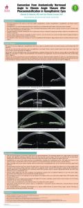
above the aortic bifurcation. The diameter of the aorta above and below the aneurysm was 16 mm and 15 mm respectively ( Fig. 1A) The C-reactive protein (CRP) level was 124 mg/L. The erythrocyte sedimentation rate (ESR) was 110 mL/hour. The white blood count (WBC) was 11,000/mm 3 and it was assumed that the AAA was a complication of BD. An urgent repair was performed using an off the shelf 20 mm Endurant Il iac tube limb endoprosthesis (ENEW2020C80EE) (Medtronic, Minneapolis, MN) inserted percutaneously via the right common femora l artery. The abdominal pain was resolving and the patient was discharged home 5 days later. Follow up CT scans performed three and six months after the procedure demonstrated a decrease in the size of the aneurysm sac without endoleak, migration, or kinking of the stent graft (Fig. 1B). The patient had regular follow up visits and controlled his BD with oral prednisolone (60 mg/dL) and colchicine (60 mg/dL). Figure 1 (A) A computed tomography (CT) scan showing an infrarenal saccular abdominal aortic aneurysm (AAA) 5 cm in diameter, 5 cm below the right renal artery and 3 cm above the aortic bifurcation. The diameter of the aorta above and below the aneury sm is 16 and 15 mm respectively. (B) Six month post-procedure
follow up CT scan demonstrating no endoleak, migration, or kinking of the stent graft. (C) CT scan demonstrating migration and kinking of the stent graft, which takes up space in the sac causing migration of the distal landing zone. The proximal landing zone and distal landing zone become aneurysmal, caused by an oversized stent graft and fragile aortic tissue. (D) A CT scan with oral contrast showing close contact and loss of the fat streak between the bowel and the aneurysm with air in the aneurysm sac. alt-text: Figure 1
Thirteen months after repair, the patient was admitted as an emergency with haematemesis and melaena. A CT scan showed migration and kinking of the stent without endoleak (Fig. 1C). A CT scan with oral contrast showed close contact and loss of the fat streak between the bowel, and air in aneurysm sac (Fig. 1D). The positive findings in laboratory data were as follows: leukocytosis (WBC, 12,000/mm3) with predominantly polymorphonuclear leukocytes; ESR of 40 mg/dL and CRP of 35 mg/dL. Endoscopy was performed, but the findings were unremarkable. A diagnosis of stent graft infection secondary to AEF was confirmed by CT scan (Fig. 1D). Laparotomy revealed an inflamed aneurysm sac adherent to the jejunum. After proximal infrarenal and distal iliac control was obtained, the aneurysm sac was opened. It was filled with purulent material that was collected for bacteriological examination and culture. A jejunotomy was done to expose the active fist ula with the exposed stent graft in the first part of the jejunum. The stent graft was not well incorporated in the aortic wa ll and was totally explanted. Treatment included extraanatomical axillobifemoral bypass, explantation of the infected stent graft, aneurysm sac debridement, jejunal resection with end to end anastomosis, and omental interposition. The blood cultures were negative. Bacteroides fragilis and Escherichia coli grew on the intra-operative cultures taken from the endograft; these were treated with intravenous antibiotics (amoxicillin, clavulanic acid, and
metronidazole) for 14 days. Histopathology of the AAA specimens showed a fibrotic inflammatory cellular infiltrate with blood clot in the media. There was evidence of an old haemorrhage within the granulation tissue (Fig. 2A). The thrombus associated with the fistula wall contained several foci of neutrophil and histiocyte aggregation (Fig. 2B).
Figure 2 Histopathological analysis of abdominal aortic aneurysm specimens showing showing fibrotic inflammatory cellular infiltrate with blood clots in the media. (A) Granulation tissue with many haemosiderin laden macrophages (H&E, × 40). (B) Many neutrophils and histiocytes within the thrombus (H&E, × 400). alt-text: Figure 2
The post-operative course was uncomplicated. The patient was discharged five weeks later in a stable condition on oral prednisolone (6 0 mg/dL) and colchicine (60 mg/dL) with a regular follow up 3, 6, and 9 months later without any vascular complications. Consent was obtained from the patient to publish these images and history.
Discussion Secondary AEF is a rare complication after open AAA repair, occurring in 0.5–1.5% of cases.2,3 Regarding management of AAA in BD patients, only a few cases have been reported with AEF after open AAA repair. The incidence is poorly defined (Table 1).3–7 AEF after endovascular repair has not been described.
Table 1 Summary of published reports detailing secondary AEF after open surgical and endovascular management of AAA in Behcet's disease patients. alt-text: Table 1 First author
Year
Case
Anastomotic aneurysm
AEF
Operations
Results
Koike 1,a
1988
M. 45
–
21 m
Aortic reconstruction, bowel repair
Dead, 36 d. PO
Qzeren2,a
2000
M. 35
15 m
5 yr
Aortic reconstruction, bowel repair
Dead, MOF
Alkim3,a
2008
M. 35
–
2 yr 3 m
1. Aortic reconstruction, bowel repair
Alive, FU 15 m.
2. ABF, graft excision
Ogve 4,a
2008
M. 34
–
8 m.
Suturing of aortic graft with omental wrapping, bowel repair
Alive
Gullu5,a
2011
M. 28
–
7 yr
Suturing of aortic graft, bowel repair
Dead, 5 d PO. Aortic graft ruptured
Arwornb
2016
M. 42
–
13 m.
ABF, Graft excision, bowel repair
Alive
AEF = aortoenteric fistula; AAA = abdominal aortic aneurysm; M = Male; m = month; yr = year; d = day; PO = post-operative; MOF = multiple organs failure; ABF = axillobifemoral bypass; FU = follow up. a
Open surgical management.
b
Endovascular management. Urgent endovascular repair of an AAA was performed in the patient 13 months previously using the only available off the shelf Endurant iliac tube limb endoprosthesis (Medtronic, Minneapolis, MN, USA), which was 25–30% oversized. The excessive oversizing of the stent graft
caused a discrepancy between the aorta above, below, and at the level of the endograft (Fig. 1C). The pathogenesis of AEF development after endovascular abdominal aortic repair was due to mechanical failure of the stent graft and kinking of the device in the aneurysm sac, which led to aortic graft erosion and pressure necrosis of the bowel wall. 5–7 In addition, aorto-enteric fistula was precipitated by inflammatory aortitis after endovascular repair in BD ( Fig. 2A). Although the aneurysm was focused in one area and the tube graft seemed to be adequate for coverage, the involvement of the aortic wall may have bee n more extensive than appeared, and more extensive coverage might have been considered with minimal oversizing. Endovascular AAA repair for a BD patient is less invasive with immediate technical success and anastomotic pseudoaneurysm formation can be avoided.8 There are also reports of pseudoaneurysm formation at the access site or a recurrent aneurysm at the edge of an inserted stent graft.8,9 The patient was treated as an emergency and no pre-operative steroid was administered during either the first endovascular repair or the second surgical operation. Adjunctive immunosuppressive therapy may help to prevent recurrence of aneurysm. 9 Some believe immunosuppressive therapy may be useful during the acute inflammatory period and before an operation to reduce the operative complications. 9 Immunosuppressive therapy is not recommended for urgent conditions and severe stent graft infections. Immunosuppressants may increase the risk of complications if not administered properly, and the length of time required for immunosuppressive treatment after surgery remains to be determined. Medical treatment should be tailored to the patient based on their condition and the risk of adverse effects. No guidelines are available regarding immunosuppressant use in BD patients, and there is no consensus; further prospective studies and evaluation on a larger scale are warranted. Published articles from 1998 to 2016 that report secondary AEF in BD patients after open surgical repair were reviewed along with the patient described here following endovascular repair. The search found five articles with five patients (all men; between 28–45 years of age) (Table 1). Secondary AEF was diagnosed from 8 months to 7 years post-operatively (average 35.2 months). Two patients (patients 3 and 6) survived after stent graft explantations and axillobifemoral (ABF) bypass. Two patients (patients 4 and 5) underwent graft repair with omental graft: one died from graft rupture and one survived. Two patients who underwent direct aortic reconstruction died (patients 1 and 2). Secondary AEF is a serious complication in infected stent grafts.10 Direct aortic reconstruction was associated with a high mortality rate compared with patients who underwent ABF bypass and graft excision. Conventional surgical treatment options are associated with high morbidity and mortality and include graft removal with prima ry or secondary axillobifemoral bypass and graft removal and in situ reconstruction, depending predominantly on the infection status of the s urgical site. Secondary AEF is managed by graft explantation, aortic stump closure, and lower limb revascularisation by axillobifem oral bypass. Primary closure of the aortic defect is considered inadequate, because the infection is not eradicated, and the infected prosthetic graft is not removed. Revascularisation by axillobifemoral bypass has been challenged by the possibility of aortic stump blowout, a risk that also remains during long-term follow up. In situ bypass grafting using homografts, prosthetic or vein grafts should not to be used in grossly infected fields. Prosthetic grafts can have wider application after extensive debridement of the retroperitoneal space, impregnation of the graft with antimicrobials (silver or rifampicin), and omental wrapping.10
Conclusion This case demonstrated that AEF is a potential complication of endovascular treatment of AAA for a BD patient . Explantation of the infected graft and axillobifemoral bypass was performed.
Funding None.
Conflicts of interest None.
References 1. Y. Fei, X. Li, S. Lin , X. Song, Q. Wu, Y. Zhu, et al., Major vascular involvement in Behcet's disease: a retrospective study of 796 patients, Clin Rheumatol 32, 2013, 845– 942. 2. T.W. Kwon , S.J. Park, H.K. Kim, H.K. Yoon, G.E. Kim and B. Yu, Surgical treatment result of abdominal aortic aneurysm in Behcet's disease, Eur J Vasc Endovasc Surg 35, 2008, 173– 180. 3. S. Koike, K. Matsumoto , M. Kokubo, Y. Mori, S. Murakawa and M. Hirose , A case of aorto-enteric fistula after reconstruction of an abdominal aortic aneurysm associated with Behcet's disease and special reference to 95 reported cases in Japan, Nippon Geka Gakkai Zasshi 89, 1988, 945– 951.
4. M. Ozeren, I. Mavioglu , O.V. Dogan and E. Yucel, Reoperation results of arterial involvement in Behç et’ s disease, Eur J Vasc Endovasc Surg 20, 2000, 512– 519. 5. H. Alkim, E. Parlak, N. Sasmaz , M. Akdogan, S.O. Kuran , M. Bayazit , et al., Secondary aortoenteric fistula in Behcet's disease, Turk J Gastroenterol 19, 2008, 49– 53. 6. H. Ozguc , N.B. Topal and E. Topal, Secondary aortoenteric fistula in a patient with Beh çet disease: successful surgical treatment by direct suture and use of omental flap, Vascular 16, 2008, 300– 302. 7. B.E. Gullu , A.U. Gullu and M. Ates, Prosthetic graft erosion complicating an aorto-duodenal fistula in a young patient with Behcet's syndrome: case report, Turk Klin Cardiovasc Sci 23, 2011, 283– 286. 8. S.W. Kim, Y. Lee do , M.D. Kim, J.Y. Won, S.I. Park, Y.N. Yoon, et al., Outcomes of endovascular treatment for aortic pseudoaneurysm in Behcet's disease, J Vasc Surg 59, 2014, 608– 614. 9. O. Balcioglu , S. Ertugay, H. Bozkaya , M. Parildar and H. Posacioglu, Endovascular repair and adjunction immunosuppressive therapy of aortic involvement in Behcet's disease, J Vasc Endovasc Surg 50, 2015, 593– 598. 10. S.K. Kakkos, C.D. Bicknell, I.A. Tsolakis and D. Bergqvist, Hellenic Co-operative Group on Aortic Surgery, Editor's choice – management of secondary aorto-enteric and other abdominal arterio-enteric fistulas: a review and pooled data analysis, Eur J Vasc Endovasc Surg 52, 2016, 770– 786.