7th Annual International IEEE EMBS Conference on Neural Engineering Montpellier, France, 22 - 24 April, 2015
Investigation of the Spatial and Spectral Patterns of Hand Extension/Flexion using High-density ECoG Tianxiao Jiang, Nuri F. Ince* Senior IEEE Member, Tao Jiang, M.D., Taylor Wang, Shenshen Mei, Yunlin Li, M.D, Xiaofei Wang, M.D., Sujit Prabhu, M.D. ,Zhiyi Sha, M.D. 1
Abstract— High density electrocorticogram (ECoG) of motor cortex was recorded during awake surgeries of two subjects with respectively epilepsy and brain tumor. Subjects were asked to execute spontaneous hand movements: extension and flexion during the experiments. We investigated the ECoG in time frequency plane during these hand movements and computed event-related desynchronization (ERD) and event-related synchronization (ERS) levels of beta (8-32 Hz) and gamma frequency bands. The distribution of ERD and ERS over channels provided an idea about the spatial distribution of the cortical activity within each task. In both subjects, consistently, the scale of power changes in beta and gamma band was larger in hand flexion compared to hand extension. In addition, the spatial distribution of ERD in beta and ERS in gamma band during flexion was larger than extension but highly overlapped between these tasks. Gamma band was more spatially localized than beta band activity and cortical patterns were more distinct between the tasks. Our preliminary results indicate that the high density ECoG can be effectively used in awake surgery for functional mapping.
I. INTRODUCTION Electrocortical stimulation mapping (ESM) is considered as the gold standard to delineate eloquent cortex (motor and language regions) of human brain [1][2]. By placing subdural electrodes on the relevant cortical areas, a subject-specific electrical current is applied, generating an outcome in the form of stimulation or inhibition in motor and language functions. Despite of its great clinical importance in aiding brain surgeries for many years, it still suffers from major drawbacks such as after-discharges, time consuming and vague spatial scale [3][4]. Previous studies on electrocorticography (ECoG) brain mapping provided evidences that significant overlaps existed between ESM responsive sites and ECoG contacts with power alteration in specific frequency bands [5]. These studies confirmed ECoG to be a useful supplement to ESM in functional brain mapping. It has been known that sensorimotor behaviors can result spectral modulations in specific frequency bands of ECoG. Typical modulations are in the form of event-related desynchronization (ERD), an amplitude decrease, in (713Hz) and (13-32Hz) bands and event-related synchronization (ERS), an amplitude increase, in (40200Hz) band [6]. Functional brain mapping based on spectral changes in different frequency bands of ECoG has been explored and it has been shown that the highest sensitivity and specificity relative to electrical stimulations mapping (ESM) sites can be achieved in high frequency band (65-95 Hz) [7].
Recent advancement in micro electrode technology now enables us to record cortical surface potential in an unparalleled spatial-temporal scale. Traditional ECoG electrodes used in human brain functional mapping had either large inter-contact distance (10 mm) [5] or small inter-contact distance (4 mm) with limited channel number [8]. In this study, we use a customized high-density 120-channel micro-ECoG electrode with an inter-contact distance of 4mm and contact diameter of 2mm. We focus on functional mapping of two crucial daily hand movements: extension and flexion. We investigate spectral- spatial patterns of ECoG and explore the between these tasks. We show that while spatial patterns in low band greatly overlap, high frequency provided distinct spatial patterns between hand flexion and extension. II. MATERIALS AND METHODS A. Subjects Micro-ECoG electrode was placed on the perirolandic cortex in two adult patients during craniotomy. The first patient (S1) underwent epilepsy surgery and the second patient (S2) underwent brain tumor surgery. Intraoperative ECoG recordings were carried out in two institutions: Chinese PLA General Hospital and Beijing Haidian Hospital. TABLE I listed detailed information. The study protocol was approved by the institutional review board (IRB) of each hospital. Informed consent forms were provided to and signed by each patient before their participation. TABLE I. Case
S1 S2
Epilepsy Tumor
Sex (F/M) F F
Age 36 40
Hand (R/L) R R
Flexion (EN/TN) 23/27 25/30
Extension (EN/TN) 31/37 22/25
Time (min) 6 7.7
F=female; M=male; R=right; L=left; EN=eligible trial number; TN=total trial number.
B. Micro-ECoG recording and behavioral system The platinum Micro-ECoG used in this study was 12 by 10 grid electrode (Ad-Tech). The diameter of the contacts was 1.2 mm and the inter-electrode distance was 4 mm. Intraoperative ECoG data sampled at 2 kHz and video were simultaneously collected by XLTEK EMU 128FS system. As the frequency of interest in current functional brain mapping studies only reaches up to 200 Hz [9][10], we further down-sampled the data to 500 Hz. To minimize the aliasing problem during downsampling, a FIR low-pass filter with a cut-off frequency 220 Hz was applied in advance.
*Corresponding author. Tel.: 713-743-4461; Fax: 713-743-0226; Email:
[email protected]
978-1-4673-6389-1/15/$31.00 ©2015 IEEE
ID
SUMMARY OF SUBJECTS AND EXPERIMENTS
589
A digital glove with a Bluetooth interface (DG5 VHand 2.0) was used to capture individual finger movements of the subjects during hand flexion and extension movements (See Figure 1). Finger positions were captured by five bending sensors with 10-bit resolution. Three axis accelerometers with 0.5° resolution captured the roll and pitch angles. The finger positions provided by the glove is saved by a laptop computer. A synchronization signal was generated by behavioral system and recorded by the neural system and behavioral system simultaneously. Neuronal data and behavioral data were synchronized later by detecting the rising and falling edges of the pulses of synchronization signal. The initial sampling rate of the digital glove was 25 Hz. Then behavioral data and synchronization signal were stored by behavioral system at a rate of 200 Hz. The aligned finger position data and the video were used in movement onset annotations.
Figure 2. (a) High-pass filtered (60 Hz) noise segments within channel 1 to 10 of S2’s recording. (b) 10 PCA components computed from averaged covariance matrix, sorted by eigenvalues in a descending order. (c) Original data of S2, high-pass filtered by 3 Hz. Noise components were denoted as ‘A’ (green). (d) Denoised data using spatial principle component analysis (SPCA).
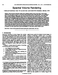
Figure 1. (a) A high-density ECoG grid electrode with 120 channels was placed on the hand representational cortex of the patient in operating room. (b) Hand flexion. (c) Hand extension.
C. Experiment paradigm Micro-ECoG electrodes were placed onto the primary motor cortex to cover the hand representational area. Patients were instructed to perform hand extension and flexion in a repeated meaner at a self-paced rate of less than 0.5 Hz. Movements are intermittently performed and separated by arbitrary interval (relax). Eligible movements lasting 1 to 2 seconds with an interval of more than 2 seconds were extracted for analysis. D. Preprocessing All ECoG data were visually scrutinized and bad channels were excluded from analysis. Furthermore, in order to exclude the 50 Hz power line frequency component and its harmonics, a series of customized FIR notch filters with stop-bands covering 50 Hz, 100 Hz, 150 Hz and 200 Hz were applied.
matrix of all channels. Noise components were observed in the first two PCA components (Figure 2-b). Then the subspace spatial projection matrix was applied on the entire recording of S2. The first two PCA components were eliminated. The remaining PCA subspace components were projected back to original data space. The noise components were denoted in Figure 2-c. In the denoised data, the periodic noise was successfully eliminated (Figure 2-d). E. ERD/ERS The ERD/ERS is defined as the relative power decrease (ERD) or power increase (ERS) of activity period compared against reference period several seconds before the trigger onset [11]. In this study, a second-order Butterworth band-pass filter was used to filter the ECoG data in 8-32 Hz and 60-200 Hz ranges. ERD in low frequency band (LFB: 8-32 Hz) and ERS in high frequency band (HFB: 60-200 Hz) were evident in a series of channels during movements. Epochs of raw data with 1.7 seconds of baseline data before the onset of movement and
In addition, a periodic high frequency noise with uniform spatial distribution was found in the entire recordings of S2, probably due to the electromagnetic noise of a nearby equipment. Spatial principle component analysis (SPCA) was applied to eliminate this noise. Original data were first high pass filtered with a cut off frequency of 60 Hz in order to align the noise components. Then, 30 noise segments were manually annotated (Figure 2-a). The subspace spatial projection matrix was obtained by performing PCA on the averaged covariance 590
Figure 3. Band-pass filtered data segments during S1’s flexion with 1.7 second of baseline period and 1 second of movement period. LFB=low frequency band (8-32 Hz), HFB=high frequency band (60-200 Hz). The scale of ERD is 500 uV while the scale of ERS is 50 uV.
one second of movement data were concatenated and visualized in Figure 3. F. Time-frequency analysis Four seconds of neural data starting two seconds before the movement onset and extending two seconds after were used in time-frequency analysis. In each trial, time-frequency maps were computed by short-time Fourier transform (STFT) with a 200-sample Hanning window and shifted by 20 samples. By averaging over trials and channels, a generalized time frequency map was obtained for each movement type (extension/flexion). We normalized the time-frequency map ̅ with the mean of the 0.5-second baseline spectrogram ̅ to obtain a centered spectrogram ̅ . ̅
10
log
The spectrogram of flexion showed steeper changes in high frequency band at around 80 Hz. ERD in LFB started approximately 500 milliseconds before onset and ERS in HFB started around 200 milliseconds before onset. The power rations (movement versus baseline) of hand flexion and extension in both frequency bands are provided in Fig.5. Our results indicate larger power changes in both LFB and HFB during hand flexion compared to hand extension (Figure 5).
̅ ̅
G. Spatial and spectral pattern analysis In this study, in order to circumvent the notch-filtering effect of 50 Hz power line noise, we define two separate frequency ranges: 8-32 Hz band (LFB) and 60-200 Hz band (HFB). Baseline was determined at 300 milliseconds before the movement onset to exclude the motor planning phase. For each subject, spatial and spectral patterns were obtained by calculating the band power ratios (movement/baseline) in LFB (8-32 Hz) and HFB (60-200 Hz) for hand extension and flexion then averaged through all trials. Channels with significant power changes (>50%) during movements compared to resting states were defined as active channels in spatial spectral pattern analysis. The geometric center of active channels was denoted as activation center to reflect the general location of the activation patterns. III. `RESULTS Noticeable power decrease in LFB (8-32 Hz) and increase in HFB (60-200 Hz) were observed in the averaged timefrequency maps of both subjects (Figure 4). Blue regions in the map corresponded to a power decrease in specific frequency range and red regions corresponded to a power increase accordingly. Darker color indicated greater changes.
Figure 5. Mean and standard deviation of the band power ratio (movement/baseline) calculated using active channels (power change >50%) in low frequency band (8-32 Hz) and high frequency band (60-200 Hz).
The spatial patterns of LFB and HFB highly overlapped with each other in both movements, which suggested that, generally the same cortical areas were activated. For S1, the ERD pattern in LFB experienced the smallest topographic change from extension to flexion in that only one channel (C10) switched its state (activated in flexion and deactivated in extension). In ERS pattern of HFB, five channels (C8, C9, C37, C63, C67) switched their states. For S2, in both LFB and HFB, there were dramatic spatial pattern changes between hand flexion and extension, where 6 channels for LFB and 8 channels for HFB changed their states. It is also interesting to note that the most active channel (with the highest power change) in HFB spatial pattern of subject 2 changed completely from C109 (extension) to C97 (flexion), across an approximate cortical distance of 9 mm (Figure 6). Generally speaking, compared to high frequency band spatial pattern (HSP), low frequency band spatial pattern (LSP) tends to experience less topographical alteration. In addition, LSP is widely observed in the covered cortical area while HSP is more spatially localized. Specifically, there were approximate 10 less active channels in HFB than LFB for both hand extension and flexion in each subject. The detail of the number of active channels in all conditions can be found in TABLE II. TABLE II. ID
Figure 4 Average time frequency maps. (a) S1 Extension. (b) S2 Extension. (c) S1 Flexion. (d) S2 Flexion. Movement onsets were aligned to second zero. The spectrograms were shown in dB scale and smoothed by a 2-D Gaussian filter.
591
NUMBER OF ACTIVE CHANNELS
Extension
Flexion
LFB
HFB
LFB
HFB
S1
48
37
49
40
S2
31
19
33
23
HSPs were more localized compared to LSPs. Topographical changes and active channel changes in HSPs were also greater than LSPs.
Subject 2
Subject 1
ACKNOWLEDGMENT The authors are grateful to the Chinese PLA General Hospital and Beijing Haidian Hospital for providing the opportunities of intraoperative ECoG recordings. REFERENCES [1]
Figure 6. The spatial spectral patterns of hand extension and flexion for S1 and S2. (a) Hand extension in LFB of S1. (b) Hand flexion in LFB of S1. (c) Hand extension in HFB of S1. (d) Hand flexion in HFB of S1. (e) Hand extension in LFB of S2. (f) Hand flexion in LFB of S2. (g) Hand extension in HFB of S2. (h) Hand flexion in HFB of S2. Each circle denoted a contact. Active channels were marked as black triangles (upward triangle for power increase and downward triangle for power decrease). Activation center was also marked out by asterisk. Iterative interpolation was applied to fill out the missing contacts (bad channels). All figures were smoothed by 2-D Gaussian filter.
[2]
[3]
IV. DISCUSSION
[4]
Based on the anatomical discovery of column-wise arranged thalamocortical projections to the sensory cortices, it has been postulated that distinct spatial patterns of cortical activity exist between different behaviors. In an earlier study on spatio-temporal correlations in human gamma band ECoG [13], it was concluded that subdural electrode arrays must have a spacing under 5 mm in order to capture the gamma band spatial patterns (if existed) of human brain. In this study, the high-density ECoG electrode of 4 mm interelectrode distance enables us to investigate cortical activity in a very fine spatial resolution. Between hand extension and flexion in our subjects, the displacements of the center of active channels (marked as asterisks in Figure 6) were generally less than 2 mm in both frequency bands (LFB: 8-32 Hz and HFB: 60-200 Hz). It may suggest that the overall activation region remain the same across different movements (extension/flexion). While significant spatial alterations were observed when we focused on individual channels, especially in high frequency band. For both subject, the spatial pattern has more channels switched their activation states in HFB than in LFB. It might be helpful to use spatial information of HSP in distinguishing movements in addition to the power ratio (movement/baseline). However it should be kept in mind that all phase information was lost in spectral analysis thus we cannot reveal cortical circuit propagation effects. For the future work, more advanced time-frequency analysis and timedomain correlation analysis should be added to include phase information. Also, in order to make it clinically helpful, a comparison between the obtained patterns with direct cortical stimulation mapping results should be added. V.
[5] [6] [7]
[8]
[9]
[10]
[11] [12]
[13]
CONCLUSION
Significant temporal spectral modulations were found in a series of channels of our high-density ECoG electrode recordings during hand movements. Comparing between hand extension and flexion, high frequency band spatial patterns (HSPs) and low frequency band spatial patterns (LSPs) differed greatly in intensity. In the sense of spatial distribution, 592
S. Chitoku, H. Otsubo, Y. Harada, V. Jay, J. T. Rutka, S. K. Weiss, M. Abdoll, and O. C. Snead, “Extraoperative cortical stimulation of motor function in children.,” Pediatr. Neurol., vol. 24, no. 5, pp. 344– 50, May 2001. G. Ojemann, J. Ojemann, E. Lettich, and M. Berger, “Cortical language localization in left, dominant hemisphere. An electrical stimulation mapping investigation in 117 patients.,” J. Neurosurg., vol. 71, no. 3, pp. 316–26, Sep. 1989. E. C. Leuthardt, K. Miller, N. R. Anderson, G. Schalk, J. Dowling, J. Miller, D. W. Moran, and J. G. Ojemann, “Electrocorticographic frequency alteration mapping: a clinical technique for mapping the motor cortex.,” Neurosurgery, vol. 60, no. 4 Suppl 2, pp. 260–70; discussion 270–1, Apr. 2007. N. Pouratian, A. F. Cannestra, S. Y. Bookheimer, N. A. Martin, and A. W. Toga, “Variability of intraoperative electrocortical stimulation mapping parameters across and within individuals.,” J. Neurosurg., vol. 101, no. 3, pp. 458–66, Sep. 2004. K. J. Miller, M. DenNijs, P. Shenoy, J. W. Miller, R. P. N. Rao, and J. G. Ojemann, “Real-time functional brain mapping using electrocorticography,” Neuroimage, vol. 37, pp. 504–507, 2007. D. K. Su and J. G. Ojemann, “Electrocorticographic sensorimotor mapping.,” Clin. Neurophysiol., vol. 124, no. 6, pp. 1044–8, Jun. 2013. M. J. Vansteensel, M. G. Bleichner, L. T. Dintzner, E. J. Aarnoutse, F. S. S. Leijten, D. Hermes, and N. F. Ramsey, “Task-free electrocorticography frequency mapping of the motor cortex.,” Clin. Neurophysiol., vol. 124, no. 6, pp. 1169–74, Jun. 2013. W. Wang, A. D. Degenhart, J. L. Collinger, R. Vinjamuri, G. P. Sudre, P. D. Adelson, D. L. Holder, E. C. Leuthardt, D. W. Moran, M. L. Boninger, A. B. Schwartz, D. J. Crammond, E. C. Tyler-Kabara, and D. J. Weber, “Human motor cortical activity recorded with MicroECoG electrodes, during individual finger movements.,” Conf. Proc. ... Annu. Int. Conf. IEEE Eng. Med. Biol. Soc. IEEE Eng. Med. Biol. Soc. Annu. Conf., vol. 2009, pp. 586–9, Jan. 2009. N. E. Crone, D. L. Miglioretti, B. Gordon, and R. P. Lesser, “Functional mapping of human sensorimotor cortex with electrocorticographic spectral analysis. II. Event-related synchronization in the gamma band.,” Brain, vol. 121 ( Pt 1, pp. 2301–15, Dec. 1998. K. J. Miller, E. C. Leuthardt, G. Schalk, R. P. N. Rao, N. R. Anderson, D. W. Moran, J. W. Miller, and J. G. Ojemann, “Spectral changes in cortical surface potentials during motor movement.,” J. Neurosci., vol. 27, no. 9, pp. 2424–32, Feb. 2007. G. Pfurtscheller and F. H. Lopes da Silva, “Event-related EEG/MEG synchronization and desynchronization: basic principles.,” Clin. Neurophysiol., vol. 110, no. 11, pp. 1842–57, Nov. 1999. G. Pfurtscheller, B. Graimann, J. E. Huggins, S. P. Levine, and L. A. Schuh, “Spatiotemporal patterns of beta desynchronization and gamma synchronization in corticographic data during self-paced movement.,” Clin. Neurophysiol., vol. 114, no. 7, pp. 1226–36, Jul. 2003. V. Menon, W. J. Freeman, B. A. Cutillo, J. E. Desmond, M. F. Ward, S. L. Bressler, K. D. Laxer, N. Barbaro, and A. S. Gevins, “Spatiotemporal correlations in human gamma band electrocorticograms,” Electroencephalogr. Clin. Neurophysiol., vol. 98, no. 2, pp. 89–102, Feb. 1996.