Linda Rodgers, Joseph A. Beavon, and Michael Wigler$**. From the Scold ...... Charbonneau, H. (1990) in Cyclic Nucleotide Phosphodiesterases: Structure,.
Vol. 268. No. 17, Issue of June 15, pp. 12925-12932,1993 Printed in U.S.A.
THEJOURNALOF BIOLOGICAL CHEMISTRY
0 1993 by The American Society for Biochemistry and Molecular Biology, Inc.
Isolation and Characterizationof a Previously UndetectedHuman cAMP Phosphodiesterase by Complementation of CAMP Phosphodiesterase-deficientSaccharomyces cereuisiae" (Received for publication, October 1, 1992, and in revised form, March 1, 1993)
Tamar MichaeliSP, TimJ. Bloomn, Tim Martinsll, Kate Loughneyll, Ken FergusonII ,Michael Riggs, Linda Rodgers, Joseph A. Beavon, and Michael Wigler$** From the Scold Spring Harbor Laboratory, Cold Spring Harbor, New York 11 724, the 11 ICOS Corporation, Bothell, Washington 98021, and the TDepartment of Pharmacology, University of Washington, Seattle, Washington 98195
We have established a highly sensitive functional tivity to cofactors and inhibitory drugs (reviewed in Refs. 2 screen for the isolation of cDNAs encoding cAMP phos- and 3). These families are (I) Ca2+/calmodulin-dependent phodiesterases (PDEs) by complementationof defects PDEs, (11) cGMP-stimulatedPDEs, (111) cGMP-inhibited in a Saccharomyces cereuisiae strain lacking both en- PDEs, (IV) CAMP-specific PDEs, (V) cGMP-specific PDEs, dogenous cAMP PDE genes, PDEl and PDE2. Three and (VI) photoreceptor PDEs. As the amino acid sequences groups of cDNAs corresponding to three distinct hu- of members of these PDE families are being determined, it is man genes encoding CAMP-specific PDEs were isolated becoming apparent that all these PDE families containa from a human glioblastoma cDNA library using this related domain, thought to be the catalytic domain, with functional screen. Two of these genes are closely re- -30% sequence identity between families (2). Members of the lated to the Drosophila dunce CAMP-specific PDE. The third gene, which we named HCP1,encoded a novel same family are more closely related, and the available seCAMP-specific PDE. HCPl has an amino acid sequence quences suggest that they share between 6040% sequence related to the sequences of the catalytic domains ofall identity extending throughoutthe entirecoding region (2, 4). Two PDE genes, PDEl and P D E 2 , have been identified cyclic nucleotide PDEs.HCPl is a highaffinity CAMPand cloned from the yeast, Saccharomyces cerevisiae (5-8). specific PDE (K,= 0.2 pM) that does not share other properties of the CAMP-specific PDE family, i.e. exten- PDE2 is a high affinity CAMP-specific PDE related in sesive sequence homologyto the Drosophila dunce cAMP quence to PDEs of mammalian cells. PDEl is a low affinity PDE andsensitivity to rolipram andR020-1724. The CAMP-specific PDE that belongs to a different evolutionary PDE activity of HCPl is not sensitive to cGMP orother branch of PDEs. The PDEl sequence is more closely related as mil- to thesecreted PDE form of Dictyostelium discoidum than to inhibitors of the cGMP-inhibitable PDEs, such rinone. The biochemical and pharmacological proper-PDE2 and the known mammalian cell PDEs. ties of HCPl suggest thatit is a member of a previously In S. cerevisiae, RAS proteins are regulators of adenylyl undiscovered cyclic nucleotide PDE family. Northern cyclase and of cAMP production (9). The mutationally actiblot analysis indicates that high levels of HCP 1 mRNA vated RAS2 gene, RAS2v""9, leads to an increase of cellular are present in humanskeletal muscle. cAMP content and to several associated phenotypes (9-11). Among the phenotypes that result from expression of RAS2va"9is an acute sensitivity to heat shock. The yeast Cyclic nucleotides serve as second messengers that mediate PDEl and PDE2 genes were isolated as multicopy suppressors a variety of cellular responses to extracellular signals such as of the RAS2v""9heat shock phenotype. In a similar manner, hormones, light, andneurotransmitters. Cyclic nucleotide two mammalian cDNAs encoding CAMP-specific PDEs were phosphodiesterases (PDEs)' play a role in signal transduction previously isolated rat (DPD) and human (JC44) cDNAs, by regulating the cellular concentrations of cyclic nucleotides both encoding homologs of the Drosophila dunce cAMP PDE (reviewed in Ref. 1).Mammalian cells contain multiple PDEs and members of family IV (12, 13). In this study we describe that have been distinguished into six families based on their the establishment of a more sensitive functional screen for substrate affinity and specificity and on their selective sensi- isolation of cDNAs encoding cAMP PDEs and its use to isolate a human cDNA that appears to define a new family of * This work was supported by the National Cancer Institute, the PDE genes.
American Cancer Society, and National Institutes of Health. The costs of publication of this article weredefrayed in partby the payment ofpage charges. This article must therefore be hereby marked "aduertisernent" in accordance with 18 U.S.C. Section 1734 solely to indicate this fact. The nucleotide sequence(s) reported in thispaper has been submitted to the GenBankm/EMBL Data Bankwith accession number(s) L12052. 5 Present address: Dept. Devel. Biol. & Cancer, Albert Einstein College of Medicine, 1300 Morris Park Ave., Bronx, NY 10461. ** American Cancer Society Research Professor. To whom correspondence should be addressed Cold Spring Harbor Lab., P. 0. Box 100, Cold Spring Harbor, NY 11724. Tel.: 516-367-8376; Fax: 516367-8381. The abbreviations used are: PDEs, phosphodiesterases; kb, kilobase(s); PCR, polymerase chain reaction; ORF, open reading frame.
'
EXPERIMENTAL PROCEDURES
Strains, Growth Conditions, Heat Shock Assays, and Segregation Analysis-Plasmids werepropagated in Escherichia coli strains HBlOl or SCSl (Stratagene). S. cereuisiae strains TKLGI-R2V: (MATa RAS2"""' leu2 his3 u r d ade8 trpl) (9) and lODAB ( M A T a leu2 his3 ura3 ade8 p d e l A D E 8 pde2:URAJ ras1::HZSd) (12) were grown in rich medium (YPD) or synthetic medium (SC) with appropriate supplements. Heat shock assays of variable time periods were performed as previously described (12).To screen the expression library, yeast transformants were plated at approximately lo3 colonies/plate on selective medium. Colonies were allowed to grow for 3 days and then replica plated onto preheated plates. Heat shocks were carried out at 55 "C for 15 min and followed by 2-3 days of recovery at 30 "C. Surviving colonies were picked, restreaked on synthetic
12925
12926
HCP1,Affinity a High
medium plates for colony purification, and then cultured in rich medium for 2-3 days to allow for plasmid loss from some cells. ml of these cultures were then plated onto YPD plates. After 2-3 days of growth, these plates were replica plated onto synthetic medium plates (Leu+selection), YPD plates,and YPD heat shock plates. Colonies were scored to ascertain if the observed heat shock resistance was plasmid-dependent. Plasmid, DNA Manipulutions, and Sequencing-The plasmids employed in this study are expression vectors containing the ADHl promoter, the yeast 2p replication origin, and the LEU2 selectable marker: ADNS was describedpreviously (12) and AD54 is aderivative of ADNS in which the ADHl promoter is attached to an epitope derived from the influenza hemagglutinin protein. The DNA sequence encoding this epitope tag ends with a SalI restriction endonuclease site and is followed by the portion of the pUC18 polylinker residing between the Sun and the EcoR I sites. TM22A was generated by deleting the 0.5-kb BglII fragment of TM22. L22M1 through 4 were generated by inserting PCR-amplified DNA fragments of TM22, which were flanked by SalI sites, into theSalI site of AD54. The 20cycle PCR amplification was performed under standard conditions. PCR reactions for generating the SalI fragments of L22M1-4 included a common COOH-terminal oligonucleotide 5"GCTAGTCGACCTGGCTGGCATCACTCAC(1603). The NHP-terminal oligonucleotides included in these PCR reactions were: L22M1, 5'- GCTAGTCGACGATGGAAGTGTGTTACCAG (68); L22M2, 5'- CGTAGTCGACTATGCTAGGAGATGTACGTG (261); L22M3,5'- CGTAGTCGACCATGCTGGAAAAAGTTGGA (506); L22M4,5'- CGTAGTCGACCATGATGAAACTTCGTAGA (638). The numbers indicate the coordinates, as inFig. 2. The DNA sequence of the PCR-generated Sun fragment of L22M4 wasdetermined and found to be identical to the corresponding TM22 sequence. Sequencing was performed by the dideoxynucleotide chain-termination method (14, 15). Bothstrands of the TM22 4.0-kb Not1 fragment were sequenced as detailed in thestrategy shown in Fig. 2. Northern Blotting-Cytoplasmic RNA from the U118-MG cell line was isolated in a buffer containing 140 mM NaCI, 1.5 mMMgC12, 10 mM Tris-C1, pH 8.6, and 0.5% Nonidet P-40. Following a 1.5-min 12,000 X g spin, the supernatant was incubated with 0.5 mg/ml proteinase K for 30 min at room temperature, extracted with a 1:l phenol/chloroform mixture, and ethanol precipitated. Poly(A)+ RNA was selected on oligo(dT) by established procedures (16). RNA was fractionated on a 1%formaldehyde-agarose gel and transferred onto a nylon membrane (Genescreen Plus, New England Nuclear). The membrane was baked at 80 "C for 2 h. Hybridization was performed at 60 "C in 1%sodium dodecyl sulfate, 1 M sodium chloride, and 10% dextran sulfate. Following hybridization the membrane was washed at 60 "c in 0.3 M sodium chloride, 0.03 M sodium citrate, and 1% sodium dodecyl sulfate. The membrane was stripped of the radioactive probe by a 10-minute incubation in boiling water. Removal of the radioactivity from the membrane was monitored by autoradiography. RNA probe complementary to ORF2 was transcribed in vitro by T7 RNA polymerase from a template containingthe SpI-NszI fragment (base pair 3372-3646) of TM22 (Fig. 2). Hybridization of this ["PI CTP-labeled probe was performed at 42 "C in the hybridization buffer described, except with the addition of 50% formamide. Washes were performed as detailed above. Blots of size-fractionated poly(A)+ RNA from human tissues were purchased from Clonetech, Inc. Blots were hybridized to 32P-labeled DNA probes at 43 "C in 50% formamide, 0.75 M sodium chloride, 50 mM sodium phosphate monobasic, pH 7.4, 50 mM EDTA, 0.16% Ficoll, 0.16% polyvinylpyrrolidone, 0.16% bovine serum albumin, 0.16% sodium dodecyl sulfate, and 100 pg/ml denatured salmon sperm DNA. Following hybridization the blots were washed under stringent conditions for 20 min at 50 and at65 "C in 0.015 M sodium chloride, 0.0015 M sodium citrate (0.1 X SSC), and 0.5% sodium dodecyl sulfate. One blot was first hybridized to a 650-nucleotide long single-stranded DNA probe, generated by PCR, complementary to the 5'end of TM22, and then to the nick-translated double-stranded probe (1.2kb Notl-EcoRI fragment of TM22). The hybridization pattern of the single-stranded probe was identical to the double-stranded HCPl probe (not shown). Phosphodiesterase Assays-Yeast cells weregrown at 30 "c in synthetic medium (SC-leucine). Cells were harvested and resuspended in lysis buffer containing 50 mM potassium phosphate, pH 7.4, 150 mM NaCI, and 30 mM benzamidine. Cells were broken with glass beads, and cellular debris was removed by a 5-min spin at 12,000 X g. Cyclic nucleotide phosphodiesterase activity in the supernatant
CAMP-specific PDE was measured as previously described, with modifications (17, 18). Assays were performed in vinyl microtiter plates (Costar) at 30 "C. Incubation mixtures contained40 mM Tris-C1, pH 8.0, 1 mM EGTA, 5 mM magnesium chloride, 0.1 mg/ml bovine serum albumin, diluted enzyme, [3H]cyclicnucleotides (0.14 pCi/assay) (New England Nuclear), ["CIAMP (0.01 pCi/assay) (New England Nuclear), and varying amounts of unlabeled cyclic nucleotides in a total volume of 125 pl. The assays were terminated by removing a 100-pl aliquot of the reaction mixture to a microliter well containing 100 pl of stop buffer (0.2 M ethanolamine, pH 9.0, 1 M ammonium sulfate, 20 mM EDTA, and 1%SDS). The 5' nucleotide products were separated from the unreacted cyclic nucleotides by chromatography on columns (8 X 5 mm) containing Affi-Gel 601 (Bio-Rad), an acrylamide matrix containing covalently coupled aminophenylboronate. The columns were washed with 2.5 ml of wash buffer containing 0.1 M ethanolamine, pH 9.0, and 0.5 M ammonium sulfate. The columns were then eluted with 2 ml of elution buffer containing 0.25 M acetic acid. The column eluate that collected into the vials was mixed with scintillation fluid (EcoLume, ICN Biochemicals) and analyzed for 3H and "C content by dual channel liquid scintillation spectroscopy. The recovery of 3H nucleotide reaction products were corrected for the recovery of ["C] AMP. All the kinetic data points represent measurements of initial rates, determined by incubations for multiple intervals at suitable enzyme dilutions. One unit of phosphodiesterase activity is expressed as 1pmol of cyclic nucleotide hydrolyzed/min. mg protein. RESULTS
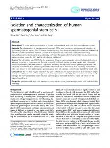
Isolation of Human cDNAs Encoding CAMPPDEs-Several mammalian homologs of the Drosophila dunce cAMP PDE have been previously isolated from cDNA expression libraries based on their ability to suppress the heat shock-sensitive phenotype of RAS2"""' (12, 13). In this study we identified two new human genes encoding cAMP PDEs. cDNAs corresponding to these genes were isolated based on their ability to suppress the heat shock-sensitive phenotype of the strain 10DAI3, a strain in which the two yeast cAMP PDE genes have be disrupted. The strainlODAB was transformed with a yeast expression library containing cDNAs derived from the human glioblastoma cell line U118-MG (Human Tumor Cell Line Bank, Human Tumor Cell Laboratory, Memorial Sloan-Kettering Cancer Institute, 13). lo6 transformants were screened for their ability to withstand heat shock a t 55 "Cby the replica plate method. Seventy-four colonies were found to contain plasmids capable of rendering lODAB cells resistant to heat shock. These plasmids contained threegroups of cDNAs. The largest group, with 70 members, contained cDNAs that were variants of JC44 and of the previously isolated high affinity rolipram-sensitive CAMP-specific PDE (13, 19). The second group, represented by TM72, contained twocDNA clones encoding an additional member of the high affinity, rolipramsensitive, CAMP-specific PDE family that will be described elsewhere.2 The amino acid sequence of these two groups are highly related to the D. melanogastet dunce cAMP PDE and to the four dunce homologs found in rat (12, 13, 19-21). Thethird group, represented by TM22, contained two cDNA clones encoding a novel PDE whose properties are detailed in this study. The ability of TM22 to render lODAB cells resistant to heat shock is depicted in Fig. 1. Representative cDNAs from allthree groups that were isolated as suppressors of deficiencies in cAMP PDEs of the strain lODAB failed to suppress the heat shock sensitivity of ~ ~ s 2 v a l l(Fig. 9 1): However, JC44, a structural variant of CAMP-specific PDEs of the firstgroup has been isolated as a G. Bolger, T. Michaeli, T. Martins, T. St. John, B. Steiner, L. Rodgers, M. Riggs,M. Wigler, and K. Ferguson, manuscript in preparation. T. Michaeli, T. Bloom, T. Martins, K. Loughney, K. Ferguson, M. Riggs, L. Rodgers, J. Beavo, and M. Wigler, unpublished observations.
HCPl,Affinity a High
A
CAMP-specific PDE
12927
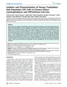
into theCOOH terminus of ORF2. The structureof this group of cDNAssuggests that TM22 is notanaberrant fusion product generatedby reverse transcriptase. A second group of 55"C,5mln cDNAsincludedseveralmembers that contained parts of ORFl and were polyadenylated at either of two adjacent Vector locations, following nucleotides 3104 and 3106. A polyadenylation AATAAA consensus sequence was found 15-17 nucleTM 22 otides upstream of these sites, and they are followed by a TTt~'22~ rich region (reviewed in Ref. 22). This putative polyadenylation site lies within ORF2. The structure of these cDNAs suggests that thetwo open reading frames might be normally found on two independent transcripts. T o confirm the relationshipsbetween the two open reading frames and the structure of the isolated cDNA insert, we have 6 RASZ tested whether the two open readingframes were indeed found on two independent transcripts. For this purpose we per55"C,5mln formed a Northern blot analysis of poly(A)+RNA from U118MG cells (Fig. 3). The blot was first hybridized to a 1.2-kb Vector NotI-EcoRI DNA fragment that containedmost of ORFl and none of the sequences downstream of it. This DNA probe hybridized to a single mRNA band estimated tobe 4.5 kb in size. The blot was stripped of this probe, and hybridized to a 1.0-kb BstXI-Not1 DNA fragment encompassing ORF2 and about 50 base pairs of the non-coding sequence downstream FIG.1. Suppression of the pdel-pde2- and the RAS2""' of its COOH terminus. This probe hybridized strongly to a heat shock phenotypes. A , lODAR cells were transformed with the 6.0-kb mRNA band andvery weakly to the samesize band to ADNS vector, with TM22, and with a deletion mutant of TM22, TM22A. R, RAS2'"1'9cells were transformed with the AD54 vector, which ORFl hybridized (-4.5 kb). A single-stranded RNA with L22M1, an expression vector of the TM22 cDNA, and with probe containing sequences complementary to ORF2 hybridYEpPDE2, a plasmid containing the yeast PDE2 gene. Two inde- ized to a 6.0-kb mRNA band as well (data not shown). The pendent transformants were patched onto SC-leucine plates, grown weak hybridization of the double-stranded ORF2 probeto the at 30 "C for 3 days and then replica plated onto a control plate and 4.5-kb ORFl mRNA band is probably due to the fact that it onto an experimental plate that was subjected to a 5-min heatshock containedabout 110base pairsupstream of theputative treatment at55 "C.Plates were photographed following a 24-h recovpoly(A) site of ORFl (HCPl). Thus, the two open reading ery period at 30 "C. See also Fig. 5. frames are found on two independent transcripts. The location of the polyadenylation sites and the size of suppressor of RAS~"""~ (13). Thus, it appears thatpdel-pde2the HCPlmRNA mightindicate that TM22 is a partial cDNA cells are more sensitive to theeffects of cAMP phosphodiesclone of H C P l which contains only 3 kb of the 4.5-kb mRNA terases than areRAS2v""9cells. detected on Northern blots. It is therefore notclear whether Sequence of TM22"The cDNA insert of TM22 was a 4.0the first ATG found in ORFl serves as the initiatormethiokb DNA fragment flanked by the restriction sitesof the NotI nine of HCP1. Attempts to isolate additional 5' sequences of endonuclease used in its cloning (13). The DNA sequence of the HCPl mRNA have met with persistent obstacles. We this fragment revealed two open reading frames(Fig. 2). The first open reading frame, called ORF1, was 498 amino acids additionally screened three cDNA libraries derived from hulong with the first ATG appearing a t codon 17. The termi- man brain, two from heart, and a second from the U118-MG nation codon of this open reading frame was followed by a cell line. Each library screen yielded HCPl cDNA clones, but 2.5-kb non-coding DNA fragment endingwith a NotI restric- their DNA sequence indicated that none extended 5' of the tion endonuclease cleavage site. An additional open reading known TM22 sequences. The difficultiesin obtaining fullframe of 293 amino acids, named ORF2, was found on the length clones may be due, in part, to the high GC content non-coding strand on the distal 1.0 kb end of the cDNA (82%) of the 5' end of TM22, or may reflect that the 5' (between nucleotides 3048-3817). The initiator methionineof sequences already cloned represent the 5' end of the tranORF2 was preceded by a termination codon. T o determine script. Similarities between HCPl and CAMPPDEs-A search for which of the two open reading frames played a role in supsequences similar to HCPl in the data banks revealed a strong pressing pdel-pde2- defects, we generated an in-frame deleof aportion of tion in ORF1, TM22A. This deletion mutant failed to sup- homology to cAMP PDEs. The alignment press the heat shock sensitivity of lODAB (Fig. 1). Thus, HCPl to representativemembers of several cAMP PDE famORFl is required for suppression of pdel-pde2- defects. The ilies whose sequences are known is shown in Fig. 4. Repregene corresponding to ORFlwas subsequently named HCPI, sented were the CAMP-specific PDEs (rat DPD), the Ca2+/ calmodulin-dependentcyclic nucleotide PDEs (61-kDa bovine for reasons discussed later. bovine cGMP-stimulated PDE (4, 12, AdditionalcDNAsisolatedfromasecondcDNA library brain form) and the derived from the U118-MG cell line and from a human heart 23). Significant homology was observed in a COOH-terminal catalytic cDNA library by screening with DNA probes encompassing region of -300 amino acids, thought to constitute the ORFl were characterized by sequencing. The structure of portion of these enzymes (24-28). The homology encompassed the region between residues 164-451 of HCP1. Sequence these cDNAs indicated that inefficient polyadenylation may account for the formation of a read-through transcript with relatedness among these PDEs was determined by the MAthestructure of the TM22 cDNA. One group of cDNAs CAW program (29) which defined withinthis COOH-terminal included several members that,like TM22, contained partsof region seven homology blocks (A-G), interspersed by short ORF1, lacked a poly(A) tail, and extended to various extents unrelated sequences.
pdel-pdeZ-
12928
HCPl, a High Affinity CAMP-specificPDE
A
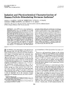
FIG. 2. Structure and sequences of TM22.A, a diagram of the structure of the TM22 cDNA includes the location and direction of the two open reading frames, ORFl andORF2, marked by arrows. The location of the polyadenylation sites of ORFl is marked by PA. The location of the restriction endonuclease cleavage sites BglrI (Bg), BstXI (Bs), EcoRI ( E ) , and Not1 (Not) are indicated. The sequencing strategy of TM22 is detailed below the diagram: the orientation and length of the fragments sequenced with a single oligonucleotide are detailed. B , the DNA sequence of TM22 and the predicted amino acid sequence of ORFl are presented. Coordinates on the right indicate nucleotide and amino acid positions. The polyadenylation sites of ORFl (nucleotides 3104 and 3106) are underlined and in bold type, and upstream of these sites is the polyadenylation consensus sequence AATAAA (underlined). The location of sequences used to generate L22M1-4 is indicated by the arrows labeled MI "4, respectively.
1
I kb
i
122
40 242 110 161 120 ,112 160
962
320 1082
360 1202
400 1322
440
1562 498 16112 1802 1922
2042 2162 2212 2402
2522 2642 2762
21112 1002
1122 1242
1362 14112
1602 3122
3842 1962 39n7
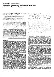
The NHa-terminal border of this conserved domain was found to extend 40 residues upstream of the boundary previously thought to demarcate homology among the PDEs (2). The extended NHp-terminalportioncontained homology block A, a poorly conserved spacer, and the first 14 residues of homology block B (Fig. 4,A and B ) . The sequences of the cGMP-stimulated PDE depicted in homology block A were located 450 residues upstream of block B. However, an additional sequence (A') of the cGMP-stimulated PDE with reduced similarity to sequences of block A was located just upstream of block B of this PDE (Fig. 4C). Although the sequence similarity within homologyblock A' is poor, its proximity to block B might be of functional importance. An additional structural element of HCP1, an 18-residue NH2-
terminal repeat sequence, was found by a double diagonal analysis (Fig. 4 0 ) . Within the highly conserved region there were several motifs that were identical among all the known mammalian PDE families and may constitute important structural elements. HCPl exhibited the highest degree of homologyto theCAMPspecific PDE (35% identity, 51% similarity), and the lowest to thecGMP-stimulated PDE (24% identity, 37% similarity). The homology within this region between members of the known CAMP-specificPDE family (family IV), varies between 85-95%, and is not limited to the region depicted in Fig. 4, but extends throughout most of the coding region (data not shown). Thus, it appears that HCPl is a novel PDE that is more closelyrelated to theCAMP-specific PDEs of family IV,
HCPl,Affinity a High
a
b
CAMP-specific PDE
12929
cells carrying an expression vector without an insert. Thus, H C P l appears to be a CAMP-specific PDE with a very high affinity for cAMP as a substrate. These resultssuggest that, kinetically, HCPl hassome resemblance to the cGMP-inhibited PDE family in that it has a similar low K, for cAMP and that HCPl also resembles the CAMP-specific PDE family in that it isspecific for CAMP. To determine whether H C P l is pharmacologically related to the CAMP-specific or the cGMP-inhibited PDE families, we tested whether its PDE activitywas sensitive to competi28s tive inhibitors selective to these PDEfamilies (Fig. 7). These compounds are thought to inhibit cAMP hydrolysis by interacting at the catalytic site (31). As expected, cAMP was a potent inhibitor with an IC50of 0.2 p~ (Fig. 7A). Unlike the cGMP-inhibited PDEs, the PDE activity of H C P l was virtually unaffected by cGMP.Inaddition,thecAMPPDE activity of H C P l was not significantly affectedby two specific inhibitors of the cGMP-inhibited PDEs, amrinone and milrinone (Fig. 7B). Although a 50% inhibition of HCPl was observed inthe presence of 60 p~ milrinone, an even stronger inhibition of TM72 was observed a t these high concentrations of milrinone. TM72is ahighaffinitydunce-like CAMPspecific PDE that has been isolated in this screen, and, as a member of this PDEfamily, is notexpected to be particularly FIG. 3. Northern blot analysis of ORFl and ORF2. Two pg sensitive tomilrinone. Apartially purified bovine lung cGMPof poly(A)+ RNA prepared from U118-MG cells werefractionated on inhibited PDE was, however, sensitive to milrinone with an a formaldehyde agarose gel and transferred onto a nylon membrane. , that the drug was effective during The blot was hybridized to the 32P-labeled EcoRI-Not1 1.2-kb DNA IC50of 1 p ~ indicating fragment of TM22 ( a ) .Following the removal of this probe the blot experimental manipulations (data not shown). Thus, it apof H C P l is not especially was hybridized again to the 32P-labeled BstXI-Not1 l-kb DNA frag- pearsthatthecAMPactivity ment of TM22 ( b ) . sensitivetothe known inhibitors of thecGMP-inhibited PDEs. The cAMP activity of H C P l was not affected bytwo potent but is not a member of this family or of any of the other inhibitors of dunce-like CAMP-specific PDEs, R020-1724 and known cAMP PDE families. Deletion Analysis of HCP1-To improve the production of rolipram (Fig. 7C). TM72, a new member of this dunce-related HCPl and to enable ourbiochemical analysis, we constructed PDE family, was sensitive to these drugs. Thus, HCPl apa new expression vector, L22M1. In thisvector the firstATG pears to be pharmacologically distinct from the dunce-like codonwasfused to an epitope derivedfrom the influenza CAMP-specific PDEs. Expression of HCPl mRNA in Human Tissues-The abunvirus hemagglutinin transcribed from the strong yeast promoter, ADHl (30). L22M1 drastically improved the suppres- dance of H C P l mRNA in human tissues was determined on sion of p d e l p d e r heat shock phenotype (Fig. 5). This im- Northern blotsprobedwith the 1.2-kbNotI-EcoRI DNA provement may be due, in part, to theremoval of the highly fragment encompassing residues 1-399of HCPl (Fig. 8). A GC rich area upstream of the first ATG that might impede 3.8-kb H C P l mRNA was abundant in skeletal muscle and transcription and translation. Three additional NHz-terminal detectable in kidney and heart. An additional 4.0-kb HCPl deletions of H C P l were generated by sequential deletion of mRNA band was detectable in the brain, kidney, and pansequences from the first ATG codon to residue 81 (L22M2), creas. The significance of the two HCPl transcripts is not 163 (L22M3), and 207 (L22M4). While L22M2 and L22M3 known. An additional Northern blotwithmRNAsamples derived from different individuals had a similar HCPl mRNA were eachcapable of efficient suppression of pdel-pde2defects, the largest deletion mutant, L22M4, failed to do so distribution in the heart and skeletal muscle, but its abunkidney, and pancreaswas greatly reduced. (Fig. 5). All 10 independentclones of L22M4 that were tested dance in the brain, failed to suppress the heatshock sensitivity of 10DAB. West- These blots were subsequently hybridized to an e-tubulin ern blot analysis indicated that equivalent amounts of epi- cDNAprobe as a control forRNA concentrations.These tope-tagged peptides were produced from all four expression results indicate that HCPlencodes a novel cAMP PDE that plasmids (L22M1, 63 kDa; L22M2, 50 kDa; L22M3, 43 kDa; is expressed more abundantly in skeletal muscle than in the L22M4, 39 kDa). Thus, this functional deletion analysis de- other tissues we examined. fined the NHz-terminal border of the minimal fragment of DISCUSSION H C P l required for cAMPhydrolysis to reside between amino acids 163-207. Interestingly, this border coincides with the We have cloned a human cDNA encoding a novel cAMP NH2-terminal border of the conserved domain of cAMP PDEs PDE, HCP1, that is structurally, biochemically, and phardetailed above (Fig. 4). macologically distinct from known cAMP PDEs. HCPl is a Biochemical Analysis of HCPl-Substrate and inhibitor cAMP PDE with a very high affinity for cAMP (K,,, = 0.2 kineticanalyses were conductedusingcrude homogenates p ~ )a ,property used to derive its name,high affinity CAMPfrom lODAB cellsoverexpressing HCP1. The K,,, deduced specific PDE. Although the sequence of HCPl is related to from the Lineweaver-Burke plots was 0.2 p~ CAMP, and the those of the Ca"/calmodulin-dependent, the cGMP-stimucalculated Vmaxwas 0.025 nmol/min. mg total protein(Fig. 6). lated, and the CAMP-specific dunce-like families of cAMP No cGMP PDE activitywas detectable in theoverexpressing PDEs, thehomologies are not as close as those shared between cells, and no cAMP PDE activity was detected in extracts of family members, and thus HCPl is not a member of these
i) 8
HCPl, a High Affinity CAMP-specificPDE
12930
A
E n . A
HCP 1
Ca++/Cam d e p .
cGMP s t i m .
-
6 C D E F
G
"4 98
529
[D
A'
[r
1 1 1
1 0
I nI-lt
92 1
B 6 HCP 1 DPD Ca'YCam
ril
dep.
cGMP s t i m .
. . C
HCP I DPD Ca++/Cam d e p . cGMP s t i m .
hsqn hsdv skyk rdp.e.
lfdrltngnslvsl------nvagyshnrpltci------alneasgehslkfm------ekhtlvalkrvqalqqr/../Sm 176 5 9 -2 p= 10
..
M4
. D
p= 0
.
0
22 1 225 210 630
.. .
epklan tpalda gtgimh nleltn
.
285
289 275 693
HCP 1 DPD Ca++/Cam d e p . cGMP s t i m .
HCP 1 DPD Ca++/Carn d e p . cGMP s t i r n .
44 1 453 4 28 85 1
HCP 1 :%Cam dep cCMP s t i m .
.
.
C HCP 1 DPD Ca++/Cam d e p . cGMP stirn.
"""""""""-
.. Alp= 4.5
. IO-1
"""""""""" " " " " " " " "
yhmkvsddeytkllhdgi
&)tvtprsipeddtsmdILSMLQ 604 I "
D HCP 1 HCP 1
"_""" gaisfssssalfg
alfgcpn qtalyir
48 72
FIG. 4. Alignment of the amino acid sequences of the HCP1,DPD, Ca'+/calmodulin-dependent and cGMP-stimulated PDEs. The alignment was obtained by the MACAW program (29). A, a schematic representation of the alignment of the cyclic nucleotide PDEs. Blocks of homologyare boxed and marked A-G. Gaps mark the gaps introduced to maximize homology. B , amino acid sequences that constitute homology blocks A-G. Blocks of homology are boxed and cdpitalited. The probabilities of chance occurrence associated with each block were derived by the MACAW program and are indicated above the blocks. A probability of zero is indicated when the probability of chance occurrence is less than Dashes indicate gaps introduced to maximize the homology. Coordinates on the right indicateamino acid are depicted above these positions. The location of the NHz-terminal border of the peptides expressed from L22M3 ( M 3 ) and L22M4 (M4) residues. Residues that are similar among a t least three of the four PDEs presented are shaded. Fully conserved residues are under marked with an asterisk (*). The grouping of similar amino acidsare: ( V,L,I),(K,R),(E,D),(Q,N),and ( S , T ) .C, amino acid sequences that constitute homology blockA'. Sequences in this block are identical to those of homology block A except those of the cGMP-stimulated PDE which are located close to homology block B. D, a repeat sequence of HCPI. The boxed sequence depicts an 18-residue repeat sequence found by a double-diagonal analysis.
HCPl,Affinity a High
CAMP-specific PDE
12931
A
0
FIG. 5. Deletion analysis of HCP1. lODAR cells were transformed with AD54 (uector), with TM22 and with four deletion mutants of HCPl named L22M1-4. Two independent transformants were patched onto SC-leucine plates, grown a t 30 “C for 3 days, and then replica plated onto a control plate and onto an experimental plate that was subjected to a 5-min heat shock treatment at 55 “C. Plates were photographed following a 24-h recovery period a t 30 “C.
0
L+”.L.,ta>,, ”... “ L ” u u l _ u U u l 0 I OE-07 I OE-06 IO€-05 IO€-04 I OE-03
P
0
L4 .L- # I” ,”I,
0
IO€-8
I OE-7
I.OE-6
,,...”I._“ !OE-5
..&l__
IO€-4
.....”I IO€-3
[Inhibitor] M I CE-09
I OE-OR
I OE-07
IOE-06
IO€-05
[CAMP] M
FIG.6. Kinetic analysis of HCP1. cAMPPDEassays were performed on all extracts as described under “Experimental Procedures.” cAMPconcentrations rangedfrom 3.10-$ to M. Each data point shown is a measurement of the initial rate of hydrolysis a t a suitable enzyme dilution. The inset on the upper left corner of the graph depicts the double-reciprocal Lineweaver-Burke plot derived from the kinetics curve.
FIG.7. Effects of various inhibitors on the cAMP PDE activity of HCPl and of TM72. cAMP PDE assays were performed on cell extracts asdescribed under “Experimental Procedures.” SamM [‘HICAMP andthe indicated concentrations plesincluded 3 . of inhibitor. A, cAMP and cGMP. The indicated concentrations of unlabeled cAMP (+, M) or cGMP (0, 0 ) were added to assays. B, milrinone and amrinone. The indicated concentrations of milrinone (+, W) or amrinone ( 0 , O ) were added to the assays. C, rolipram and R020-1724. The indicated concentrations of rolipram (*, W) or R020-1724 (0,0 ) were added to the assays. Squares are extracts harboring HCP1, anddiamonds are extracts harboringTM72.
families. Recently, the sequence of the cGMP-inhibited PDE has been published and HCPl is not more closely related to Expression of HCPl was studied by Northern blot analysis this PDE family than to others (32). The kinetic and phar- of poly(A)+ RNAs obtained from various human tissues. Two macological data also suggestthat HCPlrepresents a member different sized mRNAs were found, 3.8 and 4.0 kb. We do not of a new family of cyclic nucleotide PDEs. With respect to know if these arise from differential splicing or termination, substrate specificity, H C P l most closely resembles the CAMP- or if they even arise from the same locus. We have been specific PDE family (3). However, the apparent affinity for hampered in these studies by our inability to isolate fullsubstrate is substantially higher than for any other CAMP- length cDNAs. Low levels of mRNAs were detected in a specific PDE,andthe enzyme isnotinhibited by either variety of tissues, including brain and heart. Consistentwith rolipram or R020-1724, potent inhibitors of all known mem- this, cDNAs for H C P l were isolatedfrom cDNA libraries bers of this family. The affinity for cAMP is most similar to derivedfrom these tissues. We have found high levels of that of the cGMP-inhibited PDEs, but it is not selectively expression of HCPl transcripts in human skeletal muscle. inhibited by either cGMP or milrinone, a drug that inhibits Very little is known about the expression of cAMP PDEs in all the known members of this family. Thus, the HCPl cAMPskeletal muscle, and thiswould seem to be the tissue in which PDE is structurally, kinetically and pharmacologically dis- to undertake further studiesof this cAMP PDEisoform. tinct from the currently known high affinity cAMP PDEs. It The alignment of the HCPlsequence with those of cAMP is possible, of course, that since the5’ end of the HCPlclone PDEs of different families reveals homology to theconserved may be truncated that both the substrate and inhibitor spec- COOH-terminal catalyticdomain of cAMP PDEs. TheNHpificity could be altered compared to the nativeenzyme. How- terminal border of this domain, as defined by the MACAW ever, it seems ratherunlikely that truncation would increase program, extends 40 residues upstream of the previously the apparent affinity for cAMP but decrease the affinity for delineated border. OurNHz-terminal deletionanalysis of the drugs and for cGMP. Since the sequence indicates that HCPl indicated thatthe minimal fragment required for H C P l is a separate andhighly distinct gene product, it seems cAMP hydrolysis encompasses this expanded domain of homore likely that the kinetics reflect intrinsic differences in mology. A peptide lacking thisNHz-terminal region, and the properties of the enzyme. H C P l therefore appears to be containing thepreviously delineated conserved domain almost a representative of a previouslyunknown cAMP PDEfamily, in its entirety, is notcatalytically active. These results are in which we now designate as family VII. agreement with previous studies of proteolytic fragments of
HCPl, a High Affinity CAMP-specific PDE
12932
A
a b c d e f g
6.
type of RAS2"""9cells may depend onmore than theirelevated cAMP content, and therefore may be refractory to theeffects of weak cAMP PDEs. The isolation of cAMP PDEs by complementation of defects in yeast asdescribed in this study hasyielded a member of a new cAMP PDE family. The complete sequence of this PDE, theexistence of additional members of this family, their distribution, andphysiological roles remain to be determined. Additional functional screens in this yeast system may yield additional previously undiscovered PDEs. Acknowledgments-We thank Dr. Paul Feldman, Glaxo Research Institute, Inc., for supplying the rolipram used in this study and the Sterling Drug Company, Inc. for supplying milrinone, Sonja Kalbfleisch and Kim McCaw, ICOS Corporation, for excellent technical assistance, and P. Bird for herhelp in preparingthis manuscript. REFERENCES
FIG.8. Tissue distribution of HCPl mRNA. Two pg of poly(A)+RNA prepared fromhuman pancreas ( a ) ,kidney ( b ) ,skeletal muscle (c), liver ( d ) ,lung (e), brain ( f ) ,and heart ( g )were fractionated on a denaturing formaldehyde agarose gel and transferred onto a nylon membrane. The blot was hybrididzed to the 32P-labeledNotIEcoRI 1.2-kb DNA fragment of TM22 (A) and to the 3ZP-labeledatubulin cDNA ( B ) .Blots were washed under stringent conditions for 20 min at 65 "C in 0.1 X SSC. Molecular weight RNA markers are 9.5, 7.5, 4.4, 2.4 and 1.35 kb long, and their migration on the gel is depicted on the right hand side of panel A.
1. Beavo, J. (1988) in Advances in Second Messenger and Phosphoprotein G. A., eds.) Vol. 22, Raven Press, Research (Greengard, P., and Robinson,
New York
2. Beavo, J., and Houslay, M. D. (eds) (1990) Cyclic Nucleotide Phosphodiesterases: Structure. Regulation and D r w Action. Vol. 2. John Wiley and
Sons, Ltd., Chichester
3. Bentley, J. K., and Beavo, J. A. (1992) Curr. Opin. Cell Biol. 4,233-240 4. Novack. J.. Charbonneau. H.. Bentlev. - J... Walsh. K.. and Beavo, J. (1991) B i o c h i m h y 30,7940-7947 Londesborough, J., and Suoranta, K. (1983) J. Biol. Chem. 258,2966-2972 5. J., Sass, P., and Wigler, M. (1987) Mol. Cell. Riol. 7,3629-3636 .; Nikawa, Sass, P., Field, J., Nikawa, J., Toda, T., and Wigler, M. (1986) Proc. Natl. Acad. Sci. (1. S. A. 83,9303-9307 Suoranta, K., and Londesborough,J. (1984) J. Biol. Chem. 269,6964-6971 8. 9. Toda, T., Uno, I., Isbikawa, T., Powers, S., Kataoka, T., Broek, D., CamBroach, J., Matsumoto,K., and Wigler, M. (1985) Cell 40, 2ieron,
s.,
2fi thecGMP-stimulatedandthe Ca2+/calmodulin-dependent 10. KaGoka, T., Powers, S., Cameron, S., Fasano, O., Goldfarb, M., Broach, PDEs that assigned a catalytic role to a 36-kDa fragment J., and Wigler, M. (1985) Cell 40,19-26 encompassing this conserved domain (24-28). The NHp-ter- 11. Marshall, M., Gibbs, J., Scolnick, E., and Sigal, I. (1987) Mol. Cell Biol. 7. 2309-2315 minal border of the minimal active fragment of H C P l is 12. Colicelli, J., Birchmeier,C., Michaeli, T., ONeill,K..Riggs, M., and Wigler, located 17-18 residues downstream of the NHp termini of the M. (1989) Proc. Natl. Acad. Scr. U. S. A. 86,3599-3603 J., Nicolette, C., Birchmeier, C., Rodgers, L., Riggs, M., and 36-kDa proteolytic fragments. The catalytically active 36-kDa 13. Colicelli, Wigler, M. (1991) Proc. Natl. Aead. Sci. U.S. A. 88,2913-2917 fragment of the cGMP-stimulated PDE containedhomology 14. Biggin, M. D., Gibson, T. J., and Hong, G. F. (1983) Proc. Natl. Acad. Sei. C'. S.A. 80,3963-3965 block A' as detailed in Fig. 4C, and not homology block A 15. Sanger, F., Nicklen, S., and Coulson, A. R. (1977) Proc. Natl. Acad. Sci. which was found by the MACAW program. The role of U. S. A. 74,5463-5467 16. Maniatis, T., Fritsch, E., and Sambrook, J. (1982) in Molecular Cloning, a homology blocks A and A' in catalysis remains to be deterLaboratory Manual,Cold Spring Harbor Laboratory, Cold Spring Harbor, mined. NY Davis, C., and Daly, J. (1979) J. Cyclic Nucleotide Res. 5, 65-74 We have established a highly sensitive screen for the iso- 17. 18. Martins, T., Mumby, M., and Beavo, J. (1982) J. Biol. Chem. 255, 19731 a70 lation of cAMPPDEs based ontheirabilitytosuppress 19. Livi, G., Kmetz, P., McHale, M., Cieslinski, L., Sathe, C.. Taylor, D., Davis, defects in yeast lacking endogenous cAMP PDEs. We have R:, Torphy, T., and Balcarek, M. (1990) Mol. Cell. Biol. 10,2678-2686 previously isolated yeast and mammalian cAMP PDEs in the 20. Daws, R., Takayasu, H., Ebenwne, M., and Myres, J. (1989) Proc. Natl. Acad. Sci. U. S.A. 86,3604-3608 mS2val19 strain that, like the pdel-pde& strain, contains 21. Swinnen, J., Joseph,D., and Conti,M. (1989) Proc. Natl.Acad. Sci. U. S. A. elevated intracellular CAMP. Comparisons of the frequency 86,5325-5329 Proudfoot, N. (1991) Cell 64,671-674 of isolation of one of the CAMP-specific PDEs (JC44) from 22. 23. Sonnenburg, W., Mullaney, P., and Beavo, J. (1991) 266, 17655-17661 the library employed in this study indicated that the PDE24. Krinks, M. H., Haiech, J., Rhoads, A,, and Klee, C. B. (1984) Adu. Cyclic Nucleotide Res. 16,31-47 deficient yeast strain (10DAB) is almost a hundred-fold more 25. Kincaid. R. L.. Stith-Coleman.. I. E... and Vauehan. .. . M. (1985) J. Biol. Chem. sensitive than the RAS2va'19strain (70/105 in pdel-pde2260,9009-9015 26. Charbonneau, H., Novack, J. P.. MacFarland, R. T., Walsh, K. A,, and uersus 415. lo5 in RAS2'e"9) for the detection of PDEs. Beavo, J. A. (1987) in Calcium-Binding Protein8 in Health and Disease cDNAs encoding the three different PDEsisolated in this (Norman, A. W., Vanaman, T. C., and Means, A. R., eds) pp. 505-517, Academic Orlando, FL screen failed to suppress the heatshock-sensitive phenotype 27. Strooo. S. D..Press, Charbonneau. H., and Beavo, J. A. (1989) J. Biol. Chem. of RAS2ve'19.This seems a t first a surprising result sincetwo 264,1371&13725 28. Charbonneau, H. (1990) in Cyclic Nucleotide Phosphodiesterases: Structure, mammalian CAMP-specific PDEs, rat DPD and human JC44, Regulation and Drug Action (Beavo, J., and Houslay, M. D. eds) Vol. 2, are capable of suppressing RAS2ve1'9,and since the intracelpp. 267-296, John Wiley and Sons, Ltd., Chichester G. Altschul. S.. and Lioman. . . D. (1991) Proteins Struct. Funct. lular levels of CAMP in RAS2'e"9 cells are actuallylower than 29. Schuler. Genet. 9, 180-190' those of pdel-pde2- cells (33). This apparent paradox canbe 30.. Field, J., Nikawa, J., Broek, D., MacDonald, B., Rodgers, L., Wilson, I., Lerner. R., and Wigler, M. (1988) Mol. Cell. Biol. 8, 2159-2165 understood only in the context of the function of RAS in S. 31. Weishaar, R. E., Cain, M. H., and Bristol, J. A. (1985) J. Med. Chem. 28, cerevisiae. The cAMP PDE activity of these threenew human 537-545 E., Taira, M., Moos, M., Jr., Smith, C., Movsesian, M., Degerman, cAMP PDEs are weaker than those of DPD and JC44when 32. Meacci, E., Belfrage, P., and Manganiello,V. (1992) Proc. Natl.Acad. Set. U S. A. assayed inpdel-pde2- cells (see "Results"). Poor cAMP PDE 89,3721-3725 J., Cameron, S., Toda. T., Ferguson, K., and Wigler, M. (1987) activity may account for the inability of the newly isolated 33. Nikawa, Genes & Deo. 1,931-937 PDEs to suppressRAS2ve"9.RAS appears to control not only 34. Toda, T., Broek, D., Field, J., Michaeli, T., Cameron, S., Nikawa. J., Sass, P., Birchmeier, C., Powers, S., and Wigler, M. (1987) Oyogene and the cAMP pathway in S. cerevisiae but an additional signaling Cancer (Aaronson,S. A,, et al., eds) pp.253-260, Japan Scientlfic Society pathway as well (34). Thus, the heat shock-sensitive phenoPress, Tokyo I . ,
'
0 Y
'