Isolation and Characterization of Human Factor VI1. ACTIVATION OF FACTOR VI1 BY FACTOR X,*. (Received for publication, February 20, 1980, and in revised ...
THEJOURNALOF BIOLOGICAL CHFXISTRY Vol 256, No. 1, Issue of January 10, pp. 2.57-259. 1981 Prmtedm C1.S.A.
Isolation and Characterizationof Human Factor VI1 ACTIVATION OF FACTOR VI1 BY FACTOR X,* (Received for publication, February 20, 1980, and in revised form, July 21, 1980)
S . Paul Bajaj, Samuel I. Rapaport, and Stephen F. Brown From theDepartment of Medicine (H-811@,University of California a t S o nDiego, La JoEta, CaZifornia 92093
A procedure has been developed for the isolation of human Factor VII to apparent homogeneity as judged by the analytical disc electrophoretic system of Davis sulfate (SDS)-polyacryl(pH 8.9) and by sodium dodecyl amide gel electrophoresis. The isolation procedure involves adsorption of Factor VI1 onto barium citrate, ammonium sulfate fractionation, DEAE-Sephadex chromatography, and preparative polyacrylamide gel electrophoresis. The overall yield of Factor VII is 15 to 20% of starting plasma and the purified protein has a specific activity of 1800 to 2200 units/mg in a clotting assay. Factor VI1 protein obtained by this method has only 1.3 to 1.5 times more activity in a one-stage clotting assay than in a coupled a m i d o l ~ i cassay {Seligsohn, U., (bsterud,B., and Rapaport, S . I. (1978)Blood 52,978988), which is taken to mean than itcontains less than 5 % of activated Factor VII. Human Factor VI1 is a single chain glycoprotein with an apparent molecular weight of 50,000 f 2,000 as determined by SDS-polyacrylamide gel electrophoresis. It has alanine as anNH2-terminalamino acid resii c residues/ due and contains 8.8 y - c ~ b o x y g l u t ~acid mol of protein. Incubation of purified Factor VI1 with Factor X, in the presence of Ca(U) and phospholipid results in a rapid up to 25-fold increase in its clotting activity. Factor VI1 activity in the coupled amidolytic assay remains unchanged throughout the incubation, Activation of Factor VI1 byFactor X, is associated with cleavage of the M, = 50,000 native protein to a protein consisting of two chains of M, = about 26,000 and 22,000, as determined by SDS-polyac~lamidegel electrophoresisinthe presence of 2-mercaptoethanol. Other properties of human Factor VII, including its amino acid composition and its reactions with an antiFactor VI1 antibody, are described.
dependent clotting factors, which are inert in their zymogen form, native bovine Factor VI1 has been reported to possess a low level of inherent clotting activity (6) and has been shown to hydrolyze several syntheticesters (7). Incubation with thrombin f4), Factor X, (4, 6 ) , Factor XII, (8-lo), or Factor IX, (10) can strikingly enhance the clotting activity of native Factor VII. For bovine Factor VI1 incubated with Factor X, (4,6) or Factor XII, (8,9), the enhanced clotting activity has been shown to be associated with a cleavage of the native single chain molecule to yield a two-chain molecule. A method for the partial purification of human Factor VI1 has been described briefly from this laboratory earlier (2). In the present communication, we describe a s u b s t a n t i ~ ymodified procedure for the isolation of human Factor VI1 to apparent homogeneity. Partial characterization and activation of this zymogen by human Factor X, are also reported. EXPERIMENTALPROCEDURES
MateriaZs-Tissue factor was a saline extract of human brain tissue (11) which clotted recalcified normal human plasma in 14 to 16 S. The phospholipid was a rabbit brain extract (12) and was obtained from Sigma. The concentration of total phospholipid was determined by analysis of organic phosphate (13). Purified human Factor X, was prepared as described.’ The preparation contained approximately 90% Factor X, and 10%Factor X*(+The chromogenic substrate BzIle-Glx-Gly-Arg-p-nitroanilide (S-2222)2was obtained from Ortho Diagnostics, Raritan, NJ. Hereditary Factor VII-deficient plasma was from George King Biomedical, Overland Park, KS. Chromatographically purified soybean trypsin inhibitor (STI) was purchased from Sigma and was used without further purification. Benzamidine-HCI was from Aldrich. DEAE-Sephadex A-50 was a product of Pharmacia. ~ m m o n i u mpersulfate, 2-mercaptoethanol, acrylanide, A‘, N, N’, N” tetramethylethylenediamine, and glycine were all of electrophoretic purity grade and were products of Bio-Rad. All other reagents and chemicals were of the best commercially available grade. Assays-Factor VI1 clotting activity (VII,) was measured by a onestage assay (14) in which 50 pl of an equal part mixture of Factor VIIdeficient plasma and BaSOI adsorbed bovine plasma was incubated with 50 pl of tissue factor for 3 min at 37OC. Then, 25 pl of the test sample and 50 p1 of 0.035 M CaCL were added and the clotting time noted. Test samples were diluted in 0.05 M Tris. HCI and 0.15 M NaCl, pH 7.5, containing I mg/ml of bovine serum albumin (NaCliAlb buffer). Citrated human plasma pooled from 10 healthy donors was used as a reference standard, which was arbitrarily defined as containing 1 unit of Factor VI1 activity. Factor VI1 activity was also measured by a two-stage coupled amidolytic assay (VII,,) (15) in which Factor X, was generated over 3 min in a mixture of purified Factor X, tissue factor, Ca(II), and Factor VI1 as provided by the test sample (15).The Factor X, activity
Factor VI1 is a traceplasma protein that plays a key role in the ~itiation of blood coagulation. It has no coagulation activity in the absence of tissue factor. However, in the presence of tissue factor and Ca(II), Factor VI1 rapidly activates both Factor X and Factor IX (2). Investigators fromtwo laboratories (3-5) have recently described the isolation of bovine Factor VIJ. It is a single chain glycoprotein with a minimum molecular weight of 45,000 as determined by sedimentation equilibrium (5).An apparent molecular weight of 54,OOO was estimated by sodium dodecyl ’ S. P. Bajaj, S. I. Rapaport, C . Prodanos, and W. A. Russell, sulfate gel electrophoresis (4, 5). Unlike the othervitamin K- submitted for publication. * This work was supported by National Institutes of Health Program Project Grant HL 18576. A preliminary account of this work has been reported (1). The costs of publication of this article were defrayed in part by t,he payment of page charges. This article must, therefore be hereby marked “aduertisement” in accordance with 18 U.S.C. Section 1734 solely to indicate this fact.
’The abbreviations used are: S-2222, Bz-Ile-Glx-Gly-Arg-p-nitroanilide; STI, soybean trypsin inhibitor; NaCl/Tris buffer, 0.05 M TriseHCl and 0.15 M NaCl, pH 7.5; NaCI/Alb buffer, 0.05 M Tris. HCI and 0.15 M NaC1, pH 7.5, buffer containing 1 mg/ml of bovine serum albumin; SDS, sodium dodecyl sulfate; VIIc, Factor VI1 clotting activity; VIIam, Factor VI1 amidolytic activity: Gla,y-carboxyglutamic acid; dansyl, 5-dimethylaminonaphthalene-1-sulfonyl.
253
254
Human Blood Coagulation Factor VII
was then measured with the specific chromogenic substrate S-2222. In contrast to the clotting assay, the coupled amidolytic assay measures total Factor VI1 level independent of the activity state of Factor VI1 (15). Therefore, the ratio of the Factor VI1 activity of a test sample in the clotting assay to the Factor VI1 activity of the test sample in the amidolytic assay (VIIJVIIam) reflects the activation state of the FactorVI1 protein in that testsample. Assays for Factors X, IX, and prothrombin were performed as described elsewhere.' Anti-Factor VZZ Antibody-Antibody to Factor VI1 was prepared in a goat with purified Factor VI1 as the antigen. Subsampfes of a single preparation containing 25 to 30 pg of protein in NaCI/Tris, pH 7.5, were emulsified with anequal volume of Freund'scomplete adjuvant and injected intramuscularly on day 1 and intradermally on Days 8 and 16. One week later, blood was drawn, clotted, and the serum was made 0.01 M in sodiumoxalate. The serum was then adsorbed with BaS04 (100 mg/ml) and heated a t 56°C for 30 min. Solid ammonium sulfatewas added to 35% saturation, thesuspension was centrifuged, and the resulting precipitate was washed twice with 35% saturated solution of ammonium sulfate. The precipitate was then dissolved in 0.01 M K2PO4buffer, pH 8.0, and dialyzed overnight against the samebuffer. The dialyzed sample was applied to a DEAEcellulose column (7 X 10 cm) equilibrated with 0.01 M K2P04, pH8.0. T h e column was washed with the samebuffer and thewash fractions containing y-globulin-neutralizing Factor VI1 activity were pooled. The protein in the pooled fractions was precipitated by the addition of solid ammonium sulfate to 35% saturation. The precipitate was dissolved in NaCl/Tris, pH 7.5, dialyzed extensively against this buffer, and stored frozen in small aliquots a t -20°C. Immunodiffusion a n d Immunoelectrophoresis-Double immunodiffusion and immunoelectrophoresis was carried out in 1.5%agarose in 0.06 M sodium barbital buffer, pH 8.6, on microslides. In immunoelectrophoresis experiments, electrophoresis was carried out for approximately4h a t 8 mA/2.5 cm width of the microslides. The antibody was then added to the centertrough and allowed to diffuse for 24 to 48 h. Polyacrylamide Gel Electrophoresis-Polyacrylamide gel electrophoresis in the presence of sodium dodecyl sulfate (SDS) was performed by themethod of Weber and Osborn (16) utilizing 10% acrylamide gels. SDS-gel electrophoresis was also performed according to the method of Laemmli (17) Disc gel electrophoresis was performed according to the method of Davis (18). SDS gels were stained with Coomassie blue and disc gels were stained with Amido Black. Gels were destained electrophoretically. For determination of apparent M,by the SDS gel electrophoretic technique, reduced and S-carboxymethylated standards (bovine serum albumin, 68,000; ovalbumin, 43,000, pepsin, 34,700, trypsinogen, 24,000, and cytochrome c, 12,500) were utilized. Gels were scanned a t 640 nm using a linear transparent assembly (Gilford model 24015) and a recorder attached to a Gilford spectrophotometer 250. Activation of Factor VIZby Factor X,-Purified Factor VI1 was incubated with purified Factor X. in the presence and in the absence of Ca(II), phospholipid, or both. The buffer employed forthese studies was NaCl/Tris, pH 7.5. The final concentration of Factor VI1 was 50 pg/ml and of Factor X, was 1.0 pg/ml. When phospholipid and Ca(I1) were added to the reaction mixtures, their final concentrations were 30 p~ and 10 mM, respectively. Reaction mixtures were incubated a t 37°C and 10-pl subsamples were removed a t different times and added to 200 p1 of NaCl/Alb buffer, pH 7.5, containing6 mM disodium EDTA. This stopped activation of Factor VII. The sample was then diluted at least 100-fold in buffer and assayed for Factor VI1 activity in both the clotting and amidolytic assays. Samples were also withdrawn from the reaction mixture a t different times for analysis by SDS-gel electrophoresis as described elsewhere (2). Amino Acid Analysis-Theamino acid composition of human Factor VI1 was determined by hydrolyzing the protein in 6 M HCI for 24.48, and 72 h a t 110°C and performing the analysis on a Beckman 119C amino acid analyzer. Half-cystine was determined as cysteic acid according to the method of Moore (19). Tryptophan was determined spectrophotometrically (20).y-Carboxyglutamic acid (Gla)was estimated after hydrolyzing the samplein 2.5 M KOH in plastic tubes (21). Alkaline hydrolysate was desalted on Dowex 50W-X4 resin prior to analysis (22). E n d Group Analysis-Qualitative NHS-terminal amino acid analysis of Factor VI1 was performed by the dansyl chloride method as described by Gray (23). Dansylated protein (-2 nmol) was made saltfree by dialysis and lyophilized. Acid hydrolysis was carried out in tubes (4 X 30 mrn) for 18 h a t 110°C. The sample was dried over sodium hydroxide and the dansylated amino acid identified by chro-
matography on polyamide sheets (23, 24). Extinction Coefficient-E&, of Factor VI1 protein was determined in duplicate from the absorbance (0.10 to 0.15) of the protein a t 280 nm in 0.025 M citrate and 0.1 M NaCI, pH 6.0, buffer. The absorbance was corrected for Rayleigh scattering using theequation AZHo= A m oh* - (1.7 X A320 ,,hi). The concentration of the protein portion of the molecule was determined from the amino acid analysis of the sample utilizing norleucine as an internal standard. Purification of Factor VZI-Human blood was collected from normal healthy volunteers into citrate anticoagulant containing benzamidine. HC1 and STI. Theplasma, obtained by centrifugation, contained 0.01 M citrate, 0.01 M benzamidine. HCI, and 40 pg of STI/ml. Four tofive liters of pooled plasma were used as the starting material. All steps were performed a t 4'C. The plasma was adsorbed with barium citrate as described previously for bovine plasma (25) except that thediluted citrate-saline contained 0.01 M benzamidine. HCI and 50 p g of STI/ml. The thudbarium citrate precipitate was suspended in 150 ml of 35% saturated ammoniumsulfatecontaining 0.05 M benzamidine.HC1 and stirred for 15 min. The suspension was centrifuged at 3000 X g for 20 min. The supernatant was saved. The precipitate was resuspended with 150 ml of 35%saturated ammonium sulfate containing 0.05 M benzamidine. HC1, the suspension centrifuged, and the precipitate discarded. Both supernatants were combined and adjusted to 70% saturation by the slow addition of solid ammonium sulfate. The suspension was centrifuged a t 8000 X g for 30 min and the precipitate dissolved in approximately 30 ml of 0.025 M citrate, 0.1 M NaCI, and 1 mM benzamidine. HCI, pH 6.0, containing 0.05 mg/ml of STI. The protein solution was then dialyzed twice against 2 liters of 0.025 M citrate, 0.1 M NaC1, and 1 mM benzamidine, pH 6.0. A small amount of insoluble protein which appeared after dialysis was removed by centrifugation ai. 8000 X g for 20 min. Thesupernatant, containing the vitamin K-dependent clotting factors, was then applied to a DEAE-Sephadex column (5 X 45 cm) equilibrated with the starting buffer (0.025 M citrate, 0.1 M NaC1, and I mM benzamidine.HC1, pH 6.0). The column was washed with the starting buffer until the Aan0was then less than 0.05. A linear gradient consisting of1200 ml of 0.025 M citrate, 0.1 M NaCI, and 1 mM benzamidine. HCI, pH 6.0 in the mixture chamber and0.025 M citrate, 0.6 M NaCI, and 1 mM benzamidine. HCI, pH 6.0, in the reservior was then applied. Fractions containing 2 units/ml or greater of Factor VI1 clotting activity (see Fig. 1) were pooled and brought to 80%saturation by the slow addition of solid ammonium sulfate. The suspension was centrifuged a t 8000 X g for 30 min and theprotein dissolved in 0.025 M Tris, 0.2 M glycine, and 50 mM benzamidine.HC1, pH 8.3, and dialyzed against 2liters of the samebuffer. The dialysis was allowed to proceed overnight and then the dialysis buffer was changed to 0.025 M Tris, 0.2 M glycine, and 10 mM benzamidine. HC1 and the sampledialyzed for 6 h. The protein sample was then subjected to preparative disc electrophoresisina BuchlerPoly-Prep 200 apparatus at 4°C for approximately 30 h. The temperature was maintained by a forced circulating coolant (10% ethanol in water, v/v). The separating gel (64 ml) contained 10 g of acrylamde, 0.27 g of N , N-methylenebisacrylamide, 55 pl of N , N , N', N"tetramethylethylenediamine, and 0.18 g of ammonium persulfate in 100 ml of 0.375 M Tris-HCI, pH 8.9, buffer. After polymerization a t 4"C, the stacking gel solution (30 m!) was layered on top of the separating gel. The stacking gel solution contained 2.5 g of acrylamide, 0.625 g of N,N"methylenebisacrylamide, 1 mg of riboflavin, 5 g of sucrose, and 10 pl of N , N, N', N" tetramethylethylenediamine in 100 ml of 0.059 M Tris-H:jPO1,pH 6.9, buffer. The stacking gel solution was photopolymerized at 4°C by the use of daylight fluorescent lamp. The upper buffer was 0.025 M Tris, 0.2 M glycine, and 10 mM benzamidine-HC1 and the lower buffer was 0.043 M Tris-acetate, pH 8.1. The eluting buffer was made by diluting the lower buffer with an equal volume of distilled water containing enough benzamidine to give a final concentration of 10 mM. Electrophoresis was conducted a t a constant current (45 rnA) and a flow rate of 60 ml/h was maintained. It is important to emphasize that a 10% acrylamide concentration was used in the separating gel. In preliminary experimentsin which a 7.5% acrylamide concentration was used, Factor VI1 was incompletely separated from several minor contaminants with a slower mobility than Factor VI1 protein on SDS-acrylamide gel electrophoresis. Fractions containing 3 units/mI or greater of Factor VI1 clotting activity were pooled, diluted with an equal volume of cold, distilled watercontaining 10 mM benzamidine.HC1, and passed through a small DEAE-cellulose polyethylene column (1.5 X 0.4 cm) at the rate of 1 ml/min. The adsorbed Factor VI1 was then eluted with 2 ml of
255
Human Blood Coagulation Factor VII
3.0
t
:
R
-
6
I
5
v
0
CD
N
4
U
3
2 1
120 140
160 180 200 220 240 260 280 300 320 340 360 380 FRACTION NUMBER
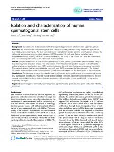
FIG. 1. DEAE-Sephadexcolumn gradient elution profile (see VII,. Column was initially “Experimental Procedures”). 0,Azm, 0, washed with 0.025 M citrate, 0.1 M NaCI, and 1 m~ benzamidine. HC1, pH 6.0, buffer until the absorbance a t 280 nm was less than 0.05. A linear gradient of 2 X 1200 ml from 0.1 M NaCl to 0.6 M NaCl in 0.025 M citrate and 1 mM benzamidine. HCl, pH 6.0, was then applied and 7-ml fractions were collected. The arrow on the descending limb
of the last protein peak indicates where the gradient buffer was depleted and additionalbuffer, 0.025 M citrate, 0.6 M NaCl, and 1 mM benzamidine-HCl, pH 6.0, was applied to complete the elution of prothrombin, Factor X, and Factor IX. The horizontal arrows show the fractions which were pooled for further purification of Factor VI1 and for further purification of Factor IX, X,and prothrombin.
0.025 M citrate, 0.375 M NaCI, and 10 mM benzamidine. HCI, pH 6.0. A total of four 0.5-ml fractions was collected. The second and third
fractions containedalmost all of the activity and were combined and stored at -2OOC in 50% glycerol containing 10 mM benzamidine. HC1. RESULTS
Factor VIIIsolation-Factor VI1 was purified some 140,000-fold over starting human pooled plasma. Approximately 70% of the activity was recovered after barium citrate adsorption and ammonium sulfate fractionation. DEAESephadex column chromatography effectively separated Factor VI1 from prothrombin, Factor X, and Factor IX (Fig. 1). A protein peak (Fractions 270 to 290of Fig. 1) was always noted between the Factor VI1 activity peak (Fractions 220 to 250 of Fig. 1) and the prothrombin, Factor X, and Factor IX activity peak (Fractions315 to 355 of Fig. 1). Fractions 270 to 290 contained protein C when assayed by the method described by Kisiel (26). Elution of prothrombin, Factor X, and Factor IX was never complete at the end of the gradient buffers and was achieved by applying 0.025 M citrate, 0.6 M NaCl, and 1 m~ benzamidine HCI, pH 6.0, buffer at the end of the gradient. Further purification of these proteins is reported elsewhere.’ It is emphasized that a long DEAE-Sephadex column is necessary to achieve the desired resolution. We have recently applied as much as 600 mg of protein (obtained from 6 to 7 liters of starting plasma)without affecting the resolution. However, if the length of the column is reduced to 30 cm instead of 45 cm, protein C elutes with Factors 11, IX, and X peak and Factor VI1 is not effectively separated from other vitamin K-dependent factors. Inclusion of 0.7 M NaCl in the limit buffer and utilizing the presently described method also gives poor resolution of Factor VI1 from other vitamin Kdependent proteins and protein C co-elutes with Factors 11, IX, and X. Most of the protein (-95%) in pooled Factor VI1 fractions from the DEAE-Sephadex column was ST1 as judged by SDS-gel electrophoreticanalysis. Preparative disc electropho-
80
100
120
140
FRACTION NUMBER
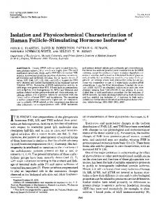
FIG. 2. Elution profile of human FactorVI1 from preparative disc electrophoresis (see “Experimental Procedures”). Fraction numbers are plotted against Factor VIL,. Five-milliliter fractions were collected at a flow rate of 1 ml/min, and those fractions containing 3 or greater VII, units/ml were pooled and concentrated.
resis separated the ST1 and other contaminating proteins from the Factor VI1 protein. Factor VI1 activity was eluted as a sharp symmetrical peak (Fig. 2) from the preparative electrophoresis gel. When assayed at 0.1 mg of protein/ml, this peak contained undetectable levels of prothrombin, FactorX, Factor IX, and protein C activity. The purification scheme is summarized in Table 1. Activation of Factor VI1 during purification was monitored by the VIIc/VIIa,,, ratio, which is 0.9 for Factor VI1 in plasma from normal health donors (15). As noted in Table I, no evidence of Factor VI1 activation was found through the stepof DEAESephadex column chromatography. However, after the step of preparative polyacrylamide electrophoresis, the value for VIIJVIIa,,, rose. In seven of the ten preparations made to
Human Blood Coagulation Factor VII
256
TABLE I Purification of human Factor VII Values given are for a typical preparation. Purification stage
Volume
Total units
units (vII,.cl?tting actlvlty)
I Recovery activity Ratio VIIJ ~ ~ ~ o~ t a protein" l ; ;Specific ~ VII. ~ VII.. VII., ~ activity itv)
ml
3555 Plasma Ammonium sulfate fractionation DEAE-Sephadex column1400 Preparative polyacrylamide gel electrophoresis
mR
3950 48
3702520
284.4 3995 2755
280 165
640
1505 490
x IO:' 50 0.324
units/mX
0.0139 6.8 28 1970
0.89 0.91
100 71
0.93 1.3
36 18
disc electrophoretic systemof Davis (18).However, when the gels were overloaded with the protein, one ortwo additional bands could be detected. The concentration of the contaminants could not be determinedaccurately because of the heavy loading of the gels but usually appeared to beless than 2% of the totalprotein. To determine that the protein band in the SDS and disc gels in fact represented Factor VI1 protein, a duplicate disc gel under nondenaturing conditions (18) was also run. The protein was eluted and assayed for FactorVI1 clotting activity. The peak of clotting activity coincided with the absorbance peak of the stained protein band(Fig. 3B). Immunodiffusion and Immunoelectrophoresis of Human Factor VII-To obtain further information on the purity of Factor VI1 preparations, thepurified VI1 protein was analyzed G ~ LINGI*(nm) L DYE by immunodiffusion against our anti-Factor VI1 antibody. A FIG. 3. Polyacrylamide gel analysis of Factor VII. A , exami- single precipitin line was obtained (Fig. 4 ) . Immunoelectrophoresis also yielded a single precipitinline (Fig. 4 ) . A precipnation of human Factor VI1 by polyacrylamide gel electrophoresis. itin line could not be detected when plasma wasused in place (From left to right) Gel 1, disulfidebonds unreduced, Weber and Osborn (16) method; Gel 2, disulfide bondsreduced,Weberand of the purified Factor VI1 preparations (Fig. 4). Osborn (16) method; Gel 3, disc (Davis (18) method, pH 8.9). In each Neutralization of Factor VII Activityby Anti-Factor VII instance, approximately10 pg of protein in 20 pl of sample was applied Antibody-Normal citrated pooled plasmawas incubated on the gel. B, elution of Factor VI1 activity from disc gel. A duplicate with an equal volume of either various dilutions of Factor VI1 gel (comparable todisc gel in A ) was slicedinto 1.3-mm segments and antibody or an inert control material (NaCl/Albbuffer or an each slice was soaked in 0.1 ml of NaCI/Alb buffer, pH 7.5, in plastic immunoglobulin fraction from normal goat serum) for 30 min tubes overnight. Factor VII, was determined as described under "Experimental Procedures." The disc gel of A was scannedand migration of protein band was corrected due to expansion of the gel during staining anddestaining.
date, the VIIc/VIIamratio rose to between 1.3 and 1.5; in the three preparations, the VIIc/VIIam ratio rose to 2.0 to 2.5. Studies presented later in this section demonstrate that fully activated FactorVI1 gives a VII,./VIIamratio of approximately 25. Thus, a VII,./VIIam ratio of 1.5 reflects the presence of approximately 5% of activated Factor VI1 and the VIIc/VIIam ratio of 2.5 reflects the presence of approximately 10% activated Factor VII. Factor VI1 preparations consistently gave approximately 1600 units of amidolytic activity/mg of protein. The overall yield ranged from 15 to 20%. Purified Factor VI1 preparations did not activate further during storagein the presenceof 10 mM benzamidine HCI at pH 6.0. However, when preparations were dialyzed to remove benzamidine prior to use in further studies, some preparations FIG. 4. Immunodiffusion and immunoelectrophoresis of huunderwent a slow further activation, e g . from a VIIJVIIam man Factor VII. In the double immunodiffusion experiment (top). ratio of 1.5 to a VIIc/VIIa,,, ratio of 2.5 after a period of 8 h a t each of the two wells on the left contains 15 pI of plasma. The two wells on the right contain 15 pl of Factor VI1 (-3 pg) from two pH 7.4. Polyacrylamide Gel Electrophoresis-An electrophoretic different preparations. The center well contains 15 pI of anti-Factor VI1 antibody. In the immunoelectrophoresis experiment (bottom),the analysis of human Factor VI1 is depicted in Fig. 3A. Prepa- two wells contained 15 pl of Factor VI1 (-3 pg) from two different rations were effectively homogenous in SDS-polyacrylamide preparations. After electrophoresis (anode on the right),100 pl of the gels, both in the presence and absenceof a reducingsubstance. anti-Factor VI1 antibody was added to the center trough and allowed Preparations were also homogenous when analyzed by the to diffuse for 48 h.
-
Human Blood Coagulation Factor VII
257
at room temperature. Aliquots were then diluted andassayed by SDS-polyacrylamide gel electrophoresis inthe absence and for clotting factor activities. The effect of incubation with presence of 2-mercaptoethanol. Alanine was the only NH2dilutions of antibody upon the FactorVI1 clotting activity of terminal amino acid residue identified for human Factor VII. plasma is shown inFig. 5. A 300-fold dilution of the antibody The amino acid composition of human FactorVI1 is shown in preparation neutralized 50% of the Factor VI1 activity in the Table I1 along with data for bovine Factor VI1 taken from the literature (5).A hexosamine peak was seen in the aminoacid plasma. The antibody preparation alsoreadilyneutralized analysis, indicating that human FactorVI1 is a glycoprotein. purified Factor VII. Factors IX, X, and prothrombin activities could not be carried out due were not neutralized when plasma was incubated with the A detailed carbohydrate analysis to thelimited material available. Accordingly, the aminoacid undiluted antibody preparation. Characterization of Human Factor VII-Human Factor composition, listed inTable 11, is for carbohydrate-free protein VI1 is composed of a single polypeptidechain (Fig. 3A);it had only. Bovine Factor VI1 has a minimal molecular weight of an apparentmolecular weight of 50,000 & 2,000 as determined 45,500, contains 13% carbohydrate by weight (5), and would, therefore, have a molecular weight of 40,000 for the carbohydrate-free protein. For comparison, we have shown in Table o r I1 the aminoacid compositionof the carbohydrate-freeprotein z 100of human FactorVI1 based upon a molecularweight of 50,000 z4 and also of 40,000 for thecarbohydrate-free protein. The B y 80extinction coefficient E& of Factor VI1 was determined tobe t 13.9. Since the concentration of the protein in this experiment c was determined from the amino acid analyzer, this value is > 60alsobased uponcarbohydrate-freeprotein. Accordingly, a U 4 value of 13.9 is slightly higher than the correct E& for the total glycoprotein and will need minor correction when the 40carbohydrate composition becomes available. E Activation of Human Factor VII with Factor X,-When Factor VI1 was incubated with Factor X, in the presence of i 20phospholipid and Ca(II), its clotting activity increased rapidly. Y A new plateau of activity, representing a 22- to 25-fold increase I I I I I I I 0.1 0.2 0.3 0.4 0.5 0.6 over the initial clotting activity, was reached within approximately 10 min. Factor VI1 activity, as measured in the amiX ANTIBODYCONCENTRATION FIG. 5. Neutralization of Factor VI1 in plasma clotting activ- dolytic assay, was unchanged throughout theincubation (Fig. itybyanti-Factor VI1 antibody. Citrated pooled plasma was 6). When Factor VI1 was incubated for up to 1 h with Factor incubated with an equal volume of various dilutions of the antibody in NaCl/Alb buffer, pH 7.5 as given on the abscissa for 30 min at X, in the absence of phospholipid and Ca(II), noincrease in room temperature. The sample was then diluted and assayed for clotting activity could be measured. When Factor VI1 was Factor VI1 coagulant activity as described under "Experimental Proincubated with Factor X, in the absence of phospholipid but cedures." in the presence of 10 mM Ca(II), a 2- to &fold increase in TABLEI1 clotting activity was observed after 1 h. Amino acid compositions of human and bovine Factor VI1 Activation of Factor VI1 by Factor X,, phospholipid, and Data for the bovine system is from Kisiel and Davie (5). , , Ca(I1) was associated with conversion of single chain Factor Human VI1 to a two-chain molecule without loss of peptide material Bovine (resiResidue detectable by SDS-polyacrylamide gel electrophoretic analy(residues,w,OOO") (residues/ dues/40,000") 413 n ~ ) sis (Fig. 7 ) .The apparentmolecular weights of the two chains
e
Asx Thr Ser Glxb Pro Gly Ala %z Cys' Val Met Ile Leu Tyr Phe Trp" His LYs Arg Gla Carbohydrate
42.2 24.6 33.5 55.3 23.6 41.7 25.4 30.5 23.8 3.3 16.1 49.6 13.2 18.2 6.1
12.9 20.4 25.7 10.5
present
33.8 19.7 26.8 44.2 18.9 33.4 20.3 24.4 19.0 2.6 12.9 39.7 10.6 14.6 4.9 10.3 16.3 20.6 8.5
28.4 14.2 17.0 47.5 23.5 39.8 27.7 28.0 25.8 1.3 7.7 33.1 7.3 16.0 5.0 8.7 9.5 25.4 9.4' 138
'' Molecular weight given is of the protein mass only(excluding the carbohydrate portion). Includes the Gla content also. Determined as cysteic acid (19). Determined spectrophotometrically (20). e From DiScipio and Davie (28).
2500
> u
1
1
1500
Q
1000 (L
0 I-
500
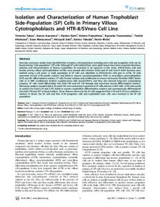
15 20 25 30 TIME (MINI FIG. 6. Activation of humanFactor VI1 with Factor X,, Ca(II), and phospholipid. The buffer used was NaCl/Tris, pH 7.5. The Factor VI1 concentration was 50 pg/ml and Factor X., concentration was 1.0 pg/ml. Concentration of Ca(I1) was 10 mM and of phospholipid was 30 PM. Per cent increase of Factor VI1 activity both in the clotting assay (0)and in the amidolytic assay (0) is plotted as a function of incubation timeat 37°C (see "Experimental Procedures").
5
10
Human Blood Coagulation Factor
258
-
VII
thepresence of phospholipid and Ca(I1)converted single chain native Factor VI1 to an activated two-chain Factor VI1 within approximately 10 min. This was associated with a 22II to 25-fold increase in Factor VI1 activity in the clotting assay but not in the coupled amidolytic assay. The rapidity with which Factor X, can activate Factor VI1 probably explains why the coupled amidolytic assay gives identical values for native and activated FactorVI1 in a test sample (15). In this -* assay, Factor X is incubated in a great molar excess with Factor VI1 in the presenceof tissue factor and Ca(I1). Enough Factor X. should be generated during theinitial period of the I incubation to convertall of the Factor VI1 in the test sample to Factor VII. and, thus, to make the amount of Factor X. generated over the total 3-min incubation period of the assay independent of the initial activity state of Factor VI1 in the 5.110 test sample. lo I I Human FactorVI1 had a similar apparent molecular weight FIG. 7. A time courseof activation of human Factor VI1 with of 50,000 as determined by SDS-polyacrylamide gel electroFactor X., as evaluated by SDS-polyacrylamide (126) gel elec- phoresis both in the Tris buffer system of Laemmli (17) and trophoresis in Tris buffer system of Laemmli (17). V I I , single in the phosphatebuffer system of Weber and Osborn (16). In chain Factor VII; H, heavy chain of Factor VII,,; L, light chain of contrast, the apparentmolecular weights of the two chains of Factor VII.. Gels on the right are in the presence of 2-mercaptoethanol. The numbers on the bottom of the gels represent incubation activated Factor VI1 differed in these two systems. Apparent molecular weights of 31,500 and 24,000 for the two chains times. were obtained in the Laemmli (17) systemandapparent of Factor VII. were 31,500 and 24,000 when SDS-polyacryl- molecular weights of 26,000 and 22,000 were obtained in the amide gel electrophoresis wasperformedaccording to the Weber and Osborn (16) system. Since the molecular weights method of Laemmli (17) and were 26,000 and 22,000 when the of glycoproteins obtained by SDS-gel electrophoresis are usuelectrophoresis was performed according to the method of ally anomalous (29), the valuesof molecular weights of native Weber andOsborn (16). Since theLaemmli system gave better Factor VI1 and of the constituent polypeptide chains of actiseparation of the chains, only the gels run in this system are vated Factor VI1 must be considered very approximate. Bepresented in Fig. 7. cause the molecular weights of the two chains of activated Factor VI1 obtained in the Weber andOsborn system (26,000 DISCUSSION and 22,000) when added give a molecular weight of 48,000, Human Factor VI1 was purified approximately 140,000-fold which is approximately the apparentmolecular weight of the over starting plasma. Preparations were effectively homoge- single chain FactorVII, we believe that themolecular weights nous as judged by electrophoretic andimmunological studies. of the two chains of activated Factor VI1 obtained by the Human Factor VI1 is a single chain glycoprotein and has an Weber and Osborn (16) system are more accurate than the estimated molecular weight of 50,000. It has alanine as an molecular weights obtained in the Laemmli (17) system. NH2-terminal residue, which is the same as the NH2-terminal A preliminary report dealing with the isolation and activaresidue of bovine Factor VI1 (5, 6). The amino acid composi- tion of human Factor VI1 has been published by Broze and VI1 is a tion of human Factor VI1 (Table 11) is very similar to thatof Majerus (30):' They reported that human Factor bovine Factor VII. Human FactorVI1 contains approximately single chain molecule of 47,000 daltons, which, upon activation with Factor X,,, is converted toa two-chain molecule. Molec9 y-carboxyglutamicacidresidues/mol of protein.Bovine 9.4 y-carboxyglutamic ular weights forthe two chains of the activatedmolecule were Factor VI1 has been reported to contain not given. acid residues/mol of protein (28). As described earlier by Radcliffe and Nemerson (6) for the The overall yield of Factor VI1 was about 15 to 20% of starting plasma. About 300 pg of protein containing approxi- activation of bovine Factor VI1 by bovine Factor X,, activation mately 500 amidolytic units of total activity was obtained of human Factor VI1 by human Factor X, required both from 4 liters of human plasma. If one assumes no denaturation Ca(I1) and phospholipid. Radcliffe and Nemerson (6) found of the molecule during its isolation, this would represent 0.5 that two-chain bovine Factor VII, is further cleaved byFactor to 0.6 pg of Factor VI1 protein/ml of starting plasma. Final X, to yield a three-chain molecule with reduced clotting preparations had no detectable prothrombin, Factor IX, Facactivity (6). In some of our experiments on the activationof tor X, or protein C activity as measured in biological assay human Factor VI1 by Factor X., Factor VI1 clotting activity, systems. after reaching a plateau, declined to a value approximately An anti-Factor VI1 antibody wasraised in a goat using one-half of the initial plateau value. However, no degradation purified Factor VI1 protein as antigen. The antibody gave a of the two-chain human Factor VII, molecule was observed single precipitin line against purified Factor VI1 in immuno- by SDS-polyacrylamide gel electrophoresis even when the diffusion and immunoelectrophoresis experiments.A precipi- incubation period was prolonged to 3 h.4 Phospholipid has tin line was not visible with normal human plasma, which been reported to inactivate bovine Factor VII, (8) and it is probably reflects the low concentration of 0.5 pg/ml of the After submissionof this manuscript,a full report on human Factor antigen in plasma. The antibody, in a 300-fold dilution, inhibited 50% of the Factor VI1 activity of plasma. Undiluted VI1 by these authors appeared in print (Broze. G . J., and Majerus, P. antibody had no detectable inhibitory effect upon the Factor W. (1980)J. Biol. Chem. 255,1242-1247). The purification procedure developedby these authors is quite different from the purification IX, X, or prothrombin activity of plasma. This provides fur- procedure described in the present manuscript. However, the yield, ther indirect evidence that the purified Factor VI1 antigen isolation time, and quality of the final product are comparable. The was free of these contaminating proteins. amino acid composition of both preparations is also very similar. Factor X. in an enzyme to substrate ratio of 1 to 50 and in ' S. 1'. Bajaj, unpublished results.
I
ii
u
I
!
Human B&oodCoagulation Factor VI1 possible that the loss of Factor VI1 clotting activity observed in OUT experiments couldreflect inactivation of human Factor VI1 by phospholip~din the reaction mixture. Factor XII, and Factor IX, have also been identified as direct activators of Factor VI1 (10). Factor XIL, which does not require the presence of added Ca(I1) or phospholipid to activate Factor VII, yields the same two-chain activated bovine Factor VI1 molecule as does Factor X, @). It is very liieiy that Factor XII, and Factor IX, will yield the same cleavage productsof human FactorVI1 as those obtained with Factor X., but detailed studies are as yet not available. Ackno~~ed~ent-W thank e Dr. R. F. Doolittle of the Department of Chemistry, University of California, San Diego, for his help with the amino acid analysis. I. 2. 3. 4. 5. 6. 7. 8. 9.
REFERENCES Bajaj, S. P., Rapaport, S. I., and Brown, S. F. (1979) Blood 54, 269a (Abstract 740) gsterud, B., and Rapaport, S. I. (1977) Proc. Natl. Acad. Sci. U. S. A. 14,5260-5264 Jesty, J., and Nemerson, Y. (1974) J. Biol. Chem. 249, 509-515 Radcliffe, R., and Nemerson, Y.(1975) J. Biol. Chem. 250, 388395 Kisiel, W., and Davie, E. W. (1975) Biochemistry 14,4928-4934 Radcliffe, R., and Nemerson, Y.(1976) J . Biol. Chem. 251,47974802 Zur, M., and Nemerson Y. (1978) J. Biol. Chem. 253,2203-2209 Kisiel, W., Fujikawa, K., and Davie, E. W. (1977) Biochemistry 16,4189-4194 Radcliffe, R., Bagdasarian, A., Colman, R., and Nernerson, Y.
259
(1977) Blood 50,611-617 10. Seligsohn, U., Bsterud, B.,Brown, S. F., Griffin, J. H., and Rapaport, S. I. (1979) J. Clin. Invest. 64, 1056-1065 11. Owren, P. A. (1949) Scand. J. Clin. Lab. Invest. 1,81-83 12. Bell, W. N., and Alton, H.G. (1954) Nature 174,880-881 and Lopez, J . A. (1946) J. Biol. Chem. 162,421-428 13. Lowry, 0.H., 14. Hall, C. A,, Rapaport, S. I., Ames, S. B., and De Groot, J . A. (1964) Am. J. Med. 37, 172-181 15. Seligsohn, U., 0sterud, B., and Rapaport, S. I. (1978) Blood 52, 978-988 16. Weber, K., and Osborn, M. (1969) J. Biol. Chem. 244, 4406-4412 17. Laemmli, U. K. (1970) Nature 227,680-685 18. Davis, B. J . (1964) Ann. N. Y. Acad. Sci. 121,404-427 19. Moore, S. (1963) J. Biol. Chem. 238,235-237 20. Goodwin, T. W., and Morton, R. A. (1946) Biochem. J. 40,628632 21. Price, P. A., Otsuka, A. S., Poser, J. W., Kristaponis, J.,and Raman, N. (1976) Proc. Natl. Acad.Sci. U.S. A . 72, 1447-1451 22. Fernlund, P., Stenflo, J., Roepstorff, P., and Thomsen, J. (1975) J. Biol. Chem. 250,6125-6133 23. Gray, W. R. (1972) Methods Enzymol. 25, 121-138 24. Woods, K. R., and Wang, R. T. (1967) Biochim. Biophys. Acta 133,369-370 25. Bajaj, S. P., and Mann, K. G. (1973) J. Biol. Chem. 248, 77297741 26. Kisiel, W. (1979) J. Clin. Invest. 64, 761-769 27. Lowry, 0.H., Rosebrough, N. J., Farr, A. L., and Randall, 12. J. (1951) J. Biol. Chem. 193,265-275 28. DiScipio, R. G., and Davie, E. W. (1979) Biochemistry 18, 899904
29. Segrest, J. P., and Jackson, R. L. (1972) Methods Enzymol. 28, 54-63 30. Broze, G. J., Jr., and Majerus, P. W. (1979) Clin. Res. 27,459A