FXG Dosimeter Response for Three-Dimensional Conformal Radiotherapy Using Different Evaluation Techniques Christianne C. Cavinato1, Benedito H. Souza2, Henrique Carrete Jr.2, Kellen A. C. Daros2, Regina B. Medeiros2, Adelmo J. Giordani3, and Letícia L. Campos1 1
Gerência de Metrologia das Radiações, Instituto de Pesquisas Energéticas e Nucleares – IPEN-CNEN/SP, Av. Prof. Lineu Prestes, 2242, Cidade Universitária, São Paulo, 05508-000, Brazil
[email protected] and
[email protected] http://www.ipen.br 2 Departamento de Diagnóstico por Imagem, Universidade Federal de São Paulo – UNIFESP, Rua Napoleão de Barros, 800, Vila Clementino, São Paulo, 04024-002, Brazil
[email protected],
[email protected] and
[email protected] http://www.unifesp.br/ddi 2 Serviço de Radioterapia, Universidade Federal de São Paulo – UNIFESP, Rua Napoleão de Barros, 715, Vila Clementino, São Paulo, 04024-002, Brazil
[email protected] http://www.unifesp.br/centros/gmo
Abstract. This work aims to compare the dose-response of the Fricke xylenol gel (FXG) dosimeter developed at IPEN using 270 Bloom gelatin from porcine skin made in Brazil evaluated using the magnetic resonance imaging (MRI) technique with the dosimetric response evaluated using the optical absorption (OA) spectrophotometry technique, in order to verify the possibility of quality assurance (QA) and reproducibility of FXG dosimeter to be carried out routinely using the OA technique for three-dimensional conformal radiotherapy (3DCRT) application using a 6 MV photons linear accelerator. The response in function of the absorbed dose of FXG dosimeter developed at IPEN presents linear behavior in clinical interest dose range when irradiated with 60Co gamma radiation and 6 MV photons and evaluated using the MRI and OA techniques. The results indicate that the optical technique can be used for QA of FXG dosimeter when used in the possible application in QA of 3DCRT.
1 Introduction To ensure effective treatment and avoid deadly mistakes to patients is required a rigorous quality assurance (QA) of the complex tumors treatment techniques such as
three-dimensional conformal radiotherapy (3DCRT) and intensity modulated radiotherapy (IMRT) [1]. A dosimetric system that has been extensively studied for this purpose is the Fricke gel dosimetry, considering the ability to produce three-dimensional phantoms of various shapes and sizes [2]. Its dosimetry principle is based in ferrous (Fe2+) to ferric (Fe3+) ions oxidation caused by action of ionizing radiation [3]. This study was performed with the aim of to compare the dose-response of the Fricke xylenol gel (FXG) dosimeter developed at IPEN using 270 Bloom gelatin from porcine skin made in Brazil evaluated using the magnetic resonance imaging (MRI) technique with the dosimetric response evaluated using the optical absorption (OA) spectrophotometry technique, in order to verify the possibility of quality assurance (QA) and reproducibility of FXG dosimeter to be carried out routinely using the OA technique for three-dimensional conformal radiotherapy (3DCRT) application using a 6 MV photons linear accelerator, considering that OA is a cheap, ease and fast optical evaluation technique.
2 Materials and Methods The FXG samples were prepared at High Doses Laboratory (LDA) of IPEN. The gamma and photon irradiations were carried out in the Radiotherapy Service of the Sao Paulo Hospital (HSP) of UNIFESP, the magnetic resonance images were achieved in the Resonance Magnetic Service at Diagnostic Image Department of HSP (UNIFESP), and the optical absorption measures at LDA. 2.1 FXG Solution Preparation The FXG solutions were prepared according to Olsson [4] using 5% by weight 270 Bloom gelatin from porcine skin, ultra-pure water, 50 mM sulphuric acid (H2SO4), 1 mM sodium chloride (NaCl), 1 mM ferrous ammonium sulphate hexahydrate or Morh’s salt [Fe(NH4)2(SO4)2.6H2O] and 0.1 mM xylenol orange ferric ions indicator (C31H28N2Na4O13S); the chemicals are analytical grade. The samples were conditioned in polymethylmethacrylate (PMMA) cuvettes 10 x 10 x 45 mm3 and sets of three samples each were prepared for the irradiations and measurements. The sample sets were packed with polyvinyl chloride (PVC) film to prevent evaporation of the water contained in the Fricke gel solution and to ensure the reproducibility of the dosimetric response of the three FXG samples. The FXG samples were maintained under low temperature ((4 ± 1) ºC) and light protected during 12:30 h before irradiation. 2.2 FXG Samples Irradiation Considering the homogeneity and precision of absorbed dose delivery of 60Co gamma sources and that the effective photon energy of 6 MV photon beam is approximately 2 MeV, the FXG samples were irradiated with 60Co gamma radiation, as reference
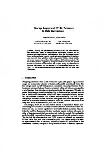
system, in the dose range of 2 to 20 Gy, dose rate of 74.98 cGy/min, 40 x 40 cm2 field size and source-surface distance (SSD) of 80 cm using a GENERAL ELECTRIC COMPANY® Alcyon II 60Co gamma radiation (HSP/UNIFESP) using a PMMA plates phantom (build-up and backscattering PMMA plates 1.5 cm thick). All FXG sample sets were positioned together in the PMMA phantom (Fig. 1a) and each set was removed when the radiation exposure time needed to obtain the desired absorbed dose was completed. Three sample sets were irradiated with 6 MV photons (one at a time), with absorbed doses of 5 to 15 Gy, dose rate of 300 cGy/min, 30 x 30 cm2 field size and SSD of 92 cm using a VARIAN® Clinac 600C linear accelerator (HSP/UNIFESP) and the same PMMA phantom, to compare the obtained results with the dose-response curves obtained for 60Co gamma radiation. The experimental set up for FXG samples irradiation with 6 MV photons is presented in Fig. 1b. The dosimetric response in function of the dose rate was also studied, since different irradiation systems can operate with different dose rates. For this study FXG samples were irradiated with 60Co gamma radiation and 6 MV photons with absorbed dose of 10 Gy and dose rates between 74.98 and 600 cGy/min.
a Fig. 1. Experimental set up for FXG samples irradiations with 6 MV photons (b)
b 60
Co gamma radiation (a) and
2.3 FXG Samples Evaluation The dosimetric response of the FXG samples non-irradiated and irradiated with 60Co gamma radiation and 6 MV photons were evaluated using both magnetic resonance imaging (MRI) and optical absorption (AO) spectrophotometry techniques using a SIEMENS® MAGNETOM® Sonata Maestro Class 1.5 T MRI scanner (HSP/UNIFESP) and SHIMADZU® UV2101-PC spectrophotometer (LDA/IPEN), respectively. For the FXG MR signal intensity analysis in function of the absorbed dose scans in the coronal orientation of the cuvettes containing the FXG solution non-irradiated and irradiated with 60Co gamma radiation (dose range of 2 to 20 Gy) were obtained using on cranium protocol-T1 approximately 30 min after irradiation. The MR image acqui-
sition parameters are presented in Table 1. The softwares syngo fastView® version VX57F24 and ImageJ® version 1.42q were used to process the MRI scans obtained. All optical measurements were performed in the wavelength range from 190 to 900 nm and achieved about 2 h after irradiations due to the distance between the facilities where the irradiations (HSP/UNIFESP) and measurements (LDA/IPEN) were made. The absorbance values presented correspond to dosimetric wavelength of 585 nm [5,6]. Each presented value corresponds to the average of the measurement of three samples and the error bars the standard deviation of the mean. The background values corresponding to the magnetic and optical measurements of non-irradiated Fricke gel samples were subtracted from all values presented. Table 1. MR image acquisition parameters
Parameters Description Image Orientation Field of View (FOV) (mm) Slice Thickness (THK) (mm) Voxel (mm) Gap (mm) Time of Repetition (TR) (ms) Time of Echo (TE) (ms) Flip Angle (º) Matrix Size (MS) (pixels) Number of Signals Averaged (NSA) Slices Number Coil Channels
Values Coronal 256 1.0 1.0 x 1.0 x 1.0 0.5 2000 3.42 15 256 x 256 1 176 Head 8
3 Results and Discussions
3.1 Magnetic Resonance Imaging versus Optical Absorption
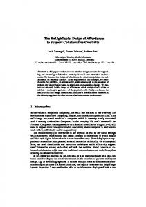
Dose-Response Curves. The PMMA cuvettes MR images that showed better resolution at the region of interest (ROI) and the corresponding MR signal intensity in function of radiation dose of studied dosimeter non-irradiated and irradiated with 60Co gamma radiation and absorbed doses of 2 to 20 Gy are presented in Fig. 2. 450
3
MR Signal Intensity (a.u.)
15 2 0 10 5
2
60
Co gamma radiation Dose rate = 74.98 cGy/min 350
300
250
200
2 0
Absorbed Dose (Gy)
PMMA Cuvettes 1
FXG dosimeter Fit linear
400
150
a
0
2
4
6
8
10
12
14
16
18
20
22
Absorbed Dose (Gy)
b
Fig. 2. Coronal MR images of the FXG solution conditioned in PMMA cuvettes non-irradiated and irradiated with 60Co gamma radiation (a) and MR signal intensity curve in function of absorbed dose of the FXG samples image (b) (modified from Cavinato et al., [7])
The optical dose-response curves of the FXG dosimeter irradiated with 60Co gamma radiation (74.98 cGy/min) and 6 MV photons (300 cGy/min) and with absorbed doses between 2 and 20 Gy are presented in Fig. 3.
1.4 60
Co gamma radiation 6 MV photons Linear range
Absorbance (a.u.)
1.2
1.0
0.8
0.6
0.4
0.2
FXG dosimeter = 585 nm
0.0 0
2
4
6
8
10
12
14
16
18
20
22
Absorbed Dose (Gy)
Fig. 3. Dose-response curves of FXG samples irradiated with 60Co gamma radiation and 6 MV photons
The FXG dosimeter response in function of absorbed dose presents linear behavior in the clinical interest dose range when irradiated with 60Co gamma radiation and 6 MV photons and evaluated with MRI and OA techniques, tending to saturation to doses equal and higher than 20 Gy. The uncertainties are better than ± 2% for OA technique and ± 5% for MRI technique because of quality of MR images. 3.2 Dose Rate Curve The optical response in function of dose rate relative to the average absorbance values of the FXG solution irradiated with 60Co gamma radiation and 6 MV photons for dose rates from 74.98 to 600 cGy/min and absorbed dose of 10 Gy is presented in Fig. 4.
Relative Response to Average Absorbance Values (a.u.)
1.07
FXG dosimeter
1.06 1.05 1.04 1.03 1.02 1.01 1.00 0.99 0.98 0.97 0.96 0.95 0.94
60
Absorbed dose ( Co and 6 MV photons) = 10 Gy
0.93
= 585 nm
0.92 0
100
200
300
400
500
600
Dose Rate (cGy/min)
Fig. 4. FXG dose rate dependent optical response curve relative to the average absorbance values irradiated with 60Co gamma radiation and 6 MV photons
The dose rate dependent response obtained is better than 3.5% indicating that the calibration of a FXG phantom developed at IPEN irradiated with 6 MV photons can be performed using a 60Co gamma radiation clinical machine.
4 Conclusions This study presents good results which indicate that the optical absorption spectrophotometry technique can be used for the quality assurance and reproducibility confirmation of the Fricke xylenol gel dosimeter developed at IPEN applied to 3DCRT.
Acknowledgments The authors are grateful to the staffs of the Radiotherapy Service and Resonance Magnetic Service of the Diagnostic Image Department of the HSP/UNIFESP to allow the FXG irradiations and MR evaluations, respectively, and CAPES, CNPq, IPEN and CNEN by the financial support.
References 1. Podgorsak, E.B., Podgorsak, M.B.: Special procedures and techniques in radiotherapy. In: Podgorsak, E.B. (ed.): Radiation oncology physics: a handbook for teachers and students, International Atomic Energy Agency (2005) 505-548 2. Schreiner, L.J.: Review of Fricke gel dosimeters. J. Phys.: Conf. Ser. (2004) 9-21
3. Gore, J.C., Kang, Y.S., Schulz, R.J.: Measurement of radiation dose distributions by nuclear magnetic resonance (NMR) imaging. Phys. Med. Biol. (1984) 1189-1197 4. Olsson, L.E., Petersson, S., Ahlgren, L., Mattsson, S.: Ferrous sulphate gels for determination of absorbed dose distributions using MRI technique: basic studies. Phys. Med. Biol. (1989) 43-52 5. Bero, M.A., Gilboy, W.B., Glover, P.M.: Radiochromic gel dosemeter for three-dimensional dosimetry. Radiat. Phys. Chem. (2001) 433-435 6. Cavinato, C.C.: Padronização do Método de Dosimetria Fricke Gel e Avaliação Tridimensional de Dose Empregando a Técnica de Imageamento por Ressonância Magnética. Instituto de Pesquisas Energéticas e Nucleares (2009) 7. Cavinato, C.C., Souza, B.H., Carrete Jr., H., Daros, K.A.C., Medeiros, R.B., Giordani, A.J., Campos, L.L.: Preliminary study of the 270 Bloom Fricke xylenol gel phantom performance for 3-D conformal radiotherapy using multiple radiation fields. In: International Conference on Medical Physics, April 17-20, Porto Alegre, Brazil (2011)