New antibiotics, decatromicins A and B were reported from culture broth of Actinomadura sp., MK73-NF4. Both inhibited the growth of gram-positive bacteria ...
INT.J.BIOL.BIOTECH.,1 (1): 49-57, 2004.
EVALUATION OF VARIOUS MICROORGANISMS FOR THE PRODUCTION OF BIOACTIVE METABOLITES M. Akhtar1, M. S. Abbassi2, S. Mehboob1, J. A. Khan, S. Nadeem, S. S. Alam and I. A. Khan Nuclear Institute for Agriculture and Biology, P.O BOX 128, Faisalabad, Pakistan 1 Government College, Faisalabad, Pakistan. 2 Islamia University Bhawalpur. ABSTRACT Bacterial and fungal strains were isolated from different soils and tested for the production of antifungal, antibacterial and insecticidal compounds. The fungal isolate F1 (Rhizopus sp.) and bacterial isolate (B1) showed maximum inhibition zone against Staphylococcus aureus. While ethyl acetate extracts of fungal isolates F3 and F1 showed 100% and 90% mortality against Tribolium estaneum respectively. Pencillium sp. (F2) exhibited 65%, Rhizopus sp. (F1) and bacterial isolate (B2) showed 60% mortality against pulse beetle Callosobruchus analis. The autoclaved culture filtrate of bacterial isolate (B 2) completely inhibited the growth of test fungus (Cladosporium cuccumerinum). The bioassay of the ethyl acetate extracts on TLC plates against Cladosporium cuccumerinum revealed that fungal isolate F3 produced one inhibition zone at Rf value 0.9, when solvent system methanol: chloroform (9:1) mixture was used to develop the TLC plates. Cytotoxicity assay against brine-shrimp larvae revealed that ethyl acetate extract of B1, B3 and F2 were 100% toxic while minimum toxicity was observed by ethyl acetate extract of B 2.
Key words: Metabolites, bioassay, chromatography, bioactive compound, biological control. INTRODUCTION Extracts from bacteria and fungi have served as a valuable source of molecular diversity in many drug discovery program and several drugs have been isolated from them. The continuously extending series of novel antibiotics of present era includes, antibiotic GR135402 was isolated from Graphium putsedinis (Kinsman et al., 1998). New antibiotics, decatromicins A and B were reported from culture broth of Actinomadura sp., MK73-NF4. Both inhibited the growth of gram-positive bacteria including methicilin-resistant Staphylococcus aureus (Momose et al., 1999). Pseudomonas sp., isolated from soil and ethyl acetate extract of this strain was found to be exhibited antifungal activity and antifungal compound purified and identified as N-butyl benzene sulphonamide (Kim et al., 2000). Two novel antifungal compounds, 1 (SCH46657) and 2 (SHH 466456), were isolated from the fermentation broth of an unidentified fungus. Both compounds were active against Candida, Dermatophytes and Aspergillus (Hegde et al., 2001). Despite all these contributions, development of antibiotic resistance not only points up but also invites up the continued need for the search of new and different antibiotics to replace those, which become ineffective. The present research work was therefore initiated to evaluate microorganisms for the production of metabolites exhibiting bioactivity. MATERIALS AND METHODS Isolation of microorganism Soil samples were collected from cotton, rice and chickpea fields of Nuclear Institute of Agriculture and Biology (NIAB) and one from roadside near NIAB office gate. Isolation of microorganism was done on nutrient agar (NA) and potato dextrose agar (PDA) media using 1/10 th and 1/100th dilutions of 10% aqueous suspensions of soil samples. The microorganisms isolated were single spore purified and stored in refrigerator for further studies. Identification of fungal isolates was performed based on colony morphology, spores and mycelial structure while gram staining led by microscopic studies was used to identify bacterial isolates. Production and isolation of metabolites The fungal and bacterial isolates were grown on liquid minimal medium and nutrient broth medium for 14 days at 25°C and 37°C respectively. The cultural filtrates were harvested by filtration through muslin cloth. The pH of the
50
M. AKHTAR ET AL.,
culture filtrates was noted and finally adjusted at 3.0 by adding 0.2M sulphuric acid drop wise, with continuous shaking. Extraction of culture filtrates was done three times into half the volumes of ethyl acetate (Merck) with vigorous shaking, in separating funnel (Alam et al., 1997). The ethyl acetate fractions were pooled together, dried over anhydrous sodium sulphate and solvent was evaporated to dryness at 30°C in rotary evaporator. They were dissolved in 3.0ml ethanol. Bioassays Antibacterial assay Anti bacterial assay against Staphylococcus aureus and a test bacterium was done by disc diffusion method as described by Jacoby and Archer (1991). Nutrient broth (100 ml) was taken, autoclaved and inoculated one with Staphylococcus aureus and other with test bacterium incubated at 37°C in dark. Ethyl acetate extract (30l) was applied to paper disc (1.0cm diameter) of whatman filter paper # 3 and after air drying, placed in the centre of preinoculated NA plates. Control paper discs impregnated with solvent and reference with streptomycin (500ppm) was also tested. All plates were incubated at 37°C for 24-48 h. The activity was determined by measuring the diameter of zones by showing complete growth inhibition. Antifungal assay Antifungal assay was done by food poisoning technique as described by Ilyas et al., (1982). Culture filtrates were centrifuged at 5000xg supernatants were removed and designated as 1.0N solution. Out of these 1N solutions, 30 ml were taken and diluted to 60ml and were designated as 0.5M solution. These 1N and 0.5N dilutions of culture filtrates were used to prepare czepek dox agar medium (oxoid). These were autoclaved at 121 oC, 15lb psi for 15min. and then poured in petri plates and in semisolid conditions placed a disc of PDA (2mm diam.) of seven days old culture of test fungus Cladosporium cucumerinum. Insecticidal assay Insecticidal assay was done by impregnation method described by Tabassum et al., (1998) against two storage pests, Tribolium castaneum and Callasobruchus analis. Paper discs of 2cm diam., were impregnated with 50µl metabolites (ethyl acetate extract), placed in petri plates in three replicates. Five young healthy insects were placed in each petri plate and then incubated at 30°C for two days. Control discs impregnated with insecticide monocrotophos (2000ppm) was also used. The experiment was repeated twice. Chromatography Thin layer chromatographic and bioassay techniques (Kim et al., 2000) were used for identification of antifungal compounds. Self made silica gel thin layer chromatographic (TLC) plates (0.5mm thick, silica gel 60 GF254 plates) were used for running the samples. The TLC plates were developed in solvent system chloroform: methanol (96:4) and ethyl acetate: n-hexane (7:63) mixture, dried at 40oC for 4 h and then sprayed with spore suspension (1 x 10⁵ spores/ml) of C. cucumerinum (test fungus) in autoclaved minimal medium and TLC plates were incubated in a humidity chamber at 25°C. After three days of incubation, the plates were removed from the chamber and the Rf values of the inhibition zones were calculated. The silica from the site of inhibition zone were scratched and dissolved in chloroform: methanol (9:1) and filtered under vacuum. Antifungal compounds were further purified after repeated TLC using solvent system benzene: acetone: acetic acid (35:5:1). Cytotoxicity assay The Cytotoxicity of ethyl acetate extracts and of purified antifungal compound was checked against brine shrimp (Artimia salina) as described by Schmidt et al., (1987). RESULTS AND DISCUSSIONS
INTERNATIONAL JOURNAL OF BIOLOGY AND BIOTECHNOLOGY 1 (1): 49-57, 2004.
51
PRODUCTION OF BIOACTIVE METABOLITES BY MICROORGANISMS
Isolation of microorganism Three bacterial isolates designated as (B1, B2, B3) and three fungal isolate designated as (F1, F2, F3) were isolated. Microscopic studies followed by gram staining revealed that B1 and B2 were gram-negative cocci forming chains and cluster, respectively, while isolate B3 was gram positive having isolated small rods cells. Microscopic studies revealed that the isolate F1 was Rhizopus sp., isolate F2 was Penicillium sp., and isolate F3 was Cladosporium sp. Production and isolation of metabolites Bacterial and fungal isolates were incubated for 7 and 14 days, respectively for obtaining culture filtrate at their maximum growth. Initial pH of nutrient broth was noted to be 7.3 and of cultural filtrate of B 2 changed to 7.5 revealing slight increase in alkalinity while decrease in pH was noted in cultural filtrate of B 1 and B2 i.e. 6.7 and 6.3 respectively. Initial pH of minimal medium was 6.2 and of cultural filtrate F2 and F3 changes to 7.3 and 7.2 respectively (Fig. 1).
7.4 7.2 7 6.8 6.6 6.4 6.2 6 5.8 5.6 NB
B1
B2
B3
F1
MM
F2
F3
Fig. 1. PH of culture filtrate of bacterial and fungal isolates and growth media.
Ethyl acetate solvent was used for the partitioning of active compounds from the culture filtrate at pH 3.0 (Alam et al., 1997: Kim et al., 2000) because ethyl acetate is intermediate polar solvent, and it is not able to extract the compounds with similar polarity but also the less polar and high polar compounds with repeated extractions. Antibacterial assay by disc diffusion method Maximum antibacterial activity against test bacteria was observed in ethyl acetate phases of F 1 (Rhizopus sp.) where growth inhibition observed was 1.95 cm diam., while F3 (Cladosporium sp.) showed highest activity against S. aureus which produced a clear inhibitory zone of 2.20 cm diam., was observed (Fig. 2). Ethyl acetate phases of F 2 (Penicillium sp.) and F3 (Cladosporium sp.) showed least antibacterial activity against test bacteria where growth inhibition was 1.20 cm diam. No antibacterial activity was produced by ethyl acetate phases of B 2 against S. aureus while it showed a zone of 1.35 cm diam., against test bacteria (Fig. 3). Isolate B 1 (Bacillus subtillus) also showed an INTERNATIONAL JOURNAL OF BIOLOGY AND BIOTECHNOLOGY 1 (1): 49-57, 2004.
52
M. AKHTAR ET AL.,
excellent antibacterial activity both against test bacteria and S. aureus and produced inhibition zones by 1.73 cm and 2.15cm diam., respectively. Average diameters of inhibitory zones produced by ethyl acetate extracts of bacterial and fungal isolates are shown in Fig. 2. No antibacterial activity against test bacteria and S. aureus was observed in control treatments while 2.50 cm diam., inhibition zone was observed in reference (streptomycin sulphate) replica against test bacteria while no effect was observed against S. aureus. The antibacterial activity was checked against S. aureus and a test bacterial (gram positive rods) isolated from soil. Test bacteria showed higher sensitivity as compared to S. aureus against streptomycin, which showed the development of resistance against the antibiotic, while the test bacteria was a soil borne bacteria which was still sensitive to that antibiotic. The ethyl acetate extracts of F3, B1 and F2 isolates showed excellent antibacterial activity against S. aureus indicated the production of potent antibiotic by these isolates against the pathogen but acetate extracts of B2 (showed 100% antifungal activity) did not show antibacterial activity against S. aureus. Antifungal assay by food poisoning technique: Cultural filtrate of B2 (rod bacteria) completely inhibited the growth of test fungus (C. cucumerinum) both in diluted and concentrated replica. Isolate F3 (Cladosporium sp.) also showed an excellent antifungal activity and produced 0.5cm diam., in diluted and 0.6 cm diam., in concentrated replica. Growth of 2.05cm diam., was produced in control treatments. The least inhibition of growth was observed by B 3 and produced 1.6cm and 1.7cm diam., in diluted and concentrated culture filtrate, respectively. The colony diameter of 0.75cm and 1.00cm were recorded in diluted and concentrated replica of F2 (Penicillium sp.). Growth diameter of test fungus both in diluted and concentrated replica of culture filtrate of F1 (Rhizopus sp.) was 1.15cm and of culture filtrate of B1 was noted to be 1.42cm and 1.30cm respectively. Diagrammatic representation of comparison of growth inhibition both in diluted and concentrated replica is shown in Fig. 4.
Test Bacteria
S. aureus
2.5
2
1.5
1
0.5
0 B1
Fig. 2.
B2
B3
F1
F2
F3
Control
Strept
Inhibitory effect of ethyl acetate phase of bacterial and fungal isolates on the growth of test bacteria and S. aureus..
INTERNATIONAL JOURNAL OF BIOLOGY AND BIOTECHNOLOGY 1 (1): 49-57, 2004.
PRODUCTION OF BIOACTIVE METABOLITES BY MICROORGANISMS
53
Fig. 3. Inhibition zone produced by the ethyl acetate phase impregnated paper disc of isolate B 1 against Staphylococcus aureus.
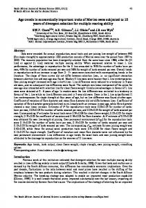
Insecticidal assay by impregnation method: Highest insecticidal activity (100% mortality) was shown by ethyl acetate phases of F 3 (Cladosporium sp) against T. castaneum. Control treatments showed 20% insecticidal activity while 50% mortality was observed in reference batch. Maximum average mortality (65 %) was produced by the ethyl acetate phase of F 2 (Penicillium sp.) against C. analis. The control treatment showed 20% mortality and 40% in reference insecticide (monocrotophos). Ethyl acetate phase F1 (Rhizopus sp.) produced 90% and 60% insecticidal activity against T. castaneum and C. analis respectively. Least insecticidal activity was observed by ethyl acetate phase of B 3 that was 40% against both insects, T. castaneum and C. analis. Isolates B1 and B2 showed 80% and 40% mortality, against T. castaneum and 40% and 60% mortality, against C. analis. Comparison of effects of ethyl acetate phases of isolates against both species of insect is given in Fig. 5. Insecticidal activity was checked against the two storage pests namely, T. castaneum and C. analis that are known to infect wheat rice and mung seed respectively. Rearing of these insects can be done easily in laboratory conditions at rice and mung bean; furthermore these can easily be obtained from the infected commercial products. Insecticidal activity of most of the isolates was even better compared to monocrotophos at 200ppm concentration indicating the production of effective insecticidal metabolites by isolates B 1, F1, F2 and F3. Mortality of the insects might be exhibited due to the production of mycotoxins (Beaver and Wilson 1990; Shelby, 1994) in (fungal isolates). It is reported that all known mycotoxin are produced by the most frequently occurring genera such as Aspergillus, Penicillium and Fursarium (Bauduret, 1990). The Penicillium sp., is well known for the production of certain mycotoxins, including penicillic acid, T-2 toxin etc (Schollenberger et al., 1999). The statement might be true in case of Penicillium sp. Chromatography Thin layer chromatographic analysis revealed better resolution of compounds in solvent systems chloroform: methanol (96:4) and ethyl acetate: n-hexane (7:63) mixture. The bioassay of the developed TLC plates in methanol: chloroform showed that the bacterial isolates B1, B2 and B3 did not produce any inhibition zone in chloroform: methanol solvent system but one inhibition zone was produced by ethyl acetate extracts of B 1 and B2 at Rf value 0.00 each. Ethyl acetate extracts of the fungal isolates F1, F2 and F3 exhibited one inhibition zone in first solvent systems at Rf value 0.00, 0.00 and 0.90 while in second solvent system R f values were 0.00, 0.11 and 0.16 respectively. Highest antifungal activity was expressed by the autoclaved culture filtrate of B2 when it was tested by poisoning food technique. But no antifungal zone was detected against the same test fungus during chromatography of TLC plates developed in methanol chloroform mixture. It might be due to the degradation of some heat labile INTERNATIONAL JOURNAL OF BIOLOGY AND BIOTECHNOLOGY 1 (1): 49-57, 2004.
54
M. AKHTAR ET AL.,
compounds, which after heating to 121°C degraded to another compound having strong antifungal property. This was confirmed by extracting the autoclaved culture filtrate of B 2 in ethyl acetate after adjusting the pH to 3.0. This ethyl acetate phase was chromatographed on TLC plate and then bioautographed against test fungus. (C. cucumerium). The bioautography revealed two inhibitory zones (Fig 6), confirming that autoclaving resulted in the degradation of certain compounds, which resulted in the formation of antifungal compounds. Cladosporium species also exhibited significant antifungal property in poisoning food assay and this property was also reflected by one antifungal compounds produced in the bioautography of the TLC plates. A good correlation was found between the two assay techniques, this is also the confirmation that ethyl acetate is a better solvent for the extraction of bioactive compounds. Concentrated C.F 2.5
Diluted C.F
2
1.5
1
0.5
0 B1
B2
B3
F1
F2
F3
Control
Fig. 4. Effect of culture filtrate of bacterial and fungal isolates on the growth of test fungus (Cladosporium cucumerinum).
Tribolium castaneum Callosobruchus analis
100 90 80 70 60 50 40 30 20 10 0 B1
B2
B3
F1
F2
F3
Control
Monocrot (500ppm)
Fig. 5. Toxic effect of ethyl acetate phase of bacterial and fungal isolates on C. analis and T. castaneum.
The autoclaved culture of bacterial isolate B1 reduced growth of test fungus more (1.30 cm) at the 0.5N concentration than (1.42 cm) 1.0N concentration of culture filtrate. It might be due to the reason that at 1.0N concentration, the culture filtrate contained higher amount of unused nutrients (broth, etc) that is why fungus grew better at 1.0N concentration as compared to 0.5N concentration, furthermore the inhibitory effect was not too significant, to overcome the nutrient factor.
INTERNATIONAL JOURNAL OF BIOLOGY AND BIOTECHNOLOGY 1 (1): 49-57, 2004.
PRODUCTION OF BIOACTIVE METABOLITES BY MICROORGANISMS
55
Silica gel 60 GF254 proved good for the resolution of low molecular weight, secondary metabolites of the fungal/bacterial isolates bioassay of the developed TLC plates also showed the resolution of antifungal compounds, which were easy to purify. The antifungal compounds produced by bacterial isolate B 2 can be proceed further for spectrophotometrically to elucidate their structure. Once the structure elucidated, it will help the chemist to find the active sites, which will lead to synthesize the structural analogue of these compounds having very low toxic effects.
Fig.6. Chromatography of TLC plate showing inhibition zones produced by the autoclaved culture filtrate extract.
Cytotoxicity assay The ethyl acetate phases of isolates B1, B2 and F2 against brine shrimp (Artemia salina) larvae, showed 100% cytotoxicity, when 25µl of each was used and incubated for 7-8 hrs. Isolate B2 showed no toxicity while ethyl acetate extract of isolate F3 (Cladosporium sp.) produced 20% toxicity (Fig. 7). When dose was decreased to 15µl and time period was increased to 17-19 hrs even then the extracts of isolates B1, B3 and F2 showed the same results. Ethyl acetate phase of B2 showed 22.95 percent toxicity, while antifungal compounds purified form ethyl acetate phases of B2 showed 29.15 and 41.65 percent cytotoxicity, respectively (Fig. 7). Cytotoxic activity (27.58 and 49.98%) was produced by F1 and F3 respectively while 25% mortality was recorded in control batches. Cytotoxicity assay against brine shrimp (Artemia salina) larvae revealed that ethyl acetate extracts of most of the isolates were toxic at 25µl concentration except of isolates B2 and F1. Toxicity was decreased at 15µl concentration of the isolates B2, F1 and F3 and both of the purified antifungal compounds. But the ethyl acetate extracts of isolates B1, B2 and F2 isolates again showed high toxicity at 15µl concentration. This might be due to the production of mycotoxins by these isolates so these isolates are not recommended to proceed further.
INTERNATIONAL JOURNAL OF BIOLOGY AND BIOTECHNOLOGY 1 (1): 49-57, 2004.
56
M. AKHTAR ET AL.,
25 ul concentration
100
15 ul conc 90
15 ul concentration
80 70 60 50 40 30 20 10 0 B1
B2
B2a
B2b
B3
F1
F2
F3
Control
Fig.7. Cytotoxic effect of ethyl acetate extract of bacterial and fungal isolates on brine shrimp (Artemia salina) larvae.
REFERENCES Alam, S.S., I.A. Khan and M.I. Chaudry (1997). Production and purification of phytotoxins from culture filtrates of Ascochyta lentis involved in the blight disease of lentil. PJP., 9: 21-25. Bauduret, P. (1990). A mycological land bacteriological survey on feed ingredients and mixed poultry feeds in Reunion Island. Mycopathologia, 109: 157-164 Beaver, R.W. and D.M. Wilson (1990). Comparison of postoculumn derivatization, liquid chromatography with thin layer chromatography for determination of aflatoxins in naturally contaminated corn. J. Assoc. Anal Chem., 73: 579-581. Hedge V.R., J. Silver, M. Patel, V.P. Gullo, R. Yarborough, E. Huang, P. Das, M.S. Puar, B.J. Didomenico and D. Loebenberg (2001). Novel fungal metabolites as cell wall active antifungals. J. Antibiotics, 54: 74-83. Ilyas M.B., M.A. Nasir and M.A. Randhawa (1982). Chemotherapy of plant disease. Lab Manual, Dept. of Plant Path. Univ. of Agri. Faisalabad. Jacoby G.A., and G.L. Archer (1991). New mechanisms of bacterial resistance to antimicrobial agents. J. Medicine, 324: 601-602. Kim K., J.G. Kang, S.S. Moon and K.Y. Kang (2000). Isolation and identification of antifungal NButylbenzenesulphonamid produced by Pseudomonas spp. ABZ. J. Antibiotic, 53: 131-136. Kinsman O.S., P.A. Chalk, H.C. Jackson, R.F. Middleton, A. Shuttleworth, B.A. Rudd, C.A. Jones, H.M. Noble, H.G. Wildman, M.J. Dawson, C. Stylli, P.J. Sidebottom, B.M. Lamont, S. Lynm, and M.V. Hayes (1998). Isolation and characterization of an antifungal antibiotic (GR 135402) with protein synthesis inhibition. J. Antibiotics, 51 (1): 41-49. Momose I., H. Iinuma, N. Kinoshita, Y. Momose, S. Kunimoto M. Hamad and T. Takeuchi (1999). Decatromicins A and B, new antibiotic produced by Actinomadura sp. MR 73-NF4. J. Antibiotics, 52: 781-786. Schmidt R., J.R. Rains and G. Kitsche (1987). Inhibition of the motility of Artemia salina (Brine shrimps) by hetrotrichothecens. Proceedings of European Seminar “Fusarium Mycotoxins Taxanomy, Pathalogy Encicity.” Warsaw. 8-10 Mycotoxin Research, Maiz.
INTERNATIONAL JOURNAL OF BIOLOGY AND BIOTECHNOLOGY 1 (1): 49-57, 2004.
PRODUCTION OF BIOACTIVE METABOLITES BY MICROORGANISMS
57
Schollenberger, S. Mergitsuchy, H.T. Lara, W. Drochner and H.M. Mueller (1999). A survey of Fusarium toxins in cereal based foods marketed in an area of Southwest Germany. Mycopathologia, 147: 49-57 Shelby, R. (1994). Comparison of TLC and competitive immunoassay methods for detecting Fumonisin on maize. T. Agric. Food Chem., 42: 2064-2067. Tabassum R., S. N. H. Naqvi, I. Ahmad and M.F. Khan (1998). Bulletin of Pure and Applied Science. 17A: 9-12.
(Accepted for publication on 28 November 2003)
INTERNATIONAL JOURNAL OF BIOLOGY AND BIOTECHNOLOGY 1 (1): 49-57, 2004.