Articles in PresS. Am J Physiol Heart Circ Physiol (October 14, 2004). doi:10.1152/ajpheart.00744.2004
H-00744-2004.R1
MRI determined left ventricular “crescent effect”: a consequence of the slight deviation of the contents of the pericardial sack from the constant-volume state Emily A. Waters1, Andrew W. Bowman2, Sándor J. Kovács PhD, MD2,1 1
Department of Biomedical Engineering, Washington University in St Louis, St Louis, MO 2
Cardiovascular Biophysics Laboratory and Cardiovascular MR Laboratories, Cardiovascular Division, Department of Internal Medicine Washington University School of Medicine, St Louis, MO
Running head: The left ventricular “crescent effect” Supported in part by the Heartland Affiliate of the American Heart Association (Dallas, TX), the Whitaker Foundation (Roslyn VA), the National Institutes of Health (HL54179, HL04023 Bethesda MD), the Alan A. and Edith L. Wolff Charitable Trust (St Louis, MO), and Philips Medical Systems (Best, The Netherlands).
Address for correspondence: Sándor J. Kovács, PhD, MD Cardiovascular Biophysics Laboratory Washington University Medical Center Box 8086, 660 South Euclid Ave. St. Louis, MO 63110 (314) 454-8146 (314) 454-5265 (FAX) E-mail:
[email protected]
Copyright © 2004 by the American Physiological Society.
H-00744-2004.R1
2
Abstract During one cardiac cycle, the volume encompassed by the pericardial sack in healthy subjects remains nearly constant, with a transient
5% decrease in volume at end-
systole. This “constant-volume” attribute defines a constraint which the longitudinal vs. radial pericardial contour dimension relationship must obey. Using cardiac MRI, we determined the extent to which the constant-volume attribute is valid from four-chamber slices (2D) compared to three-dimensional (3D) volumetric data. We also compared the relative percentage of longitudinal vs. radial (short-axis) change in cross-sectional area (dimension) of the pericardial contour, thereby assessing the fate of the 5% end-systolic volume decrease. Images from ten normal volunteers and one subject with congenital absence of the pericardium, obtained using a 1.5 T MR scanner, were analyzed. Shortaxis cine-loop stacks covering the entire heart were acquired, as were single fourchamber cine-loops. In the short-axis and four-chamber slices, relative to mid-ventricular end-diastolic location, maximum end-systolic pericardial (left ventricular epicardial) displacement was observed to be radial, occuring near end-systole. Longitudinal (apexto-mediastinum) pericardial contour dimension change and pericardial area change on the four-chamber slice were negligible throughout the cardiac cycle. We conclude the 5% end-systolic decrease in the volume encompassed by the pericardial sack is primarily accounted for by a “crescent effect” on short-axis views, manifesting as a nonisotropic radial diminution of the pericardial/epicardial contour of the left ventricle. This systolic drop in cardiac volume occurs primarily at the ventricular level and is made up during the subsequent diastole when blood crosses the pericardium in the pulmonary venous Doppler D-wave during early rapid left ventricular filling.
H-00744-2004.R1
Key Words: constant-volume heart, cardiac magnetic resonance imaging, diastolic function
3
H-00744-2004.R1
4
Introduction Anatomically, the heart consists of atria, ventricles, and the proximal portions of the great vessels, surrounded by a thin, fibrous, essentially inelastic pericardium. The pericardium is a double-walled sac, with the inner (visceral) wall adherent to the heart and the outer (parietal wall) contiguous with the mediastinum. The parietal pericardium is attached at its anterior aspect to the chest wall, and at its posterior aspect to the spinal column.
A thin film of serous fluid resides between the two layers and acts as a
lubricant, allowing them to slide freely past one another (1).
During systole, this
anatomic arrangement allows the visceral pericardium, attached to the epicardial surface of the contracting heart, to slide along the inner surface of the parietal pericardium, with the total contents of the pericardial sack maintaining a constant volume to within 5% (see Animation). This property was first noted by William Hamilton in 1932 (7) and has since been verified experimentally in dogs using the Dynamic Spatial Reconstructor (12), in humans using Magnetic Resonance Imaging (MRI) with coarse temporal resolution (6, 8, 9, 11), and more comprehensively using more current imaging modalities (3, 5). The constant-volume property of the healthy four-chambered heart is physiologically important during gestation and after delivery, because it minimizes work done in moving extrapericardial structures. Certain pathologies, such as congenital absence of the pericardium (3) and diabetes (2), may cause the pericardial volume to vary by more than the normal 5%. However, analysis of the essentially constant-volume attribute of the heart has never been extended to elucidating the kinematic mechanism and volumetric fate of the observed 5% change in the total volume encompassed by the pericardial sack (3).
H-00744-2004.R1
5
Temporally, the volume decrease occurs during systole, with a total 5% reduction at end-systole, and is regained by the next end-diastole.
Viewed from a kinematic
perspective, if the four-chambered heart were a perfect constant-volume pump, the change in pericardial volume should be 0%. The observed
5% volumetric change can
be viewed as a 5% “ejection fraction” of the passive pericardial sack, driven by ventricular contraction. Conservation of mass and volume of muscle tissue and blood (considered incompressible) requires that the
5% end-systolic volume decrease be
compensated for by global epicardial wall motion. A comparison of two-dimensional (2D) and three-dimensional (3D) measurements of MRI images of the heart and pericardial sack contour demonstrates that the 5% pericardial sack volume change is not isotropic. We assessed regional epicardial heart wall displacement in an effort to track the fate of the 5% change in pericardial sack volume over the course of a cardiac cycle. We have selected three planes for analysis: the standard four-chamber slice and two short-axis slices - one immediately below the most apical excursion of the mitral valve plane in the ventricles (hereinafter referred to as the “short-axis ventricle slice”), and a second immediately above the most basal excursion of the mitral valve plane in the atria (hereinafter referred to as the “short-axis atrial slice”). These three slices allow us to obtain both longitudinal and radial information regarding the epicardial contour, while considering separately the atria and ventricles, which exhibit a well-established reciprocal filling relationship.
Methods
H-00744-2004.R1
6
After appropriate institutional informed consent, ten normal healthy volunteers and one patient with congenital absence of the pericardium were scanned using a 1.5 T Philips Gyroscan (Release 8.1) as previously described (3). Briefly, for each study, a balanced fast field echo (bFFE) short-axis (SA) cine-stack (18-20, 9 mm thick slices, zero gap) was obtained, spanning the full pericardial range from the ventricular apex to the left atrial superior-posterior wall. The image slices were obtained during ten-second endexpiratory breath-holds, each containing 20 phases triggered from the ECG R-wave, and covering the entire cardiac cycle. Four-chamber bFFE cine-loops were also obtained, each containing 20-28 phases triggered from the ECG R-wave. Data was transferred to a remote Sun Microsystems Ultra Sparc 2 workstation running the Philips EasyVision image analysis software (Release 5.1). All image analysis was performed off-line using eFilm 1.5.3 (eFilm Medical Inc., Toronto, Ontario), Paint Shop Pro 7 (Jasc Software, Minnetonka, MN), Scion Image (Scion Corporation, Frederick, MD), EasyVision, and Matlab 6.1 (Release 12.1; The MathWorks, Inc., Natick, MA). For the 3D analyses, the pericardial contour of each slice in the short-axis cinestack was manually traced at each phase, and the segmental volumes were summed to provide an estimate of the volume encompassed by the pericardium at each phase of the cardiac cycle. Three 2D analyses were performed, on the standard four-chamber slice (Fig. 1A), the short-axis atrial slice (Fig. 1B), and the short-axis ventricular slice (Fig. 1C). The pericardial contour of each slice in the appropriate cine-loop was traced for a measure of pericardial cross-sectional area at each phase of the cardiac cycle. For the short-axis slices, epicardial chamber contours (including both myocardium and blood pool) were also manually traced.
H-00744-2004.R1
7
To evaluate longitudinal displacement of the pericardial contour, the percentage change in the distance from the base to the apex of the heart between end-systole and end-diastole was measured on the four-chamber view. Longitudinal area change was determined by comparing the four-chamber area of the heart at end-systole to that at enddiastole. Radial area change was based on a comparison of the end-systolic and enddiastolic phases of the atrial and ventricular short axis cine-loops. All 3D (volumetric) and 2D (area) data were normalized to the pericardial measurement at the first, enddiastolic, phase of the cardiac cycle as triggered by the R-wave of the ECG. Wall motion analyses were performed for each individual subject by superimposing traces of the epicardial left and right ventricular contour at end-systole over corresponding traces at end-diastole. Intra- and inter-observer variability were computed based on repeat pericardial traces of 15 randomly selected images, consisting of 5 four-chamber images, 5 short-axis atrial images, and 5 short-axis ventricular images. Reported variabilities represent the percentage difference between the two measured values.
Results Evaluation of cardiac volume over time shows a gradual decrease in volume during systole, with a maximum decrease of 5 ± 1%, with recovery of the volume primarily during early diastole (3). The pericardial cross-sectional area measured at the four-chamber slice follows a highly similar pattern, with an average maximum (i.e. endsystolic) decrease of and 5 ± 2% (Fig. 2A). However, the longitudinal change in
H-00744-2004.R1
8
dimension from apex-to-base was 0.03 ± 1.0%, consistent with previous results (5). This indicates negligible longitudinal variation of the contour of the pericardial sack over the course of a cardiac cycle and implies that volumetric changes must be accounted for by radial displacements. The pericardial contour of the short-axis ventricular slice exhibits a more exaggerated end-systolic volume decrease of 12 ± 4%, but still follows the pattern of a gradual decrease during systole and recovery primarily during early diastole (Fig. 2B). The short-axis atrial slice showed an average 1 ± 5% decrease in cross-sectional area, but the trend in cross-sectional area with respect to time was highly variable between patients (Fig. 2C). In the short-axis slices, we further examined the epicardial cross-sectional areas of individual chambers (myocardium and blood pool combined). We found an average decrease of 17 ± 7% in the left ventricle, and an average decrease of 10 ± 13% in the right ventricle. While the pattern of change in the left ventricle was a fairly smooth curve, the shape of which was highly similar across the sample population, the right ventricle was much more variable between patients (Figs. 3A-B).
Despite a slight
decrease in the average cross-sectional area of the pericardium, the cross-sectional areas of the atria on four-chamber slices increased in size during systole with the left atrium increasing by 80 ± 37% and the right atrium by 38 ± 26%. In the left atrium, there was a consistent pattern of increase during early systole, relative constancy during late systole, followed by a decrease at early diastole (Fig. 4A). Much greater variation was observed in the right atrium (Fig. 4B).
H-00744-2004.R1
9
Because the pericardium is known to provide an important constraint on cardiac wall motion (13, 14), we evaluated one patient with congenital absence of the pericardium for comparison to the normal population.
This male subject generally
exhibited higher volumetric/area changes than the normal population. His total end systolic volumetric decrease was 12% (3). On the four-chamber slice, his heart exhibited an area decrease of 8%. On the short-axis slices, ventricular and atrial changes were a 28% decrease and an 8% increase, respectively. The left ventricle decreased by 22% and the right ventricle by 36%; the left atrium increased by 37% and the right atrium decreased by 7%. We evaluated radial wall displacement by superimposing the short-axis epicardial contours at end-systole and end-diastole. Results are shown in Fig. 5. Inspection of ventricular contours shows that epicardial wall motion is significantly related to displacement of the interventricular septum, but the specific effects vary widely from subject to subject. It should be noted that in some cases there is more right ventricular wall motion than is accounted for by area change alone. Comparison of epicardial contours of the left and right ventricles also indicates bulk rotation of the chambers, a well-established consequence of torsion (15). Inter-observer variability of pericardial traces was 8% and intra-observer variability was 3%.
Discussion
H-00744-2004.R1 10 The motion (including slight rotation) of the epicardial surface of the heart which the pericardial sack adheres to in a way that allows free sliding motion between the two - physically constrains both and generates the
5% observed volume decrease
occurring between end-diastole and end-systole. The unifying theme of studies to date regarding the physiological importance of the constant-volume property of the heart has been the desirability of minimizing the cardiac energy wasted in displacing lung, liver, and other surrounding tissues (7, 10). Strictly speaking, however, the determinant of surrounding tissue displacement is not volume change, but epicardial wall motion. Prior work has focused on volume change as a measure which constrains wall motion, but this may mask physiological subtleties: if one makes the standard assumption that blood and myocardium are incompressible, then it is certainly true that volume change cannot occur without epicardial wall motion, but the converse does not hold. This is evident by the well-established degree of shape change and substantial torsion the left ventricle undergoes during both isovolumic contraction and relaxation (15-17, 19). Physiologic constraints dictate that the volumetric change and compensatory epicardial wall motion cannot be isotropic over the four-chamber heart due to the reciprocal filling arrangement between the atria and ventricles and the established helically woven nature of the myocardial wall (18). To help spatially localize wall movement, we conducted separate analyses of longitudinal motion (using four-chamber images) and radial motion at the atrial and ventricular levels (using the atrial and ventricular sets of short-axis images). One-dimensional measurements of the four-chamber view at end-systole and enddiastole showed only a 0.03 ± 1.0% difference in the linear distance between apex and
H-00744-2004.R1 11 base, indicating essentially zero longitudinal displacement of the epicardial (i.e. pericardial) surface, consistent with other findings (5). displacement responsible for the radial in nature.
As a result, the epicardial
5% volume decrease at end-systole must be primarily
This is easily understood on anatomical grounds, based on the
attachment of the pericardium at base and apex to fixed, immobile mediastinal and sternal structures. While earlier reports (5) have indicated that there is more radial pericardial motion than longitudinal pericardial motion over the course of a cardiac cycle, our highresolution images allow us to examine not just pericardial motion, but also epicardial motion of the individual cardiac chambers more precisely. In considering the radial (short-axis image) analyses, we examined separately short-axis slices from above and below the mitral valve plane. We, and others (5), had observed from four-chamber cineloops that epicardial, radially directed wall motion has its largest magnitude in the region traversed by the longitudinal motion of the mitral valve plane. Consequently, we selected for observation the first atrial slice located fully above the mitral valve plane and the first ventricular slice located below the mitral valve plane. Below the mitral valve plane, the short-axis slice contains only the ventricles. This view is the only one which consistently exhibits significant pericardial displacement, resulting in a 12 ± 4% change in area of the pericardial contour between end-diastole and end-systole. Furthermore, superimposing the end-systolic and end-diastolic epicardial contours of the right and left ventricles in this slice shows that the ~10% change in crosssectional area of the pericardial contour is primarily attributable to the left ventricle, rather than evenly distributed between the left and right ventricles (see Fig. 5).
H-00744-2004.R1 12 Specifically, the ratio of area of the epicardial contour at end-systole to end-diastole is 0.83 for the left ventricle and 0.90 for the right ventricle. However, in the case of the right ventricle, there is more subject-to-subject variation, and a ratio of unity falls within one standard deviation.
As speculated in earlier reports (3, 5), we conclude that
contraction of the left ventricle, evidenced by a systolic decrease in epicardial area, is primarily responsible for the observed
5% variation in the 3D volume encompassed by
the pericardial sack. Closer inspection of left ventricular contraction shows that, in most subjects, the wall motion responsible for its volumetric variation is also radially nonisotropic. In 9 of the 10 subjects, comparison of the epicardial contours of the left ventricle at end-diastole and end-systole demonstrate a marked “crescent effect” (see Fig. 5), wherein a portion of the contours coincide, and for the remainder of the contours, the diastolic contour expands beyond the systolic contour, creating the appearance of a crescent. Our data showed highly variable orientation of this crescent, with the greatest wall motion occurring primarily along the free left ventricular wall in some subjects, but along the interventricular septum in others. Although further study is required, we suggest that the presence of the crescent is related to interventricular coupling, and the variability in orientation reflects individual variation in fiber microstructure and mechanical interventricular coupling during systole. Radial (short-axis) displacement of the LV free wall and overlying pericardium was easily seen on the systolic and diastolic superimposed four-chamber 2D pericardial traces; this allowed overdetermination of the crescent effect (Fig. 5).
H-00744-2004.R1 13 Examination of the right ventricular epicardial contours showed evidence of wall motion related to interventricular coupling and torsion, which was not reflected in area changes. In most subjects, there was systolic radial inward motion of the free wall of the right ventricle, which was at least partially offset by radial inward motion of the interventricular septal wall due to contraction of the left ventricle. In approximately half of the subjects, this motion was primarily translational in nature; in the other half it appeared to have a significant rotational component (Fig. 5). This suggests that, despite the near constant-volume property of the heart, which should minimize epicardial radial motion, there is actually a small amount of additional radial wall motion along the free wall of the right ventricle representing, in part, bulk displacement of the chamber or through-plane motion of the dynamic right ventricular free wall. The short-axis slice above the mitral valve plane includes the atria as well as outflow tracts and roots of the great vessels. Although the atria themselves have irregular 3D geometry and are not fully represented in a single cross-sectional slice, we found no significant variation in the pericardial cross-sectional area at this level. The individual chamber cross-sectional areas, though of limited utility due to the asymmetry of the atria, appear related to the ventricles at least in terms of relative consistency: the crosssectional areas of both the left atrium and left ventricle appear to follow a highly conserved pattern across the normal subject population, while both the right atrium and right ventricle show a great degree of individual variation (Figs. 3A-B; 4A-B). Analysis of the patient with congenital absence of the pericardium provides a useful comparison to the normal data. This case emphasizes the functional role of the pericardium in maintaining normal motion. The cross-sectional (2D) four-chamber area
H-00744-2004.R1 14 varied approximately 10% between end-systole and end-diastole. Interestingly, as in the normal cases, this variation was due almost entirely to wall motion at the ventricular level. There was a 6% change in the short-axis pericardial area of the atria between endsystole and end-diastole, compared to a 1% change in normal counterparts, and an 18% change in the short-axis pericardial area of the ventricles compared to 12% in normal subjects. Both of these measurements were approximately 1.5 standard deviations from the normal average. Additionally, major differences were noted in chamber variation at the ventricular level: in this case, the variation was distributed equally between the right and left ventricles, instead of being limited essentially entirely to the left ventricle. The change in left ventricular short-axis area was within normal range, but the right ventricular short-axis area changed by 20%, compared to only 5% in normal cases. In addition to bulk translation, the left ventricle exhibited an exaggerated crescent effect as described above, sizable in width and located along the free wall of the left ventricle (see Fig. 5). There was significantly more longitudinal variation in this subject; however, some of this variation may be due to lateral displacement of the apex out of the slice plane during systole. Based on our wall motion studies, we suggest that the approximately 5% ( 40ml) decrease in the volume encompassed by the pericardial sack during the systolic portion of the cardiac cycle occurs primarily in the location of the left ventricle, and is caused primarily (but not solely) by the left ventricle. In evaluating global left heart function, we have previously demonstrated that the Doppler pulmonary vein D-wave exists only if the ideal constant-volume state of the heart is disrupted (4). Specifically, systolic deviations of the heart from the constant-volume state are rectified in diastole via the blood flow
H-00744-2004.R1 15 during the acceleration portion of the pulmonary vein D-wave, and the majority of this entering blood travels directly to the left ventricle as the left atrial conduit volume (4). Thus, the radial expansion of the ventricles observed in this study during early diastole results from the incoming atrial conduit volume (via the Doppler pulmonary vein Dwave), returning the heart to its end-diastolic total blood volume (3). In all subjects studied, this volumetric expansion (due to the left atrial conduit volume via the pulmonary vein D-wave) was accommodated by radial expansion of the left ventricle, usually in a nonisotropic fashion, but with large variation in the specific orientation of this expansion (referred to as the crescent effect). The large variation in the macroscopic motion of the left ventricular wall may reflect variation in the microscopic fiber structure between individuals, as well as interaction between the left and right ventricles during the cardiac cycle. Consequently, pending further study in various patient populations, the crescent effect could potentially be used as an easily imagable index of interventricular coupling in pathologies known to affect it, such as diabetes, left ventricular hypertrophy, and infiltrative diseases. We would also expect it to be useful in the assessment of patients with pericardial disease.
Limitations The cine images for this study were acquired during 10-second breath-holds and so contain data from multiple heartbeats. However, the breath-holds are relatively short, so subjects remain in a physiologic steady state, as evidenced by the smoothness and appearance of continuity within each cine-loop. An end-expiratory breath-hold technique was chosen because of the substantial reduction in blurring (due to respiratory motion) as
H-00744-2004.R1 16 compared to free-breathing scans. Although it was not possible to precisely monitor transpulmonary pressure, patients were equipped with a respiratory monitor which allowed the technologist to ensure that images were acquired consistently at endexpiration. Image slices were acquired with a 9 mm thickness, which could potentially result in partial-volume artifacts; however, this thickness is actually slightly smaller than that reported in many previous MR studies.
Further, the individual slices under
consideration here have overall large cross-sectional areas (which should minimize the impact of any errors around the boundary), and were acquired in regions where the heart has relatively small epicardial curvature in the long axis direction. One limitation of our wall motion analyses is that radial (short-axis) measurements of the atria and ventricles were obtained only at one representative plane each (immediately above and below the mitral valve plane). This does raise some concerns pertaining to the effect of throughplane motion, which were mitigated by the ability to observe the epicardial displacement in the four-chamber 2D slices, which further localized the area of LV epicardial radial displacement to the ventricles below the atrioventricular (A-V) plane, rather than the pericardial contents (atria and great vessel roots) above the A-V plane. Additionally, since systolic LV shortening is primarily accomplished by apical displacement of the mitral valve plane, we would expect any systemic error to manifest as an overestimation of systolic diameter - meaning that any clear trend to the contrary cannot be explained by systemic error. A final limitation of the atrial measurements is that the exact epicardial boundary is difficult to determine on some of these images, and may result in greater errors for these slices. Limitations of the acquisition sequence itself have been discussed previously (3).
H-00744-2004.R1 17
Conclusion Comparison of four-chamber and short-axis 2D slices suggests that the fate of the 5% decrease in the volume encompassed by the pericardial sack at end-systole is primarily generated by the left ventricle through radial (short-axis) decrease in epicardial cross section. This is achieved in a radially nonisotropic manner such that a “crescent” is seen in most cases when short-axis, end-systolic and end-diastolic epicardial contours are superimposed. There is virtually no change in pericardial sack dimension in the longitudinal (apex-to-mediastinum) direction from end-diastole to end-systole. recovery of the
The
5% end-systolic volume occuring during early diastole is consistent
with our previous findings demonstrating how the left atrial conduit volume (entering via the Doppler pulmonary vein D-wave) rectifies the slight systolic deviation of the heart from the constant-volume state. Assessment of this physiologic scenario in selected pathologic cases is planned.
Acknowledgements The authors thank Shelton Caruthers for technical direction; Mary Watkins and Todd Williams for image acquisition; Amr el Shafei, LaTish McKinney, and Katherine Lehr for subject recruitment.
H-00744-2004.R1 18 References 1.
Axel L. Assessment of pericardial disease by magnetic resonance and computed
tomography. J Magn Reson Imaging 19: 816-826, 2004. 2.
Bowman AW, Caruthers SD, Watkins MP, and Kovács SJ. The effect of
diabetes on the constant-volume attribute of the four-chambered heart. Society for Cardiovascular Magnetic Resonance Scientific Sessions, Lake Buena Vista, FL, 2002. 3.
Bowman AW and Kovács SJ. Assessment and consequences of the constant-
volume attribute of the four-chambered heart. Am J Physiol Heart Circ Physiol 285: H2027-2033, 2003. 4.
Bowman AW and Kovács SJ. Left atrial conduit volume is generated by
deviation from the constant-volume state of the left heart: a combined MRIechocardiographic study. Am J Physiol Heart Circ Physiol 286: H2416-2424, 2004. 5.
Carlsson M, Cain P, Holmqvist C, Stahlberg F, Lundback S, and Arheden H.
Total heart volume variation throughout the cardiac cycle in man. Am J Physiol Heart Circ Physiol, 2004. 6.
Fogel MA, Weinberg PM, Fellows KE, and Hoffman EA. Magnetic resonance
imaging of constant total heart volume and center of mass in patients with functional single ventricle before and after staged Fontan procedure. Am J Cardiol 72: 1435-1443, 1993. 7.
Hamilton W and Rompf H. Movements of the base of the ventricle and the
relative constancy of the cardiac volume. Am J Physiol 102: 559-565, 1932.
H-00744-2004.R1 19 8.
Hoffman EA. Constancy of total heart volume: an imaging approach to cardiac
mechanics. In: Imaging, Measurements and Analysis of the Heart, edited by Sideman S and Beyar R. New York: Hemisphere Publishing Corporation, 1991, p. 3-19. 9.
Hoffman EA, Ehman RL, Sinak LF, Felmlee J, Chandrasekaran K, Julsrud
P, and Ritman E. Law of constant heart volume in humans: a non-invasive assessment via X-ray, CT, MRI, and echo. J Am Coll Cardiol 9: 38A, 1987. 10.
Hoffman EA and Ritman EL. Heart-lung interaction: effect on regional lung air
content and total heart volume. Ann Biomed Eng 15: 241-257, 1987. 11.
Hoffman EA and Ritman EL. Intracardiac cycle constancy of total heart
volume. Dyn Cardiovasc Im 1: 199-205, 1987. 12.
Hoffman EA and Ritman EL. Invariant total heart volume in the intact thorax.
Am J Physiol 249: H883-890, 1985. 13.
Kroeker CA, Shrive NG, Belenkie I, and Tyberg JV. Pericardium modulates
left and right ventricular stroke volumes to compensate for sudden changes in atrial volume. Am J Physiol Heart Circ Physiol 284: H2247-2254, 2003. 14.
Maruyama Y, Ashikawa K, Isoyama S, Kanatsuka H, Ino-Oka E, and
Takishima T. Mechanical interactions between four heart chambers with and without the pericardium in canine hearts. Circ Res 50: 86-100, 1982. 15.
Matter C, Nagel E, Stuber M, Boesiger P, and Hess OM. Assessment of
systolic and diastolic LV function by MR myocardial tagging. Basic Res Cardiol 91: 2328, 1996.
H-00744-2004.R1 20 16.
Nagel E, Stuber M, Burkhard B, Fischer SE, Scheidegger MB, Boesiger P,
and Hess OM. Cardiac rotation and relaxation in patients with aortic valve stenosis. Eur Heart J 21: 582-589, 2000. 17.
Rademakers FE, Buchalter MB, Rogers WJ, Zerhouni EA, Weisfeldt ML,
Weiss JL, and Shapiro EP. Dissociation between left ventricular untwisting and filling. Accentuation by catecholamines. Circulation 85: 1572-1581, 1992. 18.
Streeter DD, Jr. and Hanna WT. Engineering mechanics for successive states in
canine left ventricular myocardium. II. Fiber angle and sarcomere length. Circ Res 33: 656-664, 1973. 19.
Stuber M, Scheidegger MB, Fischer SE, Nagel E, Steinemann F, Hess OM,
and Boesiger P. Alterations in the local myocardial motion pattern in patients suffering from pressure overload due to aortic stenosis. Circulation 100: 361-368, 1999.
H-00744-2004.R1 21 Figure Captions
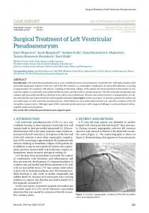
Figure 1: Typical pericardial traces for calculation of enclosed areas for images from (A) 4-chamber slice, (B) short-axis atrial slice, and (C) short-axis ventricular slice.
Figure 2: Pericardial area (normalized to end-diastole) vs. time for the three imaging planes: (A) 4-chamber slice, (B) short-axis ventricular slice, and (C) short-axis atrial slice.
Figure 3: Individual chamber area (normalized to pericardial end-diastolic area) vs. time for the left (A) and right (B) ventricles.
Figure 4: Individual chamber area (normalized to pericardial end-diastolic area) vs. time for the (A) left and (B) right atria.
Figure 5: Comparison of systolic and diastolic contours for two representative normal subjects (NL1, NL2) and one patient with congenital absence of the pericardium (NP). Contours from all three slices (4-chamber, short axis atrial, and short axis ventricular) are shown. Systolic contours are shaded in grey; diastolic contours are outlined in black. Note “crescent effect” present on free wall of left ventricle in NL1 and on the interventricular septum in NL2. Note also torsional effects on right ventricle in NL1.
H-00744-2004.R1 22 Animation Caption
Animation. Typical four-chamber bFFE cine-loop of a normal subject. Note how throughout the cardiac cycle, the pericardial boundary remains virtually fixed. See text for details.
H-00744-2004.R1 23 Table
Subject # Gender Age Height Weight Heart Rate F 1 22 5'3" 130 41 2 M 26 6'2" 185 55 3 M 26 5'11" 150 47 4 F 27 5'3" 110 78 5 F 47 5'5" 165 64 6 F 38 5'6" 175 67 7 F 21 5'6" 130 78 8 M 21 6'2" 165 63 9 M 21 5'8" 125 75 10 M 22 5'9" 135 75 NP
M
17
6'0"
230
76
Demographic data of the ten normal subjects (1-10) and patient with congenital absence of the pericardium (NP).
H-00744-2004.R1 24
Figure 1
H-00744-2004.R1 25
Figure 2A
Figure 2B
Figure 2C
H-00744-2004.R1 26
Figure 3A
Figure 3B
H-00744-2004.R1 27
Figure 4A
Figure 4B
H-00744-2004.R1 28
Figure 5