ARTICLE IN PRESS doi:10.1510/icvts.2006.149625
Interactive CardioVascular and Thoracic Surgery 6 (2007) 505–507 www.icvts.org
Proposal for bail-out procedures - Valves
Submitral left ventricular pseudoaneurysm after mitral valve replacement: early diagnosis and successful repair Charan Lanjewara,*, Bhavesh Thakkara, Prafulla Kerkara, Jagdish Khandeparkarb a Department of Cardiology, King Edward VII Memorial Hospital, Mumbai, India Department of Cardiothoracic Surgery, King Edward VII Memorial Hospital, Mumbai, India
b
Received 29 December 2006; received in revised form 5 April 2007; accepted 5 April 2007
Abstract Late left ventricle (LV) rupture with pseudoaneurysm after mitral valve replacement is rare. We report its early diagnosis by advanced technologies, e.g. MRI and successful repair of a type I AV rupture through left atrial approach. 䊚 2007 Published by European Association for Cardio-Thoracic Surgery. All rights reserved. Keywords: Pseudoaneurysm; Late left ventricular rupture; Mitral valve replacement; MRI to diagnose postoperative complications
1. Introduction Rupture of the posterior wall of the left ventricle (LV) after mitral valve replacement (MVR), although infrequent, may be a highly lethal complication. However, pseudoaneurysm formation due to incomplete or late rupture is a very rare complication. Spellberg and O’Reilly w1x first reported the pseudoaneurysm after MVR that was diagnosed using left ventriculography. Detailed surgical anatomy of sac and its communication can be delineated with transesophageal echocardiogram, cardiac MR or contrast CT scan. With these advanced diagnostic tools, early diagnosis and vigilant repair of pseudoaneurysm can reduce the operative mortality and prevent potentially lethal complication. This report describes the diagnosis and successful repair of a false aneurysm of atrioventricular groove in a patient who had undergone MVR. 2. Case report A 36-year-old Asian farmer who had three months prior undergone successful MVR with 25 mm bileaflet prosthesis for severe mitral valve stenosis presented with acute onset dyspnea NYHA class IV and acute onset severe chest pain. Clinical examination revealed normal prosthetic valve heart sounds and harsh high frequency grade 3y6 holosystolic murmur at apex that radiated to the back. In his past history, at the age of 18 years, he had undergone chordal shortening with mitral annuloplasty for chronic severe mitral regurgitation of rheumatic etiology. ECG showed first-degree heart block with non-specific STT changes, chest roentgenogram revealed an abnormal bulge along the left lateral border at left atrial appendage. *Corresponding author: C.O. Quarters Building No. 2, Flat 2, King Edward VII Memorial Hospital, Mumbai, India. E-mail address:
[email protected] (C. Lanjewar). 䊚 2007 Published by European Association for Cardio-Thoracic Surgery
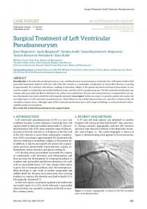
Transthoracic and transesophageal echocardiography (Fig. 1a–c) confirmed a large annular aneurysmal sac posterior to left atrium (LA) communicating to the LV. Cardiac MR (Fig. 2a,b) and contrast CT scan delineated the thin-walled large posterior sac compressing the LA. There was a welldemarcated long tortuous narrow tunnel from LV running laterally and then posterosuperiorly to the sac. Thus, MRI and CT helped us to give exact anatomic delineation and approach to such a complicated case. Coronary angiogram revealed normal coronaries with normal venous return and anatomy of coronary sinus. LV angiogram (Fig. 2c) showed simultaneous opacification of an oval cavity during LV systole through narrow tract. The sac was located posterosuperiorly to the LV or lateral heart border. 3. Procedure After securing all the monitoring lines the patient was operated under general anesthesia and femoro-femoral bypass and multidose warm blood potassium cardioplegia. Adhesiolysis was performed via parasternal left atriotomy, interior of the left atrium and left ventricle was inspected. No obvious tear in the left ventricle was noticed until the valve prosthesis was explanted. Then a 1.5 cm=1 cm tear just beneath the posteroinferior mitral annulus was observed. Thus, all anatomy was the same as shown by the MRI and CT. On exploration, a probe entered the pseudoaneurysm sac located posterior to the left atrium via a tortuous track. The tear was closed using a woven Dacron fabric patch sutured with a 3-0 polypropylene continuous suture. The valve prosthesis was re-implanted using pledgeted 2-0 polyester mattress sutures and free disc movements were confirmed. Left atriotomy was closed with a 3-0 polypropylene continuous suture. He had reasonably smooth recovery and was discharged after 10 days. His
ARTICLE IN PRESS 506
C. Lanjewar et al. / Interactive CardioVascular and Thoracic Surgery 6 (2007) 505–507
Fig. 1. (a–c) Transthoracic and transesophageal echocardiographic views showing pseudoaneurysm with tortuous neck and LV rupture site in AV groove.
follow-up echocardiographic study revealed no flow between LV and the repaired posterior sac. 4. Discussion LV posterior wall rupture after MVR was first described by Roberts and Marrow w2x in 1967. In a review by Karlson and colleagues w3x, an incidence averaged to 1.2% (0.5–2.0%) with mortality rate of approximately 75% LV rupture can be classified according to either location or timing of rupture. First, Treasures, Miller and co-workers classified the complication on the basis of the location of the tear w4, 5x.
Fig. 2. (a,b) Horizontal and coronal sections of cardiac MR, showing the complete anatomy of the pseudoaneurysm and nearby structures. (c) Left ventriculogram showing retrograde filling of posterosuperiorly located sac through a small communication at the inferior edge of mechanical valve.
Type 1-rupture located in the posterior atrioventricular groove. Type 2-rupture in the posterior wall of the LV at the base of the papillary muscle. Type 3-rupture in the area between the AV groove and papillary muscle. Karlson et al. classified the rupture according to the timing of rupture w3x. Early rupture is defined as occurring in the operating room any time after discontinuation of cardiopulmonary bypass. Delayed rupture manifests in the recovery room usually hours to days postoperatively. Late
ARTICLE IN PRESS C. Lanjewar et al. / Interactive CardioVascular and Thoracic Surgery 6 (2007) 505–507
rupture occurs days to years after the valve replacement and presents as pseudoaneurysm. The early rupture comprises two-third of LV ruptures following MVR. The appearance of excessive amounts of bright red blood in the pericardial cavity as the cardiac activity resumes strongly suggests the diagnosis. The mortality rate in these patients approaches near 50% despite early treatment w6x. Patients with delayed rupture comprise the remaining one-third of patients. Because attempts to suture a ventricular rupture against pressureloaded, beating heart are always unsuccessful and frequently extend the laceration, rapid reinstitution of bypass circulation is a prerequisite for repair. This explains the very high (approximately 90%) mortality due to mediastinal hemorrhage in delayed rupture w5x. In patients with late rupture, or occasional patients with delayed rupture, a thin layer of heart wall – usually the epicardium with fibrosed pericardium – withstand the pressure load of beating heart and escape rupture. In view of the thousands of mitral valve operations performed, it is surprising that very few cases of pseudoaneurysm have been reported w1, 7–9x. From a clinical perspective, late rupture has been reported much different from early and delayed rupture and presents as false aneurysm of LV without any significant symptoms. A large sac of pseudoaneurysm or its expansion can compromise the lumen of the circumflex artery and produce a myocardial infarction w7x. Other potential lethal complications of pseudoaneurysm include LV failure, thrombus embolization or rupture of aneurysm and death. In Type 1 rupture, regardless of the cause, the pathologic result is the separation of the annulus from the fibrous skeleton of the heart with the resultant extravasations of blood into the myocardium and ultimately frank perforation and rupture. The pathogenesis in type 1 rupture is worse than type 2. This high mortality is because of the anatomical location of rupture requiring specific surgical approach and nearby course of the left circumflex coronary artery, which can be sutured and produce myocardial infarction. The etiology in each of these types is probably different and each may have several technical causal factors in same cases; a clear separation into the classification is not possible w5, 8–10x. The exact clinical circumstances will dictate the management but generalized recommendations can be made. Once
507
the diagnosis is made, surgical repair should be done on CPB. In type 1 the preferred approach is internal repair, in that the left atrium should be reopened and buttressed sutures inserted from the outside through the valve-sewing ring in lateral fashion avoiding the circumflex artery. In types 2 and 3, defect is limited to myocardium and external repair by direct buttressed suture should be attempted w5, 6x. Regardless of the technique, the circumflex artery must be preserved in the repair of aneurysm. 5. Conclusion LV rupture following the MVR is encountered more often than reported. Diagnosis by newer techniques makes it amenable for early surgery and good prognosis. With careful precaution the incidence can be minimized. Pseudoaneurysm can be repaired promptly following precise localization with low mortality if the lesion is detected before the development of myocardial infarction. References w1x Spellberg RD, O’Belly RJ. Pseudoaneurysm of left ventricle following mitral valve replacement. Chest 1982;62:115–117. w2x Roberts WC, Morrow AG. Causes of early postoperative death following cardiac valve replacement. Clinico-pathologic correlations in 64 patients studied at necropsy. J Thorac Cardiovasc Surg 1967;54:422– 437. w3x Karlson KJ, Ashraf MM, Berger RL. Rupture of left ventricle following mitral valve replacement. Ann Thorac Surg 1965;46:590–597. w4x Miller DW, Johnson DD, Ivey TD. Does preservation of the posterior chordae tendineae enhance survival during mitral valve replacement. Ann Thorac Surg 1979;28:28. w5x Treasure RL, Rainer WG, Strevey TE, Sadler TR. Intraoperative left ventricular rupture associated with mitral valve replacement. Chest 1974;66:511–514. w6x Bjork VO, Henze A, Rodriguez L. Ventricular rupture complicating mitral valve replacement: surgical experience of six cases and review of literature. J Thorac Cardiovasc Surg 1977;73:14–22. w7x Diethrich EB, Koopot R, Kinard SA. Pseudoaneurysm of the left ventricular groove: a late complication of mitral valve replacement. J Thorac Cardiovasc Surg 1977;74:47–50. w8x MacVaugh H, Joyner CR, Pierce WS, Johnson J. Repair of subvalvular left ventricular aneurysm occurring as a complication of mitral valve replacement. J Thorac Cardiovasc Surg 1969;58:291–295. w9x Waller BF, Talierico CP, Clark M, Pless JP. Rupture of the left ventricular free wall following mitral valve replacement for mitral stenosis: a cause of complete (fatal) or contained (false aneurysm) cardiac rupture. Clin Cardiol 1991;14:341–345. w10x MacVaugh H, Joyner CR, Johnson J. Unusual complications during mitral valve replacement in the presence of calcification of the annulus. Ann Thorac Surg 1971;11:336.