Because dominant-negative. Ras17 has been shown to block the effects of PMA on pp42 MAP kinase phosphorylation, we assessed its effect on c-Jun ...
Vol.
4, 377-385,
May
1993
Cell
Multiple Signal Transduction c-Jun Protein Phosphorylation1
Christopher C. Franklin, Andrew S. Kraft2
Tino Unlap,
Victor
Adler,
and
Pathways
tion
Division of Hematology/Oncology, Birmingham, Birmingham, Alabama
University 35294
of Alabama
the
stimulate
esters) Abstract A variety of protein kinases, including pp42 and pp54 mitogen-activated protein (MAP) kinases, p342, and a partially purified protein kinase from 4$-phorbol 12myristate 13a-acetate (PMA)-treated U937 cells have been shown to phosphorylate the NH2-terminal activation domain of c-Jun in vitro. To investigate the role of pp42 MAP kinase in mediating c-Jun phosphorylation in vivo, we have treated U937 monocytic leukemia cells with a variety of pharmacological agents, including PMA, cycloheximide, AIF4, and okadaic acid. Although all of these agents stimulated c-Jun phosphorylation, cycloheximide and okadaic acid had no effect on pp42 MAP kinase phosphorylation, suggesting that MAP kinase activation was not necessary for c-Jun phosphorylation in vivo. Because dominant-negative Ras17 has been shown to block the effects of PMA on pp42 MAP kinase phosphorylation, we assessed its effect on c-Jun phosphorylation by cotransfection with a truncated c-Jun construct (c-Jun234). We found that cJun234 was expressed only in the cytosol and was inducibly phosphorylated with kinetics similar to those of endogenous nuclear c-Jun. Furthermore, we found that Ras17 had no effect on PMA-induced phosphorylation of c-Jun234. Because Ha-Ras requires isoprenylation for membrane binding, we examined the effect of the isoprenylation inhibitors lovastatin and perilhic acid on PMA-induced c-Jun phosphorylation. Pretreatment of U937 cells with these agents had no effect on PMA-induced c-Jun or pp42 MAP kinase phosphorylation. These data suggest that there are multiple pathways mediating c-Jun NH2-terminal phosphorylation in vivo, some of which are independent of MAP kinase activation, and that phorbol ester-mediated c-Jun and pp42 MAP kinase phosphorylation in U937 cells does not appear to be mediated by members of the Ras family. Introduction The c-Jun nuclear phosphoprotein of the AP-1 transcription factor
dimer can
or as a heterodimer bind
to AP-1
DNA
with sequences
is a major (1, 2). c-Jun
other and
component as a homo-
Jun or Fos proteins activate
cells contain
low levels of c-Jun c-jun gene is greatly by hormones (e.g., epidermal growth necrosis factor) and by compounds that
the
of
activity
the
of protein
(4, 5). Because
the
kinase
upstream
by the posttranslational
preexisting
protein
(6). c-Jun
appears
to be regulated
by both
terminal
phosphorylation.
boxy-terminal ity of c-Jun
to bind
to phenylalanine,
modification
transcriptional
amino-
to DNA
mimicking
of
activity
and
Phosphorylation
serine/threonine
phorbol
of the c-jun
and induction of cby protein synthetranscription of this
gene is regulated c-Jun
C (e.g.,
region
gene itself contains AP-1 sequences, jun expression is rapid and unaffected sis inhibitors, it is thought that the
carboxy-
of three
car-
residues inhibits the abil(7). Mutation of serine 243 a point mutation found in
v-Jun, blocks the phosphorylation of the other two residues, threonine 231 and serine 249, and enhances the DNA-binding activity of c-Jun (7). These three carboxyterminal sites can be phosphorylated by glycogen synthase kinase 3 (7) and CKII3 in vitro (8). Microinjection experiments using peptides that block CKII and the injection of CKII directly into cells suggest that the carboxy terminus of c-Jun is a substrate for CKII in vivo (8). c-Jun NH2-terminal phosphorylation also appears to play a role in mediating its transcriptional activity in vivo
(9-12). Phorbol stimulates the and
73 and
downstream
ester treatment phosphorylation
activates
the
of AP-1
of human of c-Jun
transcription
enhancer
leukemic cells on serines 63
of genes
elements
located
(9, 12). Studies
utilizing a fusion protein between the NH2 terminus of c-Jun and the DNA-binding domain of the yeast GAL4 protein have shown that the NH2-terminal 85 amino acids
of c-Jun
containing
these
two
phosphorylation
sites are
sufficient for transcriptional activation in vivo (1 2). Within this fusion protein, mutation of serines 63 and 73 to leucines abolishes PMA-induced transcriptional activity (12). Transfection of Ha-Ras, v-sis, v-src, or Raf-1 into fibroblasts also stimulates the phosphorylation of these serine residues (10, 1 1), suggesting that both oncogenes
and tumor promoters may modulate the same cascade of protein kinases. In contrast to these in vivo observations, in vitro transcriptional analyses suggest that NH2terminal phosphorylation does not play a significant role in regulating c-Jun transcriptional activity or DNA-binding activity (13). These discrepancies remain to be clarified. A number of protein kinases have been identified which are capable of phosphorylating the NH2 terminus of c-Jun. Both pp42 and ppS4 MAP kinases and p342
have been
shown
to phosphorylate
serines
63 and 73 of
transcrip-
The abbreviations used are: CKII, casein kinase II; PMA, 4l-phorbol 12myristale 1 3a-acetate; NGF, nerve growth factor; SDS, sodium dodecyl sulfate; PAGE, polyacrylamide gel ehectrophoresis; kDa, kihodalton(s); GAP, GTPase-activating protein; CMV, cytomegahovirus; PBS, phosphatebuflered saline; BSA, bovine serum albumin. 3
Received 1/8/93; revised 2/1 1/93; accepted 2/1 7/93. ‘ This work was supported by National Cancer institute to A. S. K. and American Cancer Society Grant IRG-66-32 2 To whom requests for reprints should be addressed.
many
transcription
stimulated both factor and tumor
at
377
& Differentiation
Mediate
(3). Although
protein,
Growth
Grant CA42533 to C. C. F.
378
c-Jun Phosphorylation
through
Multiple
Pathways
bacterial c-Jun in vitro (9, 13). A protein kinase has recently been purified from PMA-treated U937 cells, using heparin-Sepharose and affinity chromatography, that is also capable of phosphorylating c-Jun and appears to be distinct from the previously characterized protein kinases (12, 14, 15). In addition to c-Jun, MAP kinase has also been shown to phosphorylate other transcription factors, including p62TcF (1 ) suggesting that this protein kinase may have a number of important nuclear targets. Although these protein kinases have been studied in vitro, the protein kinase(s) that phosphorylate c-Jun in vivo have not been identified. We have examined whether alternative pathways independent of pp42 MAP kinase may mediate the phosphorylation of c-Jun in human leukemic cells. Here, we demonstrate that the pharmacological agents cycloheximide, a protein synthesis inhibitor, and okadaic acid, a phosphatase inhibitor, induce c-Jun NH2-terminal phosphorylation but do not stimulate the phosphorylation of pp42 MAP kinase in U937 cells. ln contrast, PMA, a protein kinase C activator, and AIF4, an activator of heterotrimeric GTP-binding proteins, stimulate the phosphorylation of both proteins. Because it has been demonstrated that, in certain cell types, PMA-induced pp42 MAP kinase phosphorylation and activation can be blocked by expression of a dominant-inhibitory Ras protein (17, 18), Ras’17, we examined the effect of this protein on c-Jun phosphorylation in U937 cells. We found that expression of this Ras mutant had no effect on PMA-induced c-Jun phosphorylation in U937 cells. We also addressed the role of Ras by utilizing the isoprenylation inhibitors lovastatin and perillic acid to inhibit Ras processing and thus membrane association and biological activity (19-21). Although pretreatment of PC12 cells with these agents abolished PMA- and NGF-induced pp42 MAP kinase phosphorylation, which have been shown to be Ras dependent (1 7, 1 8, 22), they had no effect on PMA-induced pp42 MAP kinase or c-Jun phosphorylation
in
U937
cells.
These
PMA-induced phosphorylation independent of Ras in U937 biochemical pathways, some of pp42 MAP kinase or Ras activity, phosphorylation in these cells.
results
indicate
that
of pp42 MAP kinase is cells and that multiple which are distinct from exist that regulate c-Jun
Results To evaluate the role of pp42 MAP kinase as a putative cJun NH2-terminal protein kinase in vivo, we treated U937 human monocytic cells with several pharmacological agents known to modify various signal transduction pathways. The addition of either PMA or okadaic acid to U937 cells activates a macrophage-like differentiation which is accompanied by a rapid increase in c-Jun phosphorylation, induction of c-jun mRNA expression, and activation of AP-1-mediated increases in transcription (12, 23, 24). Cycloheximide treatment of U937 cells also increases c-Jun phosphorylation as well as causing the “superinduction” of c-jun and other immediate early genes by a variety of agents (5, 6, 25). We have found that U937 cells contain only the pp42 MAP kinase (ERK2), with no pp44 MAP kinase (ERK1) being detected by immunoblot analysis with an antibody that recognizes both MAP kinase proteins (data not shown). Phosphorylation of pp42 MAP kinase on both threonine 183 and
tyrosine 185 is necessary for the activation of MAP kinase activity (26). The mobility of the phosphorylated form of pp42 MAP kinase is retarded on SDS-polyacrylamide gels, yielding a doublet band. Likewise, we have previously shown that the ladder of c-Jun protein bands on SDS-polyacrylamide gels is due to the site-specific phosphorylation of serines 63 and 73 within the NH2-terminal activation domain of c-Jun (12). U937
cells
were
labeled
or 32P and treated
with
imide.
possible
Due
to the
with
PMA,
either
okadaic
[35S]methionine
acid,
temporal
or cyclohex-
differences
in pp42
MAP kinase and c-Jun phosphorylation in response to these stimuli, we investigated the time-dependent phosphorylation of these proteins. At the indicated times, cJun
and
pp42
MAP
kinase
were
immunoprecipitated
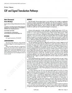
from whole cell denatured lysates. All three stimuli caused an increase in c-jun phosphorylation as judged by both an increased incorporation of 32P into c-jun and a retarded mobility of both 32P- and 35S-labeled c-Jun on SDS-PAGE (Fig. 1). However, only PMA stimulated an increase in pp42 MAP kinase phosphorylation. Cycloheximide and okadaic acid had no effect on either pp42 MAP kinase phosphorylation or mobility on SDS-PAGE (Fig. 1). The appearance of the retarded bands of protein on SDS-PAGE correlated well with the hyperphosphorylation
of both
c-Jun
and
pp42
MAP
our previous findings, which indicated in c-jun mobility is due to enhanced (12).
Although
the
apparent
sequential
kinase,
supporting
that the reduction phosphorylation phosphorylation
of pp42 MAP kinase and c-Jun in response to PMA treatment is consistent with pp42 MAP kinase-mediated c-jun phosphorylation, this hypothesis has yet to be verified. In any case, the ability of both okadaic acid and cycloheximide to induce c-jun phosphorylation in the absence of pp42 MAP kinase phosphorylation demonstrates the existence of a pp42 MAP kinase-independent pathway(s) of c-Jun phosphorylation in U937 cells. As a means of further examining the possible role of pp42 MAP kinase, we have attempted to alter the signal transduction pathway(s) that leads to pp42 MAP kinase phosphorylation and activation. Both oncogenic p21 Ras and activation of cellular p21 Ras have been shown to induce pp42 MAP kinase phosphorylation and kinase activity (17, 18, 27-29). In addition, the overexpression ofa dominant-inhibitory p21 Ras protein (Ras”7) inhibits the ability of insulin and platelet-derived growth factor in rat-i cells and of NGF in PC12 cells to stimulate the phosphorylation
of
pp42
MAP
kinase
(17,
18,
27-29).
These data suggest that Ras mediates tyrosine kinase receptor-induced MAP kinase activation. In PC12 cells, this mutant Ras protein also blocked the ability of PMA to stimulate the phosphorylation MAP kmnase; however, this effect appears to be cell type specific, as Rasnhl7 had no effect on PMA-induced phosphorylation of MAP kinase in rat-i fibroblasts (28). Because of the profound effect of this dominant-inhibitory Ras mutant on MAP kinase function, it was of interest to determine its effect on c-Jun phosphorylation. Attempts to establish a permanent U937 cell line stably expressing Ras17 were not successful, possibly due to the growth-inhibitory effects of this protein (30). To circumvent this problem, we took the approach of transiently
expressing
Rasbsr7
together
that is distinguishable from PAGE. We have previously
with
a c-Jun
construct
endogenous c-Jun on SDSassessed c-Jun posttransla-
Cell
PMA TIME
(mm)
0
5
15
O.A.
30
60
0
5
Growth
379
& Differentiation
CHX
15
30
60
0
5
15
30
60
mas*
#{149}#{149}#{149}
aaaS
S1S#{149}.
c-Jun 32
35s
*
32p
-*
-
-
-
-
.
-
MAPK
fig.
1. Time ourse of phosphorylation of c-lun and 1)1)42 MAP kinase (M.’PK( described in “Materials and Methods.” The cells were then treated with 0.2 M the indicated times, the cells were harvested, and whole cell denatured lysates and analyzed as described in “Materials and Methods.”
in U937 cells. PMA, 100 ng/ml were prepared.
tional modification ing a termination which encodes
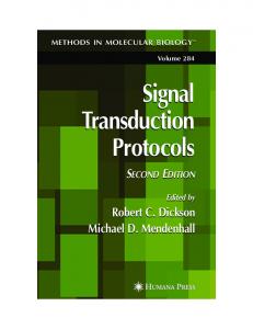
microscopy. The majority of endogenous c-Jun was found in the nucleus, although a small portion was detected in the cytosolic fraction, possibly due to low levels of nuclear lysis (Fig. 2A). In both control and PMA-treated
by utilizing a c-Jun construct codon at amino acid 234 a protein lacking both the
contain(cJun2i4), basic and
leucine zipper regions (12). This truncated protein migrates separately from endogenous c-Jun on SDS-PAGE (29 kDa versus 39 kDa, respectively). c-Jun214 is hyperphosphorylated and shifted in mobility on SDS-PAGE in response to PMA treatment of U937 cells in a manner similar to that ofendogenous c-Jun (12). However, it was recently reported that the nuclear localization signal for c-Jun resides between amino acids 268 and 284 (31),
which
are deleted
in cJun2i4,
suggesting
that the cJun2i4
protein may be cytoplasmic. To determine the cellular location of c-Jun234, cell fractionation/immunoprecipitation studies were performed. U937 cells transiently transfected with cJun2i4 were labeled with fl5S]methionine and incubated in the absence or presence of PMA for 1 h. After a gentle hypotonic lysis procedure, the lysate was separated into crude nuclear and cytosolic fractions, and c-Jun was analyzed by immunoprecipitation. The nuclei isolated by this procedure were found to be greater than 95% intact as judged by phase contrast
cells,
U937
cells were labeled with ]#{176}S]me’thionine or ‘2P, as okadaic acid (0.4.), or 100 CM cycloheximide (CIIX(. At c-Ion and pp42 MAP kinase were immunoprecipitated
c-Jun234
plasmic
protein
fraction
the phosphorylation
(fig.
was 2A).
detected Furthermore,
of both
cytosolic
only
in the
PMA
cyto-
stimulated
c-Jun2t4
and
en-
dogenous nuclear c-Jun protein as judged by the retarded mobility of the proteins on SDS-PAGE (Fig. 2A; Ref. 12). As cell fractionation studies are complicated by nuclear lysis and leakage of proteins during the fractionation process, in situ c-jun immunofluorescence staining was performed. c-Jun immunofluorescence staining of Cos7 cells transiently transfected with either vector, c-jun234 or a holo-c-jun construct, was assessed using affinity-
purified peptide in cells
Whereas
antiserum (Oncogene transfected
raised against an NH2-terminal c-Jun Science). No staining was detected with
vector
alone
(Fig.
28,
Panel
3).
holo-c-Jun expression was restricted to the nucleus (Fig. 28, Panel 1 ), c-Jun254 staining was distributed throughout the cytosol (Fig. 2B, Panel 2). Thus, although cJun2i4 is expressed as a cytosolic protein in U937 cells,
380
c-Ion
Phosphorylation
through
Multiple
Pathways
A
B
46
kDa.
30
kDa-.
Cont
PMA
cy
cy nu
nu
#{149}
phase
immunofluorescence
1
contrast
4
c-Jun
,
#{149}
.,-
.
-5 .
Jt4c4i
tF’
2
k..
.,
,
5 -‘_‘4.4-
.‘r-
c-Jun
234
-
--
.
..
. ..
.
.‘..
,
.,
3
4
‘I,’.
6
vector
fig. 2 Cellular localization of c lun2” in U937 and Cos7 ( ills. .‘\. e’ll ir, tionation (ii U9 17 c ells expressing ( -lcjiY . U9 37 c e’lls 4 X 1O) ssem translected by elec troporation with ( -(un2’4. Eighteen h l)osttransl(’c lion, the ( ells were’ starved in methii)n(’-free media or 3() miii md then lal)i’l(’d with [ methionine for 4 h. During the last hour, the cells were split. and one-half were treated xx’ith 0.2 pM PMA. Cells were’ frac tionated into e vtosolic (cy) and nuc lear ( no ( fractions as des rihed in “Materials and Methods .“ F ndogenous -I un and ( - I ‘ pr(it(’ins were’ immunoprec ipitate(l from the fractions and analyzed by S[)S-PAGE. Unphosphorvl.3ted ( -lun2 and (‘n(li)g(’n(ius ( -lun migrate ii 29 and 19 kDa, re’sp(’( tixel . B, imniunotluores e’n( i’ staining (it Cos7 ( ells expressing c-Ion (it d -lun2 ‘. Cos7 (OIls seeded on glass ove’rslips were transiec ted with hololun, c-Ion2 , or expression vector alone by the ( alcium phosphate ( oprecipitalion proc(’dur(’. ( -lun immunotluiires ence staining was analyie’d 2 days posttransf(’cii(in using aitinity-purifiecl antiserum to an NH2-terminal C -(un peptide. as desc rihed in “Materials and Methods.” Right p,im’ls, the ( (irr(’sp(inding I)hase ontrast photographs.
un2
(‘oIl
Time
0
46
0.5
cvro
(hrs)
1
2
kDa-
4
6
-
MICROGRAMS I
I
Pulse-c
h,iso
,io,ilvsis
i ii
fi
PXIr\-indu
‘(f
(
-I on
111(1
(
( Lii)’
I)(1ii-
-
t)riiteiils
sx’en’
inlnluiliit)ri’
i1)it,lt(’d
md
,inalv,’il
by
Sl)’-P’\(E.
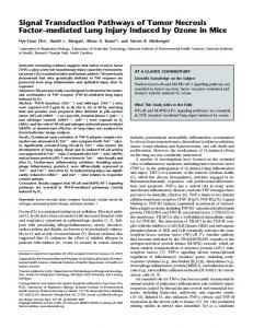
it is a substrate for PMA-inducible phosphorylation similar to that of endogenous nuclear c-Jun. These results cre not restricted to U937 cells, as similar results have also l)een observed in HepG2 cells (data not shown). To determine whether cytosolic c-lun24 was phosphorylated with similar kinetics to those of endogenous nuclear c-Jun, U937 cells were transfected with c-lun and pulse labeled with [SJmethionine. Cells were then chased with excess Linlabeled methionine in the presence of PMA, and the two c-Jun proteins were imniunoprecipitated at various times. The time course of c-Jun and c-Jun4 phosphorylation was identical, with maximal phos)hOryIation occurring between 30 mm and 1 h and decreasing thereafter (Fig. 3). PMA induces the activation of both cytosolic and nuclear pp42 MAP kinase in HeLa cells (32). We have recently isolated a protein kinase from PMA-treated U937 cells which is distinct from pp42 MAP kinase and is capable of phosphorylating serines 63 and 73 of c-Jun in vitro (14, 15). To determine whether c-Jun kinase activity is also present in an activated form in 1)0th the cytosol and nucleus of U937 cells, cell fractionation was performed in conjunction with in vitro cJun kinase assays. U937 cells were treated with PMA for 30 ruin, and crude nuclear and cytosolic fractions were isolated as described above. A fusion protein between the NH2-terminal 85 amino acids of c-Jun and glutathione S-translerase (GST-JUN) was used as a substrate for measuring c-Jun protein kinase activity as previously described (14, 15). PMA treatnient stimulated a protein kinase activity in both the cytosol and nucleus which was capal)Ie of phosphorylating the NH2 terminus of c-Jun (Fig. 4). Increasing amounts of added substrate yielded a concomitant increase in phosphor!ation. Although cJun2 is predominantly a cytosolic protein, it appears to be NH2-terminally phosphorylated in a manner identical to that of endogenous nuclear c-Jun. Thus, c-Jun2 4 appears to be a suitable substrate to analyze the effects of Ras” on PMA-induced c-Jun phosphorylation in U937 cells. To examine the role o Ras in PMA-induced c-Jun phosphorylation, U937 cells were cotransfected with c-
ii
Ii)
it
‘ri’
fli”i(
ii)
-I on
liii.itiiin
ifv
in
intl
iiiii-i,
ii iii
(
is
.15
55
,iss.ss
‘ri’
I)fi
iti
I
(,
‘iii
I -/1
10 50 100 OF GST-JUN
PPv1,\indu
iif
in
oil
.
luri
N f f-ti’rniin,il
LJ) I 7 ills. L (f 17 ((‘(Is SVi’t(’ tr(’at(’(l rudi’ iieeli’ar \t XL I ,iil vtiisiilu i)utlio(’(l “Nl,itorials md Methiicls.
1,01(1
si
ir,i( i it
phrvl.ltii)n. U9.3 7 ills sVi’t(’ i’l’ctropiiratocl svitli Iuo . md 24 h piisttransf(’ction. ( i’l(s sv(’r(’ ulS’ lal)el(’d svith nl(’thii iiluo’. Aft(’r 55 .ishiiig, the’ cells ss (‘re in iil),it(’(l in ( onipk’te nii’il a i iintaining (‘\( (‘s unlab’k’d nl(’thiiinin(’ md 0.2 xi PXIA. At the iiiiii ,it(’(l tinl(’s, ,lliclu(its sve’ri’ r(’no)v(’cI . .mnd svhiilo (‘II (l(’natur(’d lvsates \Si’t(’ Pri’t).ir’d. Both i len
-I. ((‘Ilul,ir
ic;.
Liii,isi’
kDa__#{216}I.
55
I i,’.
381
[)iff(’r(’nfialion
NUCL
10 50 100
30
Griisvth
l)Ot’\
\ I
(‘ii
for
(‘(‘0
,is
,i
in
glutathii ‘ul)str,it(’
itri
-lun pri)t(’in mc’ S-tr.insler.ise t 4 , 1 ‘i I. (
“
prot(’in
sx’ith (1.2 M PMA ) ( T( ) ) fr,utions /\liqu(its i)I e,ich
Lioasi’ ,i( .10(1 aniinii
tivity
using ,i
a
(Is S 89
Jury and the dominant-inhibitory Ras protein, Ras”’’. The ratio of Ras” to (-Jri11 DNA transfected was :1 to ensure that the cells expressing the c-Jun2 construct were also transfected with the Ras mutant. Immunopre(ipitation of Ras’ froni [ iSlnlethionine..labeled cells using an anti-Ras monoclonal antibody demonstrated that Ras” was expressed in great excess relative to that of endogenous Ras protein and was not modulated by the addition of PMA (Fig. 5B). Immunoprecipitation of cJun2 from Ras”-cotransfected cells indicated that transient expression of Ras’ did not block the PMAinduced phosphorylation of cJun2i (Fig 5A). These resuIts were not cell type specific for U937 cells, as identical results were obtained in HepG2 cells (data not shown). To further evaluate the role of Ras and other lowniolenilar-weight Ras-like GTP-binding I)roteins in mediating PMA-induced c-Jun phosphorylation, we utilized two pharmacological inhibitors of protein isoprenylation in an attenipt to block the biological function of Ras by inhibiting its ability to associate with the plasma membrane. It has been shown that several Ras proteins require isoprenylation for plasma membrane association and transforming activity (20). Although these compounds are known to inhibit Ras isoprenylation, they are also known to have other adverse effects, i.e. , inhibition of cholesterol biosynthesis and alterations in cell cycle progression (33). The effects of AIF.1 were also examined in U937 cells, as this compound is known to directly activate several heterotrimeric GTP-binding proteins which are thought to mediate its ability to stimulate pp42 MAP kinase activity (29, 34). To establish that inhibition of isoprenylation in fact inhibited Ras function, we investigated the effect of these inhibitors on PMAand NGFinduced p)42 MAP kinase phosphorylation in PCi 2 cells, which has previously been shown to be dependent on Ras lunction (17, 18). As has previously been reported, PMA and NGF stimulated the phosphorylation of pp42 MAP kinase in PC12 cells (Fig. 6A, Lanes 1-3). Pretreatment of PC12 cells with lovastatin and perillic acid, however, abolished pp42 MAP kinase phosphorylation in response to both PMA and NGF (Fig. 6A, Lanes 4-6). These results indicate that pharmacological inhibition o Ras processing can be effectively utilized to block Ras function. We utilized this phenomenon to address whether c-Jun phosphorylation in U937 cells was Ras
382
c-Ion
Phosphorylation
through
Multiple
Pathways
A
B Ras (Asnl7)
0 0
PMA
0
Ras (Asnl7)
PMA
0
PMA
0
0
Fig. S. Effect of dominant-inhibitory Ras (Ras’7( on PMA-induced c-Ion2” phosphorylation in U937 cells. U937 cells were transfected with c-Ion234 in the absence or presence ol excess Ras”7 (1:5 DNA ratio). Two days posttransfeclion, the cells were labeled with [35S]methionine, as described in “Materials and Methods.” The cells were then split, and one-half were treated with 0.2 LM PMA for 30 mm. lmmunoprecipitationof(A(c-
PMA
3044-
$‘
1
dependent. and
okadaic
234
U937
perillic
Ion2” and (B) Ras7 from whole cell denatured lysates was performed as described in Materials and Methods.”
21.5W
30
acid
acid,
cells and
5678
were
then
pretreated
treated
for
c-jun
and
or AIF4, and
with
lovastatin
30 mm
pp42
with
PMA,
MAP
kinase
ide did not stimulate the phosphorylation of pp42 MAP kinase. As threonine/tyrosine phosphorylation of pp42 MAP kinase is necessary for activation of protein kinase
phosphorylation were analyzed by immunoprecipitation (Fig. 6B). As previously shown in Fig. 1, PMA induced the phosphorylation of both proteins (Fig. 68, Lane 2), whereas okadaic acid stimulated c-Jun phosphorylation
activity (26, 35), these results suggest phorylation can occur in a pp42 MAP
in the absence
(36, 37), our results demonstrate that pp42 MAP kinase is not. The lack of an effect in response to okadaic acid is consistent with previous reports in PCi 2 cells, in which okadaic acid alone had no effect on pp42 MAP kinase but potentiated that observed in response to NGF (38). Only the time course of PMA-induced phosphorylation
of pp42
68, Lane 3). Similar
to
MAP
kinase
PMA,
AIF4
phosphorylation stimulated
(Fig.
both
pp42
MAP kinase and c-Jun phosphorylation (Fig. 68, Lane 4). Interestingly, inhibition of isoprenylation in U937 cells did not abolish the phosphorylation of either c-Jun or pp42
MAP
kinase
phosphorylation
in response
to any
of
the agonists tested, although the AIF4 response was reduced. Together, these results suggest that Ras does not play
a significant
role
in mediating
the
phosphorylation
of either pp42 MAP kinase or c-jun in U937 cells. Furthermore, it appears that PMA-induced pp42 MAP kinase phosphorylation occurs by both Ras-dependent and Rasindependent
mechanisms
in a cell
type-specific
manner.
of U937
cells
stimulates of c-Jun activation
the site-specific on serines of protein
63 and kinase
C by PMA, the exact signal transduction mechanism(s) by which PMA induces c-Jun phosphorylation remains unclear. A number of protein kinases have been identified which phosphorylate c-jun in vitro including pp42/ 44 and pp54 MAP kinases, p34(d(2 and a partially purified
enzyme from PMA-treated U937 cells which appears to be distinct from the others (9, 1 3- 1 5). However, because protein kinases promiscuously phosphorylate many subin vitro,
it is difficult
protein
kinases
to
identify
is the c-jun
which
of
these
NH2-terminal
pro-
tein kinase in vivo and which are modulated by phorbol esters. We utilized several very diverse pharmacological agents to examine the biochemical pathways responsible for c-jun phosphorylation in vivo. Whereas PMA, okadaic
acid,
cycloheximide,
phorylation
of
in U937
and AIF4 all stimulated cells,
okadaic
acid
and
c-Jun
phos-
cyclohexim-
in U937
pp42
cells.
Although
are activated
MAP
kinase
closely
Although directly address
plays
in mediating
a role
pp54
in response
phosphorylation. here did not
MAP
and
pp7O-
to cycloheximide
paralleled
that
the experiments whether pp42
PMA-induced
of
c-Jun
described MAP kinase
c-Jun
phospho-
rylation in vivo, they do suggest that there are other functional signaling pathways independent of pp42 MAP kinase that can mediate c-Jun phosphorylation in U937
both
NH2-terminal phosphorylation 73 (9, 1 2). Other than a direct
strates
manner
56 kinases
cells. We
Discussion PMA treatment
candidate
ent
that c-Jun phoskinase-independ-
have the
previously
basic
phosphorylated as those
cently Jun
and
on identical
mapped
reported resides
shown leucine
that zipper
serine
in endogenous
that the nuclear
in the
basic
protein (31), suggesting Using cell fractionation
leucine
c-jun234,
which
regions,
is inducibly
residues
(63 and
73)
It was
re-
c-Jun
localization zipper
(12).
signal region
lacks
for cof
the
that c-Jun254 might be cytosolic. and immunofluorescence stain-
ing, we have confirmed that this protein is localized to the cytosol. However, after phorbol ester treatment of U937 cells, this protein appears to be phosphorylated and dephosphorylated over an identical time course to that of endogenous nuclear c-Jun. Also, in vitro protein kinase assays demonstrate that both cytosolic and nuclear extracts contain a protein kinase activity capable of phosphorylating c-Jun. Therefore, PMA appears to stimulate both cytosolic and nuclear c-Jun NH2-terminal kinase activity in a similar manner. Serum and PMA treatment of HeLa cells has been reported to elicit a similar activation of both cytosolic and nuclear MAP kinase, which is accompanied by a translocation of MAP kinase
Cell
A .4-O
Lova.-#{248}
-04---
0
PMA
1
2
NGF
0
PMA
NGF
MAPK*II1$I
3
4
56
B 0 0
PMA
0-4---OA
AIF4
0
Lova.-* PMA
OA
AIF4
c-Jun
MAPK*
1
2345678
Fig. 6. Effect of the isoprenvlation inhibitors lovastatin and perillic acid on c-Ion and Pl42 MAP kinase (M,-\PK( phosphorylation. ‘1, effect of isoprenylation inhibitors on PMAand NGF-induced pp42 MAP kinase phosphorylation in PC12 cells. PC12 cells were’ Iett untreated or were pretreated for 20 h with 30 M lovastatin. Cells we’re then labeled for 4 h with [‘5Slmethionine in the absence’ (0) or presence of 30 pM lovastatin and 5 m perillic acid (Loca.). Cells were then treated with either 0.2 MM PMA or 100 ng/ml NGF for 15 mm, and pp42 MAP kinase was immonoprecipitated from whole cell denatured lysates as previously described. B, effect of isoprenylation inhibitors on c-Ion and pp42 MAP kinase phosphorylation in U937 cells. U937 cells were pretreated and 05 labeled exactly as described above’. Ce’lIs were then tre’ate’d with 0.2 pM PMA, 100 ng/ml okadai acid IO’\), or freshly prepare’d AIF4 as described in “Materials cipitated analyzed
and Methods.” from whole cell by SDS-PAGE.
c-Ion and denature’d
Pl42 MAP kinase were immunoprelysates as previously described and
activity from the cytosol to the nucleus (32). Attempts to address whether PMA induces the nuclear translocation of a recently identified c-Jun protein kinase were hindered by the complete lack of in vitro c-Jun kinase activity ofthis purified protein in cells nottreated with PMA (15). A dominant-inhibitory Ras mutant (Ras”7) has proved helpful in dissecting the cascade of events leading to the activation of pp42 MAP kinase by hormone receptor tyrosine kinases. Expression of this protein blocks the ability of NGF, insulin, or platelet-derived growth factor to stimulate the phosphorylation of pp42 MAP kinase in PC12 and rat-i cells (17, 18, 28). The role of Ras in mediating PMA-induced pp42 MAP kinase activity, however, appears to be cell type specific as Ras\s7 blocks the effects of PMA in PC12 cells but not in rat-i cells (17, 18, 28). A similar inhibitory effect has been reported in
Growth
& Differentiation
NIH 3T3 cells overexpressing GAP, which also acts to negatively regulate Ras activity (29). We attempted to evaluate PMA- and NGF-induced pp42 MAP kinase and c-Jun phosphorylation in the above-mentioned Ras” PC12 cells, but even under stringent control of the culture conditions, the negative regulatory effects of Ras” could be detected under uninduced conditions.4 Attempts to establish a U937 cell line which contained such an inducible mutant Ras were unsuccessful, suggesting that this gene product is highly toxic even at low levels in U937 cells (30). Similar difficulties have been overcome in NIH 3T3 cells by prior transformation with vRaf, which overcomes the growth-inhibitory effects of Ras””7 (22). This approach was not feasible for our studies, as v-Raf has been shown to activate MAP kinases, albeit indirectly (39-41). We therefore decided to examine the effect of transient transfection of Ras on c-Jun phosphorylation in U937 cells. To examine the ability of RasA57 to modulate the activity of phorbol esters, this protein was cotransfected with c-Jun214. Whereas Ras7 protein was expressed at levels far exceeding those of endogenous Ras protein, it appeared to have no effect on PMA-mediated cJun2i4 phosphorylation in U937 cells. As mentioned above, it is possible that the effects of Ras”’7 are cell type specific, since it blocked phorbol ester-induced effects in PC12 cells and not in Rat-i cells. However, we did not see any effect of this mutant on PMA-induced phosphorylation of c-jun24 in HepG2 cells (data not shown). We cannot exclude the possibility that nuclear (endogenous) and cytosolic (c-Jun2#{176}) c-Jun are phosphorylated by distinct mechanisms due to their differential compartmentalization. Many reports suggest that the Ras proteins may play a role in phorbol ester-mediated growth responses. Overexpression of GAP in mouse NIH 3T3 cells blocked the ability of PMA to stimulate MAP kinase phosphorylation (29). PMA treatment of T-cells has been shown to inhibit GAP and induce the formation of a more active GTPbound form of Ras (42). To evaluate the role of Ras proteins in the regulation of c-Jun phosphorylation, we treated U937 cells with perillic acid and lovastatin, both of which inhibit the isoprenylation of low-molecularweight GTP-binding proteins (19, 21). Both Harvey and Kirsten Ras proteins have been shown to require isoprenylation to associate with the cell membrane (20). Using PC12 cells, we have demonstrated that pharmacological inhibition of Ras isoprenylation is a viable means of inhibiting Ras-mediated cellular events. However, treatment of U937 cells with these compounds did not affect PMA-induced phosphorylation of either pp42 MAP kinase or c-Jun. These results indicate that c-Jun phosphorylation in U937 cells is independent of Ras activity and furthermore provide evidence that Ras-dependent and Ras-independent mechanisms mediate pp42 MAP kinase phosphorylation in a cell type-specific manner. It therefore appears that multiple pathways are involved in mediating c-Jun phosphorylation in U937 cells. Both genetic (Ras7) and pharmacological (isoprenylation inhibitors) manipulation of the Ras signaling path-
4
C. C.
Franklin
and
A.
S. Kraft,
unpublished
ol)servations.
383
384
c-Jun
Phosphorylation
ways
indicates
phorylation We cannot
through
that
Multiple
phorbol
Pathways
ester-mediated
in these cells is not mediated rule out the possibility that
c-Jun
phos-
by Ras proteins. other pathways
may predominate in other mouse or human cell systems that are not of hematopoietic origin. Although the exact functional role of c-Jun NH2-terminal phosphorylation in
transcriptional regulation remains to be more fully evaluated, phosphorylation of this protein in response to a variety of stimuli serves as a means for evaluating the cascade of events that regulate nuclear protein phosphorylation.
by immunoprecipitation with a v-H-ras monoclonal antibody (clone Yi 3-238; Oncogene Science), and immune complexes were collected with protein-G-Sepharose following the manufacturer’s instructions.
Cell nuclear
cells
Fractionation
and
and
fractions
cytosolic
in hypotonic
lmmunofluorescence.
Nonidet
were
insoluble
by
P-40 lysis buffer
pH 7.5-iO mM NaCI-3 mi MgCI2-0.5 ylsulfonyl fluoride-0.05% Nonidet P-40) Following a low-speed spin (500 x g), was collected and recentrifuged for iS
g to remove
Crude
obtained
material
lysing
(iO mt’i Tris,
mti phenylmethon ice for 5 mm. the supernatant mm at i2,000 x
and was designated
the
Materials and Methods Cell Culture. U937 cells were cultured in Dulbecco’s modified Eagle’s medium containing iO% iron-supplemented, heat-inactivated bovine calf serum, 100 units/ ml penicillin, and 100 zg/ml streptomycin in a humidified atmosphere of 5% C02-95% air. PCi2 cells were obtamed from the American Type Culture Collection and were maintained in monolayers in Dulbecco’s modified Eagle’s medium supplemented with iO% heat-mactivated horse serum, 5% fetal calf serum, 4 mr’i glutammne, and antibiotics. Cells were treated by direct addition of either 200 flM PMA, 100 ng/mI NGF (GIBCO), 100 M cycloheximide, iOO ng/ml okadaic acid (GIBCO), or fluoroaluminate prepared fresh daily by mixing 300 mrs’i NaF with 1 mM AICI3 at a ratio of 10:1 (final concentration, 30 mM NaF and iO LM AlCl3) (34). Lovastatin (the kind gift of Dr. A. Alberts, Merck, Sharp and Dohme) and perillic acid (Janssen Chimica) were used at 30 zM and 5 mrvt, respectively. Plasmids and Transfedions. The c-jun234 construct has been described previously (12). The dominant-inhibitory Ras expression vector, CMV-Ras”7, was prepared as follows. Plasmid pXVR (the kind gift of Dr. L. Feig) was digested with BamHl and Xbal, and a 1.i-kilobase frag-
cytosolic fraction. The crude nuclear pellet was washed immediately with lysis buffer. Nuclei were found to be greater than 95% intact as judged by phase contrast microscopy. Nuclei were lysed by boiling for S mm in denaturing lysis buffer (50 mt Tris, pH 8.0-0.5% SDS-5 mM dithiothreitol-0.5 mi phenylmethylsulfonyl fluoride),
luted
i:iO
in i%
ment
with
PBS,
blocked
of the
v-Ha-ras
gene
was
excised
and
cloned
into
pBluescript SK (Stratagene). The insert was then excised with BamHl and Notl and cloned into the pCEP4-CMV expression vector (Invitrogen). U937 cells were transfected by electroporation as previously described (i2). Approximately 2-3 x i07 cells were transfected in 0.5 ml PBS with 30-50 jzg DNA at a setting of 250 V and 250 jtfarads. When c-Jun234 and Ras17 were cotransfected, the amounts of DNA used were 10 and 50 zg, respectively. Cell Labeling and lmmunoprecipitation. U937 cells were labeled as previously described (12). PC12 cells were preincubated for 30 mm in minimal essential medium containing 0.5% dialyzed bovine calf serum and lacking either methionine or phosphate. Cells were Iabeled for 2-6 h in the same media containing either 0.i mCi/mI Trans35S-label or 0.5-i mCi/mi 32P, respectively. After
cell
treatment,
whole
cell
denatured
lysates
were
prepared as previously described (12). All lysates were precleared overnight with Protein A-Sepharose (Pharmacia). c-jun. c-Jun234, and pp42 MAP kinase were immunoprecipitated with c-jun-specific polyclonal rabbit antisera raised against a pGEX-c-jun fusion protein (1:S00 dilution) or pp42-specific antisera (1 :500 dilution; the gift of Dr. J. Ferrell, Stanford University, Palo Alto, CA), respectively.
Protein by
iO%
Immune
A-Sepharose, SDS-PAGE.
complexes
were
collected
with
washed extensively, and analyzed Expression of Ras#{176}7was analyzed
followed
by a 5-fold
dilution
with
radioimmunoprecipi-
tation assay buffer lacking SDS. c-Jun was immunoprecipitated from the nuclear and cytosolic fractions as described above. c-Jun immunofluorescence staining was performed in Cos7 cells transfected with CMV-holo-c-Jun, CMV-cJun234, or pCEP4 expression vector alone. Cells were
plated on glass coverslips and transfected with 5 jzg of plasmid DNA by the calcium phosphate coprecipitation method. Two days posttransfection, cells were washed with
PBS and
fixed
with
for 45 mm at room
PBS containing
temperature.
3% formaldehyde
Coverslips
were
washed
with PBS and incubated in ice-cold acetone for 20 mm. After rinsing, the cells were blocked in i% BSA-PBS for 20 mm at room temperature. Coverslips were then incubated for 30 mm at 37#{176}Cwith affinity-purified c-Jun polyclonal antibody (c-Jun Ab-2; Oncogene Science) di-
37#{176}C for
BSA-PBS. in
1 h with
Coverslips
1%
goat
were
then
and
incubated
BSA-PBS,
anti-rabbit
washed
fluorescein
cyanate (Southern Biotech, Birmingham, in 1% BSA-PBS. Coverslips were washed ered with Hoechst dye, and mounted fluorescence microscopy was performed.
at
isothio-
AL) diluted with PBS, on slides,
i :50 coyand
Acknowledgments The authors thank Dr. I. Ferrell for the kind gift of the pp42 MAP kinase antisera, Dr. L. Feig for the dominant-negative Ras expression vector, Dr. S. Halegoua for the PC12 cell line containing the dominant-negative Ras mutant, Dr. A. Alberts for the lovastatin, Dr. A. Morris for helpful discossions, and F. Wagner for technical assistance.
References 1. Bohmann, D., Bos, T. I., Admon, A., Nishimura, T., Vogt, P. K., and Tjian, R. Human proto-oncogene c-jun encodes a DNA binding protein with structural and functional properties of transcription factor AP-1. Science
(Washington
DC),
238; 1386-1392,
1987.
2. Rauscher, F. J., Ill, Cohen, D. R., Curran, T., Bos, T. J., Vogt, Bohmann, D., Tjian, R., and Franza, B. R., Ir. Fos-associated protein is the product 1010-1016,
of the jun 1988.
3. Chiu, R., Boyle, The c-Fos protein AP-1 responsivegenes. 4.
Dixit,
antimitogenic
V.
M.,
proto-oncogene.
W. I., Meek, interacts with Cell, 54; Marks,
action
R. M.,
of tumor
AP-1/c-jun proto-oncogene 16909, 1989.
Science
I., Smeal, c-Iun/AP-1 541-552, Sarma,
necrosis transcription.
V., factor
(Washington
R., Hunter, to stimulate 1988. and
DC),
240;
T., and Karin, transcription
Prochownik,
is associated I. Biol.
P. K., p39
Chem.,
E. V. with 264;
M. of The
increased
16905-
Cell
5. Lamph, W. W., Wamsley, Induction of proto-oncogene 334; 629-631, 1988. 6. Angel, is positively 1988.
P., Sassone-Corsi, lon/AP1 by serum
P., Hatiori, K., Smeal, autoregulated by
and
P., and Verma, I. M. TPA. Nature (Lond.(,
T., and Karin, M. The jun proto-oncogene its product, lon/AP-1. Cell, 55; 875-885,
7. Boyle, W. I., Smeal, T., Defize, L. H., Angel, M., and Hunter, T. Activation of protein kinase ation of c.lun at sites that negatively regulate Cell, 64; 573-584, 1991.
P., Woodgett, C decreases its DNA-binding
I. R., Karin, phosphorylactivity.
353; 670-674, 10.
Smeal,
B., Mercola,
D., Grover-Bardwick,
Cell. Biol., 12; 3507-3513, 1 1 . Binetroy, and stimulates 351; 122-127,
Karin, M. Ha-Ras augments of its activation domain.
12. Franklin, C. C.. Sanchez, V., Wagner, A. S. Phorbol ester-induced amino-terminal JUN but not IUNB regulates transcriptional Sci. USA, 89; 7247-7251, 1992.
c-Ion Nature
30.
15.
Adler,
V.
Nail. A.,
purified c-Ion phosphorylation 16. GilIe, transcription formation
Kraft,
of c-Ion but not Acad. Sci. USA,
v-Ion:
Pohoiskaya,
A.,
amino-lerminal for activity.
89;
A. S. Phorbol regulation
by the
5341 -5345,
31.
Wagner,
F., and
Sharrocks, factor p62T at c-los promoter.
stimulate N-terminal
Krafi,
A.
S. Affinity
Cell,
68;
proto-oncoprotein
103 1 -1040,
19. Crowell, P. L., Chang, R. R., Ren, Z., Elson, C. E., and Gould, M. N. Selective inhibition of isoprenylation of 21-26 kDa proteins by the anticarcinogen d-limonene and its metabolites. I. Biol, Chem., 266; 17679-17685, 1991. I. is
22. and
35.
regulated 6924-6928,
M.
D.
I.. Cheng, H. Evidence
protein
kinase
M., Zhen, E., Vanderbilt, for a Rasdependent
(ERKI cascade.
Proc.
Nail.
R. Differential and sierol syn-
I.. for
pp7O-S6 synthesis.
Cell.
Biol.,
Rosenberg,
R.
domain
cotransformation
K.,
of the
of rat
embryo
I. Nuclear localization kinases. Mol. Cell. Biol.,
and 12;
Naihanson,
L. 12-O-Teiradeca cell cycle arrest in G, and G2 of phosphorylation of p34Cd(2,
Siurgill,
T. W. Activation
myocytes
activates
Erikson, the
H.,
by of the
MAP
protein
kinases.
E., Alcorta,
activation
D.
of the
of mitogen-
flooroaluminate.
I. Biol.
kinase
Nature A.,
cascade
(Lond.(,
and
R. I.. kinase/
inhibition
microtubule-associated 1990.
D. W., Miyasaka, stimulates protein cells. Proc. Nail.
353;
Erikson,
RSK kinases/MAP2
kinase signaling systems are indicated by Cell Growth & Differ,, 2; 279-285, 1991.
protein
I.. Sherhine, P., and tyrosine phosphorylation Acad. Sci. USA. 88;
2
Saltiel, in 2653-
1991. U.
I. M., R., and
App.
Howe,
kinase
Acad.
and
Siorgill,
T. W.
W.,
719-723,
1990.
P., Brautigan,
MAP
N.,
of
kinase
the
D.
kinase-kinase.
L.,
Nature
and
I., Graves,
J.
p21’’
Nakielny,
S., cohen,
paihway
T. A. I.. Vincent,
of miiogen-activated
cells
of
MAP
by
P., and
the
protein
1992.
Haystead,
Activation
by v-Raf in NIH 3T3 1404-1407, 1992.
Stimulation
Banerjee,
S. I., Gomez,
71; 335-342,
P., Haser,
Downward,
X-F., activates
1992.
L. R., Leevers, Cell,
Zhang,
I. Raf-1
C. I. Activation rat.
D. A.
H.,
Avruch,
(Lond.(,358;417-421,
42.
proliferation
Mol.
transactivating
P. Dissection
factor
I., Chung,
Kyriakis,
Dent,
1992.
12;
1992.
E., and
Cohen,
38. Miyasaka, T., Siernberg. A. R. Nerve growth factor PC-12 pheochromocyioma
41.
23. Adonyah, S. E., Unlap, T. M., Franklin, C. C., and Krafi, A. S. Induction of differentiation and c-jun expression in human leukemic cells by okadaic acid, an inhibitorotprotein phosphatases.J. Cell. Physiol., 151;415-426, 1992. 24. Kim, S-I., Latyatis, R., Kim, K. Y., Angel, P., Fujiki, H., Karin, M.,
N., and
and
c. A., Feig, L. A., extracellular signal. 89;
3T3 cell
Biol.,
1991.
485-494,
3;
mechanisms
Marshall,
Sci. USA,
of teiradecaby GAP, the Cell.
for GDP.
Dosaka,
for
6286-6295,
37. Kyriakis, I. M., and Avruch, I. pp54 kinase. I. Biol. Chem., 265; 17355-17363,
40.
1992.
Mol.
of NIH
Sarnecki, C., and Blenis, and rsk-encoded protein
nerve growth 1991.
Blenis,
2657.
p2l’’’.
I. The
is required
N. G., Kilgour,
Gomez,
pp9o,,&
sites.
S. I., Marshall, of extracellular
602-604,
affinity B.,
M.
protein kinase in BC,H1 266; 10131-10135, 1991.
Rapp,
Robbins, Cobb,
Birrer,
11
& Differ.,
Anderson,
activated Chem..
39.
1990. S., and Evans, isoprenylation 1990.
preferential Bineiroy,
and
Biol.,
R-H., of erk1992.
of protein
1992.
21. Sinensky, M., Beck, L. A., Leonard, inhibitory effects of lovasiatin on protein thesis. I. Biol. Chem., 265; 19937-19941,
Cell.
32. Chen, regulation 915-917,
36.
S., and ester-
20. lackson, I. H., Cochrane, C, G., Bourne, I. R., Solski, P. A., Boss, E., and Der, C. I. Farnesol modification of Kirsten-ras exon 4B protein essential for transformation. Proc. Nail. Acad. Sci. USA, 87; 3042-3046,
regulatory
M. I. Regulation in NIH 3T3 cells
with
phorbol p42””5
of exiracellular signalEMBO I., 1 1; 569-574,
357;
G. M. Inhibition
P.,
M.,
Mol.
Distinct
kinases.
(Lond.(.
and Weber, responses
associated
with
Brown,
cells.
18. Thomas, S. M., DeMarco, M., D’Arcangelo, Brugge, I. S. Ras is essential for nerve growth of MAP
R.,
c-Ion
34.
A. D., and Shaw, P. E. Phosphorylaiion of by MAP kinase stimulates ternary complex Nature (Lond.(. 158; 414-417, 1992.
phosphorylation
Sturgill, T. W. The protein kinase
1988.
P.. Karin,
by which 170-173,
tyrosine
G.,
Cooper,
ras protein
Alani,
1 7. Wood, K. W. , Sarnecki, C., Roberts, T. M., and Blenis, I. ras mediaies nerve growih factor receptor modulation of ihree signal-transducing protein kinases; MAP kinase, RaI-1, and RSK. Cell, 68; 1041-1050, 1992.
induced
Regul.,
C. I. Activation oncoprotein.
2. Nature
protein
L. A., and
Cell Growth
serine/ihreonine -17005, 1992.
G., Halegoua, factorand phorbol
expres.
cell
and tyrosine 1992.
33, Coppok, D. L., Tansey, I. B., and noylphorhol-13-acetate induces transient in metastatic melanoma cells; inhibition
1992.
protein kinase requires I. Biol. Chem., 267; 17001
H.,
Feig,
Angel,
C. C., and
kinase
L’Allemain, acetate-induced
8; 3235-3243,
esters
the phosphorylation a domain. Proc.
27. Leevers, S. I., and Marshall, regulated kinase, ERK2, by p21’’ 1992.
by a mutant
F., Woodgeit, I. R., and Kraft, phosphorylation of human activation. Proc. Nail. Acad.
14.
Franklin,
gene
phosphatases.
J., Weber, M. I., and of the mitogen.activated
with specificity for the ihreonine Acad. Sci. USA, 89; 5221-5225,
GTPase-aciivaiing 936-945. 1992.
activity (Lond.(,
T.. Marshak, D. several protein fibroblasts.
V. A.,
of collagenase
of protein
1993.
A., Wu, activator
29. Non, M., noyl phorbol
13. Baker, S. I., Kerppola, T. K., Luk, D., Vandenberg, M. R., curran, T., and Abate, C. Jon is phosphorylated by kinases at the same sites that are modified in serum-stimulated Mol. cell. Biol., 12; 4694-4705, 1992. Adler,
8; 407-415,
is a kinase Proc. Nail.
inhibitor
B. I., Hughes, K., Franklin, C. C., Krafi, A. 5., i.eevers, S. I.. I. R. Co-purification of mitogen-activated protein kinases ester-induced c-Ion kinase activity in U937 Ieukaemic cells.
signal-regulated
cascade 73. Mol.
1992.
B., Smeal, T., and phosphorylation 1991.
Polverer, Woodgeit, phorhol
A. B. Regulation
an
& Differentiation
28. Dc Vries-Smiis, A. M. M., Burgering, B. M. T., Leevers, C. I., and Bos, I. L. Involvement of p2l’’ in activation
A., Heidecker, signalling 63 and
Roberts,
26. Rossomando, ester-dependent
I. Nikolakaki, E., and Woodgeti, by MAP kinases. Nature (Lund.),
U. R., and Karin, M. Oncoprotein-mediated c-Ion activity by phosphoryhation of serines
B., and
Oncogene,
1991.
T., Bineiroy,
G., Rapp, stimulates
M.
sion by okadaic acid, 1; 269-278, 1990. 25. and with
8. Lin, A., Frost, I.. Deng, T., Smeal, T., Al-Alawi, N., Kikkawa, U., Hunter, T., Brenner, D., and Karin, M. Casein kinase II is a negative regolalor of c-Ion DNA hindingand AP-1 activity. Cell, 70; 777-789, 1992. 9. Pulverer, B. I.. Kyriakis, I. M., Avruch, I. R. Phosphorylation of c-jun mediated
Sporn.
385
Growth
in vitro. D.,
upon
Science
Warne,
P. H.,
T-cehh
activation.
L. A., Roberts, protein (Washington Rayier, Nature
T. M.,
kinase
kinase
DC),
S., and
257;
Cantrell,
(Lond.),
346;