Endocrine-Related Cancer (2005) 12 119–134
Signal transduction pathways in androgen-dependent and -independent prostate cancer cell proliferation Paramita M Ghosh 1,4, Shazli N Malik 1, Roble G Bedolla 1, Yu Wang 3, Margarita Mikhailova1, Thomas J Prihoda 2, Dean A Troyer 2 and Jeffrey I Kreisberg 1 1
Department of Surgery, University of Texas Health Science Center at San Antonio, 7703 Floyd Curl Drive, San Antonio, TX 78229-3900, USA 2 Department of Pathology, University of Texas Health Science Center at San Antonio, TX, USA 3 Department of Molecular Medicine, University of Texas Health Science Center at San Antonio, TX, USA 4 South Texas Veterans Health Care, San Antonio, TX 78229, USA
(Requests for offprints should be addressed to P M Ghosh at the Department of Surgery, University of Texas Health Science Center at San Antonio, 7703 Floyd Curl Drive, San Antonio, TX 78229-3900, USA; Email:
[email protected])
Abstract In a previous report, we showed that increased activation of Akt, a downstream effector of phosphoinositide 3-kinase (PI3K) together with decreased activation of extracellular-signal-regulated kinase (ERK), a member of the mitogen-activated protein kinase (MAPK) family, predicted poor clinical outcome in prostate cancer (Kreisberg et al. 2004 Cancer Research 64 5232–5236). We now show that Akt activation, but not ERK activation, is correlated with proliferation in human prostate tumors as estimated by the expression of the cell proliferation antigen Ki67. We verified these results in vitro, using the androgen-dependent prostate cancer cell line LNCaP and its androgen-independent clone C4-2 as models of prostate cancer of good and poor clinical outcome, respectively. C4-2 cells expressed higher Akt activation, lower ERK activation and increased proliferation compared with LNCaP cells, similar to cases of poor clinical outcome. The PI3K inhibitor LY294002, but not the MAPK/ERK kinase inhibitor PD98059, induced growth arrest in both cell lines. Transient transfection with constitutively active Akt increased proliferation while dominant negative Akt decreased it, thus showing that Akt plays an important role in prostate cancer proliferation. Akt regulates the expression and activation of the androgen receptor. Androgen receptor inhibition with Casodex induced growth arrest in LNCaP cells, but not in C4-2 cells. Another PI3K downstream effector, p70 S6 kinase, requires prior phosphorylation by mammalian target of rapamycin (mTOR) for complete activation. Activation of p70 S6 kinase was higher in C4-2 compared with LNCaP cells. Rapamycin, an mTOR inhibitor, had a growth-inhibitory effect in C4-2 cells, but not in LNCaP cells. Our data suggest a shift from a Casodex-sensitive proliferation pathway in LNCaP cells to a rapamycinsensitive pathway in C4-2 cells. Endocrine-Related Cancer (2005) 12 119–134
Introduction Prostate cancer is the most commonly diagnosed cancer among men in the Western hemisphere, and is second only to lung cancer as a leading cause of male cancer deaths (American Cancer Society, 2004). Prostate cancer is currently diagnosed by elevated serum levels of prostate-specific antigen (PSA) and histological grading. By the Gleason grading scheme,
tumors are classified as well-differentiated (Gleason score 2–4), moderately differentiated (Gleason score 5–7) or poorly differentiated (Gleason score 8–10; Gleason & Mellinger 1974). In general, tumors with a Gleason score of ‡7 show a poor prognosis (Roach et al. 1994, Rees et al. 1997). The majority of prostate tumors are dependent on androgens for growth in the initial stages, and are effectively treated by androgenablation therapy; however, in most cases the tumor
Endocrine-Related Cancer (2005) 12 119–134 1351-0088/05/012–119 g 2005 Society for Endocrinology Printed in Great Britain
DOI:10.1677/erc.1.00835 Online version via http://www.endocrinology-journals.org
Ghosh et al.: Akt mediates proliferation in prostate cancer eventually progresses to an androgen-independent phenotype (Feldman & Feldman 2001). In spite of being insensitive to hormone-withdrawal therapy, the majority of these tumors continue to express the androgen receptor (AR), and androgen-regulated genes such as PSA, indicating that the AR pathway is active (Denmeade et al. 2003). Androgenindependent prostate cancer (AIPC) tends to progress and metastasize, and has a low survival rate (Feldman & Feldman 2001). There is currently no consensus on therapy for such tumors. In the normal prostate there is a balance between the rate of proliferation and the rate of apoptosis; however, in prostate cancer this balance is lost, leading to tumor growth (Denmeade et al. 1996). Growth factor stimulation of receptor tyrosine kinases activates different cell-signaling pathways ultimately resulting in proliferation and survival. Of the known pathways, the two most significant and well-investigated pathways are those downstream of the small GTP-binding protein p21Ras and phosphoinositide 3-kinase (PI3K). Receptor tyrosine kinase autophosphorylation results in the phosphorylation of non-receptor tyrosine kinases such as Shc and Grb2, which activate Ras. Activation of Ras triggers a multitude of signaling cascades ultimately resulting in the activation of mitogen-activated protein kinases (MAPK) including extracellular-signalregulated kinase (ERK)1/2 (p42/44MAPK; Bonni et al. 1999). Receptor tyrosine kinases also have binding sites for p85PI3K, the regulatory domain of PI3K. p85PI3K phosphorylation activates the p110PI3K catalytic domain, which catalyses the conversion of phosphatidylinositol bisphosphate to phosphatidylinositol trisphosphate. Phosphatidylinositol trisphosphate phosphorylates PI3K-dependent kinases 1 and 2, which has multiple substrates, including Akt. For complete activation, Akt requires phosphorylation at two sites, Ser-473 and Thr-308 (Burgering & Coffer 1995). Akt has often been implicated in the stimulation of both cell proliferation and survival. In the phosphorylated state, Akt promotes cell survival by phosphorylating and inactivating the pro-apoptotic proteins BAD and caspase-9 (Kandel & Hay 1999). In addition, Akt can stimulate cell-cycle progression by phosphorylating and inactivating the AFX/Forkhead family of transcription factors (Kops et al. 1999, Tang et al. 1999) that in turn suppresses AFX-mediated transcription of target genes such as p27Kip1 (Medema et al. 2000). Another substrate of Akt, glycogen synthase kinase 3b (GSK3b) is phosphorylated and inactivated by Akt, thereby promoting the downregulation of p27Kip1 (Appleman et al. 2002). ERK accumulation in the nucleus regulates transcription factors leading to
120
DNA synthesis (Pouyssegur et al. 2002). ERK is not only known to be a stimulator of cell proliferation, but also of cellular differentiation (Ochi et al. 2003, Bai et al. 2004). The role of Akt and ERK in the progression of prostate cancer to an androgen-independent state was discussed in a recent review (Ghosh et al. 2003). Another downstream target of PI3K, p70 S6 kinase, is an important regulator of cell growth (Grewe et al. 1999). p70 S6 kinase phosphorylates the ribosomal protein S6 and is involved in translational control of 5k-oligopyrimidine tract mRNAs (Pullen & Thomas 1997). The regulation of p70 S6 kinase includes phosphorylation at multiple sites (Grewe et al. 1999). p70 S6 kinase is phosphorylated by a phosphoinositide kinase-related kinase, mammalian target of rapamycin (mTOR) at Thr-389. This causes re-folding of the protein resulting in exposure of the Thr-229 site, which is then phosphorylated by PI3K-dependent kinase 1, resulting p70 S6 kinase activation. Earlier studies suggested that Akt mediated p70 S6 kinase activation, since p70 S6 kinase was stimulated by active mutants of Akt in co-transfection assays (Kohn et al. 1996, Reif et al. 1997). However, it now appears that Akt mediates p70 S6 kinase activation only as a function of constitutive membrane localization (Dufner & Thomas 1999). In the prostate, the AR is also regulated by Akt both with respect to expression (Manin et al. 2002) and transactivation (Wen et al. 2000, Lin et al. 2001, Sharma et al. 2002). Androgens are required for the normal growth and functional activities of the prostate. In men the primary androgen is testosterone. In the prostate, testosterone is converted to a more-potent form, 5a-dihydrotestosterone, which has a 10-fold higher affinity for the AR (Debes & Tindall 2002). In the normal prostate, the AR is activated by androgen binding (Ridley 2001). The AR is a transcription factor and regulates the transcription of many known genes including PSA. The AR is present in the majority of prostate cancers at both the primary and metastatic sites regardless of androgen dependence, stage or grade (Marcelli & Cunningham 1999, Culig et al. 2000). The AR gene was amplified in about 30% of androgenindependent tumors (Visakorpi et al. 1995, Koivisto et al. 1997). Concurrent overexpression of the AR was associated with a higher clinical stage, higher PSA levels and earlier relapse after radical prostatectomy (Henshall et al. 2001). While mutations in the ligandbinding domain of the AR have been identified in many cases of prostate cancer (Shi et al. 2002), the frequency of AR mutations in prostate cancers is low (Newmark et al. 1992). The commonly used androgendependent cell line LNCaP harbors a T877A mutation www.endocrinology-journals.org
Endocrine-Related Cancer (2005) 12 119–134 in the AR, which is also seen in all its androgenindependent mutants, including C4-2. The C4-2 cell line is not known to have any further mutations. We previously showed activation of Akt and inactivation of ERK in high-Gleason-grade prostate cancer (Malik et al. 2002). Akt activation was found to be an excellent predictor of poor clinical outcome in prostate cancer (Kreisberg et al. 2004). In this study we show that the PI3K/Akt cell-signaling pathway mediates proliferation in androgen-dependent and -independent prostate tumor cell lines, as well as in human prostate tumors in situ. In addition, we show that Aktinduced proliferation is mediated by AR transcriptional activity in androgen-dependent cells, whereas proliferation is mediated by p70 S6 kinase in androgenindependent cells. Our studies not only emphasize the importance of Akt in prostate cancer proliferation, but also indicate that development of the androgenindependent phenotype arises from a change in the cell-signaling pathways leading to proliferation.
Materials and methods Prostate cancer tissues A total of 74 formalin-fixed, paraffin-embedded human primary prostate cancer specimens were studied from the archival files of South Texas Veterans Health Care System, Audie L. Murphy Division, San Antonio, TX, USA. 53 samples were obtained from radical prostatectomies, and 21 samples were obtained from transurethral resections. In a majority of the cases, adjacent areas of normal prostatic epithelium, benign prostatic hyperplasia and prostatic intraepithelial neoplasia (PIN) were also available for review along with infiltrating carcinoma. The proportion of carcinoma and PIN staining, and the intensity of staining seen in different areas of the same slide were analyzed according to criteria described previously in the literature (Allred et al. 1993).
Immunohistochemistry Immunohistochemical studies were conducted as described earlier (Malik et al. 2002). Briefly, sections were heated to 60 xC, and rehydrated in xylene and graded alcohols. Antigen retrieval was performed with 0.01 M citrate buffer at pH 6.0 for 10 min in a pressure chamber at 121 xC. Slides were allowed to cool for another 30 min, followed by sequential rinsing in PBS and 50 mM Tris/HCl, pH 7.6, 150 mM NaCl and 0.1% Tween-20 (TBS-T). Endogenous peroxidase activity was quenched by incubation in TBS-T containing 3% hydrogen peroxide. Each incubation step was carried out at room temperature and was followed by three www.endocrinology-journals.org
sequential washes (5 min each) in TBS-T. Sections were incubated in primary antibody diluted in TBS-T containing 1% BSA and 0.1% sodium azide (overnight), followed by incubations with biotinylated secondary antibody for 15 min, peroxidase-labeled streptavidin for 15 min (LSAB-2 System; Dako Corp, Carpenteria, CA, USA) and diaminobenzidine substrate for peroxidase-based immunohistochemistry (Dako Corp) along with diaminobenzidine enhancer (Signet Laboratories Inc., Dedham, MA, USA) for 10 min. Slides were counter-stained with hematoxylin and mounted. The negative control was rabbit Ig fraction or mouse IgG.
Data interpretation and analysis For phospho-ERK (pERK) and phospho-Akt (pAkt), total staining was scored as previously described (Malik et al. 2002). Ki67 labeling index (Ki67-LI) was determined by counting 500 cells and determining the percentage of cells staining positively for Ki67. To compare the expression levels of pAkt, pERK and Ki67-LI, the t-test and the non-parametric Wilcoxon test were considered. Pearson’s and Spearman’s correlation coefficients were used to determine if individual expression levels were related to each other. The analyses were performed using a statistical analysis system on a PC-compatible computer with SAS 6.12 software (SAS Institute, Cary, NC, USA).
Cell culture and transfections LNCaP (ATCC, Manassas, VA, USA) and C4-2 (Urocor, Oklahoma City, OK, USA) cell lines were cultured in RPMI 1640 medium with 10% fetal bovine serum and 1% antibiotic/antimycotic solutions. LNCaP cells used were early passage only as they are known to develop spontaneous androgen independence in the late passages (Denmeade et al. 2003). The androgen dependence of these cells was tested periodically by Casodex treatment and the cells were discarded when they became resistant to this drug. Cells were transiently transfected using Lipofectamine PLUS reagent (Invitrogen, Grand Island, NY, USA) according to manufacturer’s specifications based on established protocols (Zhang et al. 2002) using 1 mg plasmid DNA plus 100 ng pEGFP-C1 (a plasmid coding for green fluorescent protein (GFP); BD Biosciences Clontech Palo Alto, CA, USA); controls received empty vector (pCMV-HA, also from Clontech) plus pEGFP-C1. GFP-expressing cells were sorted by flow cytometry 48 h post-transfection. The following plasmids were used in the transfections: constitutively active Akt (pCMV-6-myr-Akt-HA) and dominant-negative Akt (pCMV-6-Akt-K179M) were kindly provided by
121
Ghosh et al.: Akt mediates proliferation in prostate cancer Dr Thomas F. Franke, Columbia University, New York, NY, USA, while a human PSA-luciferase construct (hPSA-luc) was kindly provided by Dr Bandana Chatterjee, University of Texas Health Science Center at San Antonio.
Pharmacological agents and antibodies PD98059, LY294002, AG1478 and rapamycin were obtained from Calbiochem, San Diego, CA, USA. All these reagents were dissolved in dimethylsulfoxide (DMSO; Sigma-Aldrich) and stored at -20 xC. Epidermal growth factor (EGF) was obtained from Invitrogen. Casodex (bicalutamide) was kindly provided by AstraZeneca, Cheshire, UK. The following antibodies were used: rabbit polyclonal pAkt (Ser-473), pERK (Thr-202/Tyr-204), phospho-p70 S6 kinase (Thr-389), phospho-S6 ribosomal protein (Ser-235/-236), ribosomal protein S6 (Cell Signaling Technology, Beverly, MA, USA), Akt1/2 (H-136), ERK1 (K-23), p70 S6 kinase (C-18; Santa Cruz Biotechnology, Santa Cruz, CA, USA) and a mouse monoclonal antibody to the cell-proliferation antigen, Ki67 (clone MIB-1; Dako, Carpenteria, CA, USA).
Western blotting Cells were grown on 100 mm dishes at 2r106 cells/dish and serum starved for 48 h before the experiments. Whole-cell extracts were prepared by washing the cells twice in PBS and lysing cells in 250 ml cell-lysis buffer (50 mM Tris/HCl, pH 7.4, 150 mM NaCl, 1% Nonidet P-40 and the protease inhibitors 0.1 mM benzamidine, 1 mM phenylmethylsulfonyl fluoride and 10 mg/ml each of phenathroline, leupeptin, aprotinin and pepstatin A) and phosphatase inhibitors: 20 mM bglycerol phosphate, 1 mM sodium orthovanadate and 10 mM NaF. Proteins were quantitated using a BCA assay (Pierce, Rockford, IL, USA) and fractionated on 29 : 1 acrylamide/bisacrylamide SDS/PAGE. Electrophoresis was performed at 45 mA for 45 min using minivertical electrophoresis cells (Mini-PROTEAN II Electrophoresis Cell; BioRad Laboratories, Hercules, CA, USA). The gels were electroblotted for 1.5 h at 200 mA using a Mini Trans-Blot Electrophoretic Transfer Cell (BioRad) on to 0.2 mm poly(vinylidene difluoride) membrane (Osmonics, Westborough, MA, USA). The blots were stained with primary antibodies at a dilution of 1 : 500. The staining was detected by enhanced chemiluminescence (Pierce) after incubation with a peroxidase-labeled secondary antibody (donkey anti-mouse IgG from Chemicon, Temecula, CA, USA; goat anti-rabbit IgG, Fc-specific, from Jackson Immunoresearch Laboratories, West Grove, PA, USA).
122
Analysis of cell proliferation using flow cytometry Cells were grown under desired conditions in 100 mm dishes at 0.5r106 cells/dish as described previously (Ghosh et al. 2002). Flow cytometry was conducted on FACStar Plus (Becton Dickinson Immunocytometry Systems, San Jose, CA, USA). Cells were illuminated with 200 mW of 488 nm light produced by an argonion laser. Fluorescence was read through a 630/22 nm band-pass filter (for propidium iodide) or a 530/30 nm band-pass filter (for annexin V–FITC). Data was collected on 20 000 cells as determined by forward and right-angle light scatter and stored as frequency histograms; data used for cell-cycle analysis were analyzed further using MODFIT (Verity Software, Topsham, ME, USA).
MTT assay MTT (3-(4,5-dimethylthiazol-2-yl)-2,5-diphenyl-tetrazolium bromide) cell proliferation were performed as shown previously (Ghosh et al. 1999). Cells were plated in 24-well plates and treated as necessary. Following treatment, each well was incubated with 30 ml 5 mg/ml MTT (Sigma-Aldrich) for 1 h in a CO2 incubator at 37 xC. MTT permeates the plasma membrane and is reduced by a mitochondrial dehydrogenase enzyme in proliferative cells to yield a purple formazan product which is largely impermeable to cell membranes, thus resulting in its accumulation within healthy cells. The medium was aspirated and 1 ml DMSO added per well. The DMSO dissolves the plasma membrane, and releases the formazan, which then is dissolved in DMSO to yield a purple liquid. Proliferation rates were estimated by colorimetric assay reading formazan intensity in a plate reader at 570 nm.
AR transcriptional activity Reporter-gene activity was determined by luciferase assay (Hartig et al. 2002). LNCaP and/or C4-2 cells were transfected with 20 mg pGL3-MMTV-luc with or without co-transfection of 2 mg mutant Akt using Lipofectamine PLUS according to the manufacturer’s recommendations. After 24 h, cells were trypsinized and 100 000 cells plated in a six-well tissue-culture plate. Cells grown in phenol-free medium containing charcoal-stripped serum for 24 h were treated as required for an additional 24 h. Cells were harvested, and cell lysates prepared to perform luciferase assays using a luciferase enzyme assay system (Promega). Each transfection experiment was performed in duplicate or triplicate on at least three separate occasions. Results represent an average of independent experiments with www.endocrinology-journals.org
Endocrine-Related Cancer (2005) 12 119–134
A
Cr(2-10) = 0.47241 P < 0.0001
(8)
(12)
20
Negative control
10
(2)
(14)
(20)
(28) (3)
(40) (39)
0
(6) (3)
ia PIN2 3 4 5 6 7 al rm las Gleason Scores No perp Hy
Cr = 0.35698 (14) p = 0.003
30 Mean Ki67-LI
B
(2)
30
C
20
(5) 10
Ki67
8 9 10
(5) (11)
20
(1) (16)
10 (9)
(22) (10) 0 0
1 2 3 pAkt Staining Intensity
D
Cr = -0.2 p > 0.1
(2) (28)
Mean Ki67-LI
Mean Ki67 labeling index (LI)
40
(2)
(4)
0 0
0.5
1 1.5 2 2.5 3 pERK Staining Intensity
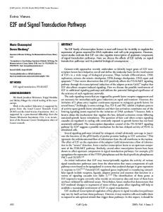
Figure 1 (A) Relationship between mean Ki67-LI and Gleason score. Ki67-LI was calculated for each tumor as described in the text. Each point on the curve represents the mean Ki67-LI for all cases of that Gleason score. (B) Immunoperoxidase staining for Ki67 using a Mib1 monoclonal antibody. Upper panel: negative control (stained in the absence of primary antibody; original magnification r40). Lower panel: poorly differentiated cancer showing strong nuclear staining for Ki67 (original magnification r40). (C) Statistical correlation between Ki67-LI and pAkt staining intensity, as measured by the expression of the phosphorylated form of Akt using a rabbit polyclonal anti-phospho-Akt (Ser-473) antibody (pAkt), that we reported earlier (Malik et al. 2002). For each tumor, pAkt was scored on a scale from 0 to 3, and the Ki67-LI for the same tumor was recorded. Each point on the curve represents the mean Ki67-LI for all cases of similar pAkt scoring. (D) Statistical correlation between Ki67-LI and pERK staining intensity, as measured by the expression of the phosphorylated form of ERK using a rabbit polyclonal anti-phospho-ERK (Thr-202/Tyr-204) antibody (pERK), that we reported earlier (Malik et al. 2002). For each prostate cancer specimen, pERK had been scored on a scale from 0 to 3, and the Ki67-LI for the same specimen was recorded. Each point on the curve represents the mean Ki67-LI for all cases of similar pERK scoring. The numbers in parentheses represent the number of cases evaluated.
data presented as relative luciferase activity using means of untreated controls as standards.
Results Proliferation in human prostate cancer correlates with Akt activation We showed recently that prostate tumors from patients with poor clinical outcome are characterized by increased expression of pAkt (Ser-473) and decreased expression of pERK (Tyr-202/Thr-204) compared with those of good clinical outcome (Kreisberg et al. 2004). Earlier we showed that high-Gleason-grade prostate cancer (‡7), which is considered to have a poor clinical www.endocrinology-journals.org
outcome, expressed higher pAkt and lower pERK compared with low-Gleason-grade prostate cancer (Malik et al. 2002). We have now investigated whether Akt or ERK phosphorylation regulated proliferation in prostate tumors. To determine proliferation, the same prostate cancer tumors analyzed previously (Malik et al. 2002) were stained with a mouse monoclonal antibody to the cell-proliferation antigen, Ki67 (n=74). The rate of proliferation was expressed as the percentage of Ki67expressing cells out of 500 cells counted (Ki67-LI). Normal and hyperplastic tissues did not express Ki67, well and moderately differentiated tumors had low Ki67-LI (£10%) while poorly differentiated tumors had high Ki67-LI (‡20%; Fig. 1A). There was a highly
123
Ghosh et al.: Akt mediates proliferation in prostate cancer
LNCaP 0 5 15 30
C4-2 0 5 15 30
A mins EGF
LNCaP C4-2 B - - + + - - + + EGF - + - + - + - + AG1478
pAkt
pAkt
tAkt
tAkt
pERK pERK tERK
tERK
Figure 2 (A) LNCaP and C4-2 cells were serum starved for 48 h and stimulated with 10 ng/ml EGF for 5, 15 and 30 min. Cell lysates were run on SDS/PAGE (10% gels). Immunoblots were stained with pAkt (Ser-473) and pERK (Thr-202/Tyr-204) antibodies as well as antibodies to the total proteins: Akt1 (tAkt) and ERK2 (tERK). (B) Serum-starved LNCaP and C4-2 cells were pre-incubated with DMSO (control) or 5 mM AG1478, and stimulated without or with 10 ng/ml EGF for 5 min. Expression of the total proteins was used as a loading control.
significant progressive increase in Ki67-LI with increasing Gleason scores (Pearson coefficient, 0.47 for Gleason scores of 2–10; P 0.1; Fig. 1D). The above results clearly demonstrate that proliferation in prostate cancer correlates with Akt phosphorylation.
LNCaP and C4-2 cells as models for good and poor clinical outcome in prostate cancer In order to study the signaling pathways characterizing tumors of good and poor clinical outcome in prostate cancer, we turned to the androgen-dependent LNCaP prostate cancer cell line and its androgen-independent clone, C4-2. Expression and activation of Akt and ERK were compared in LNCaP and C4-2 cells to determine whether they reflected the characteristics of human prostate cancer of good and poor clinical outcome, respectively. Both cell lines expressed active Akt (pAkt) under baseline conditions; with C4-2 displaying higher levels of pAkt (Fig. 2). Growth factor stimulation of both cell lines increased pAkt levels further (Fig. 2A). In contrast to Akt, ERK was not activated in the basal (unstimulated) state; however, growth factor stimulation transiently activated ERK with peak activation at 5 mins post-stimulation (Fig. 2A). The level of ERK activation in C4-2 cells
124
was reduced compared with LNCaP. Growth factor stimulated ERK phosphorylation, but not Akt phosphorylation, was inhibited by the tyrosine kinase inhibitor AG1478, which selectively inhibits EGF receptor kinase activity (Fig. 2B), further demonstrating constitutive Akt activation in these cells. pAkt and pERK levels in these cells mimicked that observed in human prostate cancer; therefore, these cells are appropriate for studying the mechanisms of prostate tumor progression.
Proliferation in LNCaP and C4-2 cells is mediated by the PI3K/Akt pathway Next we compared cell signaling pathways leading to proliferation in LNCaP and C4-2 cells (Fig. 3). Cells were plated in the presence of control (DMSO), 25 mM LY294002, an inhibitor of PI3K activity or 20 mM PD98059, an inhibitor of MAPK/ERK kinase 1 (MEK1) activity, which is directly upstream of ERK1/2. Figure 3 shows that 20 mM PD 98059 did not significantly affect cell growth in either LNCaP or C4-2 cells over a period of 3 days (Fig. 3A and B). In contrast, LY294002 significantly reduced cell growth in both LNCaP and C4-2 cells, indicating that proliferation in these cells proceeds via the PI3K, but not the Ras/MAPK pathway (Fig. 3A and B). Our previous observations indicated that LY294002 caused significant apoptosis in prostate cancer cells (Ghosh et al. 2002); hence, to ensure that the reduction in cell numbers reflected growth arrest, and not simply increased apoptosis, we further analyzed proliferation by flow cytometry following propidium iodide staining of DNA to measure the fraction of cells in the S-phase (Fig. 3C). Flow-cytometric analysis confirmed that proliferation was mediated by the PI3K pathway and www.endocrinology-journals.org
Endocrine-Related Cancer (2005) 12 119–134 C4-2 -
LNCaP
A
0.1400 control 0.1200
20µM PD
0.1200
25 µM LY
A570
A570
control
0.1400
20 µM PD
0.1000
B
0.1600
0.0800 0.0600 0.0400
25µM LY
0.1000 0.0800 0.0600 0.0400
0.0200
0.0200
0.0000
0.0000 0
1
2
3
4
0
1
2
days
1
0.5
C
D pERK tERK
*
(-92.5 %)
1.5
*
0 DMSO
4
LNCaP C4-2
(-81.4 %)
(-13.6 %)
2
(-9.5 %)
normalized S-phase
2.5
3
days
PD98059
pAkt tAkt
- + - + - + - + DMSO
PD
LY
EGF
rapamycin
LY294002
Figure 3 LNCaP and C4-2 cells were plated in the presence of one of the following: DMSO (vehicle control), 25 mM LY294002 (LY), an inhibitor of PI3K activation, or 20 mM PD98059 (PD), an inhibitor of MAPK/ERK kinase 1 (MEK1) activation, which is directly upstream of ERK1/2. (A) LNCaP and (B) C4-2 cells were plated in 24-well plates and cultured for 1–3 days in the presence of the inhibitors listed above. At the end of 1, 2 or 3 days, MTT was added to the cells for 1 h and proliferation rates estimated by colorimetric assay. (C) Cells were plated in 60 mm dishes and cultured in the presence of the inhibitors for 48 h. At the end of that period, cells were trypsinized, fixed in 70% ethanol, stained with propidium iodide and DNA content analyzed by flow cytometry. For each experiment, the percentage of cells in the S-phase was normalized to the percentage of cells in the S-phase in DMSO-treated LNCaP cells. Bars represent meanstS.E.M. from three individual experiments. * Represents changes from control that yield a statistical significance (P