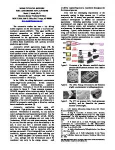
Source: Micro Electronic and Mechanical Systems, Book edited by: Kenichi Takahata, ... Case of Plasmonic Devices for Chemical and Biological Sensing ..... There are four main mechanisms of membrane-enabled separation (Baker, 2004) ...
6 Naenarbmo EnMEMS delba :sro neS CaPl foes oDe mscain Ch rof seciv dna l cime
1Institute
BigniseSlaco Zoran Jakšić1 and Jovan Matovic2
of Chemistry, Technology and Metallurgy Belgrade, 2Vienna University of Technology 1Serbia 2Austria
. 1 Innoitcudr The world is witnessing a rapidly increasing need for various types of sensing devices. The requirements are for smaller, smarter and more versatile sensors. The microelectromechanical (MEMS) devices represent a natural way to proceed along these lines and it is no wonder that the number of various device types, their complexity and the sheer number of various units are increasing at an accelerated pace (Gründler, 2007), (Martinac, 2008), (Merkoçi, 2009), (Toko, 2005). Desirable features of a MEMS sensor include high sensitivity and selectivity, low noise, high robustness, long mean time between failures, small dimensions, characteristics adaptive to the widest possible range of operating conditions, possibilities of massively parallel multisensor operation and low cost. Possibilities of self-testing and even self-repair are also advantageous. Some of these requirements are contradictory and all of them set demanding challenges to the device designers. Many of the desired properties are met in biological organisms which currently set the ultimate target in miniaturization, multiprocess operation and complexity. Thus one of the important today's paths of the MEMS development and further of the nanoelectromechanical systems (NEMS) goes toward biomimetics/bionics (Toko, 2005). Another paradigm gaining momentum these days and also connected with biomimetics are artificial nanomembranes (Vendamme et al, 2006), (Watanabe et al, 2009), (Choi et al, 2007). These may be defined as engineered quasi-two-dimensional freestanding structures (the thickness being much smaller than their width and length and belonging to a range below 100 nm – whence the prefix "nano–"). Their natural counterparts, biological lipid bilayers are the most ancient and most omnipresent natural building blocks since they envelop all living cells which critically rely on them. Artificial nanomembranes are a product of MEMS technologies which are used to produce many of today's self-supported freestanding artificial nanomembranes (Watanabe & Kunitake, 2007), (Ni et al, 2005), (Li et al, 2007) with a thickness sometimes even reaching down to an atomic monolayer (Bunch et al, 2008). Very often the fabrication of nanomembranes includes the deposition of nanocomposite precursor over a sacrificial diaphragm (Mamedov et al, 2002). Self-supported nanomembranes Source: Micro Electronic and Mechanical Systems, Book edited by: Kenichi Takahata, ISBN 978-953-307-027-8, pp. 572, December 2009, INTECH, Croatia, downloaded from SCIYO.COM
86
Micro Electronic and Mechanical Systems
fabricated today often have giant aspect ratios, readily exceeding the value of 1,000,000 – e.g. (Matovic & Jakšić, 2009). Such dimensions make them a hybrid between micro and nanosystems, even between macroscopic systems and nanosystems, since their lateral dimensions may be of the order of centimeters, while the thickness remains nanometric. Nowadays they are seen as a building block for various MEMS systems (Vendamme et al, 2006) , (Jiang et al, 2004a). Since a biological complexity is sooner or later expected to be reached by micro and nanosensors and at the same time nanomembranes are the basic natural building block, it is only obvious to merge these two concepts into a single one. In this chapter we show how such a simple fusion of two paradigms may result (and actually is already resulting) in a large multitude and variety of results. This is happening in spite of the fact that the field of nanomembrane-enabled sensors itself is extremely young, the first papers starting to appear several years ago (Jiang et al, 2004a). Here we consider only the application of synthetic/engineered nanomembranes and exclude biological structures. After a concise overview of some of promising uses of nanomembranes in microsensors generally, we concentrate to a single sensor type, that of chemical, biochemical or biological (CBB) sensors utilizing the effects of adsorption/desorption and the surface plasmon resonance (SPR) effect. We consider the possibility to use nanomembranes as a platform for long range surface plasmons. The role of self-supported ultrathin structures in improving coupling between propagating modes and surface-bound plasmons is also analyzed, as well as their application in SPR sensor selectivity boost.
2.MEMSsenarbmohgutc This Section shortly considers the use of nanomembranes in the enhancement of MEMS sensors generally. This includes various inertial, thermal and photonic devices (Gardner et al, 2001). Being ultrathin and ultra-lightweight and at the same time robust, nanomembranes appear essential for the miniaturization of sensors when scaling down from the microscale to the nanoscale. In many MEMS sensors the basic method of signal readout is to use deflection of a freestanding elastic structural part. This part is typically a microcantilever, a microbridge or a miniscule diaphragm. For instance, in a piezoresistive pressure sensor the deflecting element is a micrometer-thick diaphragm with a built-in Wheatstone bridge. Applied pressure causes the diaphragm deviation from the equilibrium and thus changes the resistance of piezoresistors. Similar situation is encountered in various inertial MEMS sensors like accelerometers and inclinometers, where the membrane or bridge deflection is caused by the movement of an inertial mass. Another elastic part whose deflection is measured in applications is the microcantilever, well known as one of the basic building blocks in MEMS and NEMS. For instance, in scanning probe microscopy, which includes Atomic Force Microscopy and represents one of the basic techniques for characterization in nanotechnologies, it is the principal element, and the readout is often based on the optical lever principle. In most of the mentioned situations either the elastic structural part is made relatively thick (the order of micrometers, which is the conventional approach) and with large lateral dimensions (millimeters) or it is made thinner and with smaller lateral dimensions. If one desires to fabricate a sensor array with a large number of elements which is at the same time as compact and as sensitive as possible, the latter appears obviously the better approach.
Nanomembrane-Enabled MEMS Sensors: Case of Plasmonic Devices for Chemical and Biological Sensing
87
The ultimate in thickness of these building blocks is posed by the mechanical properties of the material itself, and the nanomembranes whose thickness can be of the order of several atomic or molecular monolayers certainly approach that limit. Literature quotes the use of nanomembranes as the ultrathin freestanding structure to replace the conventional building blocks in deflection-based sensors (Jiang et al, 2004a). Among the obvious advantages of applying such a strategy are an increased sensitivity and a wider dynamic range. Various forms of micromachined freestanding ultrathin structures ensure much higher resonant frequencies than the conventional ones (extending into the GHz range).
O Au
Si
Cr Fig. 1. Structures of composite nanomembranes convenient for mechanical and thermal sensors. Left: polymer matrix with gold nanoparticle filler. Right: metal-composite nanomembrane. The nanomembranes for inertial sensors feature nanocomposites which may be e.g. polymer matrix filled with nanoparticles (Jiang et al, 2004b), metal-composite structures (Matovic & Jakšić, 2009), etc. Typically such structures are in a pre-stretched state. Their micromechanical properties can be readily adjusted by tuning the composition of the nanocomposite membranes. For instance, the amount of metal in a polymer-metal matrix will increase the elastic modulus of the nanocomposite. The measured elastic moduli for structures with gold nanoparticles were up to 10 GPa (Jiang et al, 2004a). The same structures can be obviously cut and formed in various ways and used in different shapes and with different anchorings as ultrathin microcantilevers and microbridges (Hua et al, 2004), (Zheng et al, 2002). Some unique properties were observed in nanomembranes for MEMS sensors. Probably the most counter-intuitive one is their autorecovery feature, actually a mechanism of selfhealing of overstretched structures (Jiang et al, 2004a). In our own experiments the metalcomposite nanomembrane driven to the range of viscoelastic deformations did not remain distorted, but returned in a matter of tens of minutes to their original unstretched state. This property ensures a safety mechanism against accidental overstretching of nanometer-thin membranes and in final instance ensures a better stability of mechanical properties of inertial and pressure sensors based on nanomembranes, as well as a longer lifetime of such products. The mechanical sensors based on nanomembranes also include acoustic imagers (Ballantine et al, 1997), (Kash, 1991). Acoustic sensitivities were reported at least an order of magnitude below the threshold of human hearing (Jiang et al, 2004a). Another approach to sensing of mechanical movements is to utilize nanomembrane-based freestanding waveguides for evanescent field sensing. This was proposed for optical
88
Micro Electronic and Mechanical Systems
measurement of deflection in micromirrors, gyroscopes, etc. and structures in the thickness range 30 nm to 100 nm were fabricated in Si3N4 (Altena, 2006). Another large field of application of freestanding nanomembranes are thermal sensors (Kruse & Skatrud, 1997). The need for large area thermal arrays of miniature detectors in infrared technology and remote sensing is large (Rogalski, 2003), (Dereniak & Boreman 1996). Various thermal detectors include bolometers (Richards, 1994), pneumatic detectors/Golay cells (Golay, 1947), (Chévrier et al, 1995), microcantilever-based devices (Datskos et al, 2004) to which bimaterial detectors belong (Djuric et al, 2007), etc. Thermal detectors are typically based on a large and thin absorbing area which reacts to thermal changes due to its irradiation by electromagnetic radiation and is sensitive in the whole electromagnetic spectrum. Nanomembranes obviously offer smaller thermal inertia and thus promise faster operation and higher specific detectivities. The assessments of polymer nanomembranes with gold nanoparticle fillers in thermal detectors show sensitivities several orders of magnitude higher than those for silicon membranes with the same diameter. For instance, temperature sensitivities below 1 mK were calculated for 55 nm thick, 200 μm diameter nanomembranes (Jiang et al, 2004a). Nanomembranes freely suspended over microfabricated cavities dedicated to infrared thermal detectors were reported in (Jiang et al, 2006). A large field of application of nanomembranes in (nano)photonics is their use in enhancement of the operation of semiconductor infrared detectors (Rogalski, 2003). These detectors are actually quantum devices whose operation is based on generation of charge carriers in semiconducting material upon illumination in a given wavelength range. Their sensitivity spectrum is much narrower than that of thermal detectors and its cutoff frequency is determined by the bandgap of the given semiconductor. One of the fields of the application of nanomembranes in such detectors is the fabrication of resonant cavity structures, which may be implemented as multilayer dielectric mirrors or one-dimensional photonic crystals (Jakšić & Djurić, 2004), (Djurić et al, 1999), (Djurić et al, 2001). In addition to their application as the building blocks for the resonator reflectors, such freestanding structures may be applied in devices with tunable resonant frequency, where electrostatic field is used to deflect the membrane and adjust position to furnish the desired resonant peak (Ünlü & Strite, 1995). Another field of application are both tunable and fixed filters for photodetectors in various wavelength ranges obtained by lamination of planar structures (Jakšić et al, 2005), (Maksimović & Jakšić, 2006). There is also a possibility to modify and tune the emissivity and absorptance by the application of such multilayers (Maksimović & Jakšić, 2005), up to the point of creating thermal antennas for visible and infrared radiation (Maksimović et al, 2008). Finally, a large field of application of nanomembranes is in chemical, biochemical and biological sensors based on plasmon resonance. The rest of this Chapter is dedicated to this important topic.
3.Plw, nihtar lu s o e c m smlif gn d at e r 3.1CBBtsmeygni A chemical, biochemical or biological (CBB) sensor may be described as a device which generates an instrument- or observer-readable output proportional to the amount of a targeted chemical, biochemical or biological analyte in a given gaseous or liquid
Nanomembrane-Enabled MEMS Sensors: Case of Plasmonic Devices for Chemical and Biological Sensing
89
environment. The output is most often electrical or optical. The most important issues regarding a CBB sensor are its sensitivity and selectivity towards a given analyte. A general CBB sensing system (Fig. 2) consists of three main blocks, (1) the unit for separation/filtering and possibly reaction enhancement, (2) the detection unit – the main part of the sensor where the signal is generated and (3) the processing unit where signals are conditioned and communicated further. Interrogating beam
Environment
Filter Separator Enhancer
Sensing surface
Readout
Signal conversion & conditioning
Readout beam Separation & enhancement unit
Detection unit
Processing unit
Fig. 2. Layout of a general CBB sensor consisting of (1) separation, filtering and enhancement unit; (2) detection unit and (3) signal conversion and conditioning unit. We analyze the use of nanomembranes in the first two blocks. In the separation unit they are useful for filtering and generally molecular recognition if functionalized by nanopores, ion exchangers, absorbing fillers, etc., since their thickness enables a more accurate control of functionalization parameters than in larger structures. In the detection unit, especially of the kind used in nanoplasmonic devices, the nanomembranes are applicable as ultrathin, fully symmetric plasmon waveguides, strongly improving the device sensitivity. 3.2opmesnlcarufS Surface plasmons polaritons (SPP) are TM-polarized surface waves propagating along a metal–dielectric interface at optical frequencies (Fig. 3). Their wavelengths are extremely short and may even enter the X-ray range (Maier, 2007). The SPPs are evanescent perpendicularly to the active surface both toward the environment and toward the metal layer. In the situation when substrate and superstrate differ, the dispersion relation will allow two different modes of propagation, one on each interface (Maier, 2007). Sensors based on the propagation of surface plasmon polaritons have become one of the most important tools in chemical, biochemical and biological sensing (Barnes et al, 2003), (Maier, 2007), (Homola, 2006), (Jung et al, 1998). They offer label-free, highly sensitive single-step measurements, real-time monitoring, require extremely small analyte samples (atomic/molecular monolayers suffice) and ensure a single generic framework for different
90
Micro Electronic and Mechanical Systems
analytes. Multichannel devices are readily implemented in such configurations. No moving parts are required and the fabrication technology is simple – the conventional SPP resonance-based sensor is a planar metal surface with the plasma frequency in the wavelength range of interest. Good metals are used to this purpose, typically gold or silver. Being fully optical, these sensors are resistant to external electromagnetic disturbances. Finally, plasmon sensors are very convenient for miniaturization and the fabrication of ultracompact sensor arrays.
z Ez
SPP at the ambientmetal interface ksp
Ex
ε2 >1
Hy
ε1 < –1 (metal or metamaterial)
SPP at the interface with the substrate
d
x
ε3 >1
Fig. 3. Basic configuration of a guide for surface plasmon polariton propagation (metaldielectric interface) It is possible to use the same structure simultaneously for guiding SPP waves and for guiding the controlling electrical signals, since the active area is made of metal (Boltasseva et al, 2005). Also, the SPP components generally have high field localizations, thus promote the use of nonlinear photonic materials, ensuring the possibility for integration of active alloptical components (Zayats & Smolyaninov, 2003). The operation of the SPP sensors is based on the modification of the propagation of surface plasmons polaritons at the sensor (metal)-environment (dielectric) interface. The analyte from the environment is bound either directly to the plasmonic surface, or (much more often) to a target-specific ligand layer. In both cases the surface refractive index is modified exactly in the position where the maximum of the SPP wave is located, since SPP waves are confined to the metal-dielectric interface and evanescent in perpendicular direction. In this way the maximum response is ensured. SPP resonance sensing is essentially thin film refractometry, where a change in the analyte concentration from c to c + Δc causes a refractive index change at the metal-environment surface n to n + Δn due to perturbed propagation conditions for the surface waves. The obvious idea here is to use a nanomembrane as a waveguide for plasmons. Since a surface plasmon polariton is a quasi-planar electromagnetic wave decaying evanescently in both perpendicular directions, it is logical to utilize as a support for it a metal or metalcomposite nanomembrane which is also quasi-planar. Plasmons in nanomembranes with metal fillers were reported in (Jiang et al, 2004b), where gold nanoparticles were used in a polymer matrix and the packing density of the gold spheres varied from below 2% to about 25%. Experimental structures are typically light blue due to a plasmon resonance peak corresponding to the plasma frequency in visible.
Nanomembrane-Enabled MEMS Sensors: Case of Plasmonic Devices for Chemical and Biological Sensing
91
3.3LoeSP rang Pomebransigutlz One of the problems in structures with conventional SPP is large signal attenuation, a consequence of a high imaginary part of the propagation constant due to the ohmic losses/absorption in metals (Zayats & Smolyaninov, 2003). In such waveguides the propagation length are typically limited to a range from tens (visible range) to hundreds of micrometers (near infrared). Another problem is their coupling with propagating modes, since typically elaborate schemes using e.g. prism couplers or diffractive gratings must be used. The way to overcome most of these shortcomings is to use long-range (LR) surface plasmon polaritons (Sarid, 1981), (Burke et al, 1986), (Charbonneau et al, 2000), (Berini, 2000). These are SPPs which propagate along metal strips with nanometric thickness (typically 10-40 nm) immersed into dielectric. Symmetric configuration: SPP are coupled
Long range SPP
ε2 >1 ε1 < –1 ε2 >1
Fig. 4. Generation of long-range surface plasmons polaritons through coupling of top and bottom modes on ultrathin metal sheets; top: metal guide is surrounded from both sides with identical dielectric; bottom: metal sheet is smaller than the decay length and LR plasmon appears In the case when the substrate and the superstrate are described by identical permittivity (the case of full immersion of the metal sheet in homogeneous dielectric), the structure is symmetrical in electromagnetic sense. The two propagating modes on the top and bottom surface then couple and propagate together, Fig. 4 top. If the metal sheet between the two identical media is sufficiently thin to make the interaction between the top and the bottom SPR non-negligible, these two modes couple and merge into a single one, Fig. 4 bottom. The degeneracy for that mode is then removed and its dispersion splits into two branches, one for low-frequency mode (odd), and the other for high-frequency mode (even). The even modes have a very short propagation path. The propagation constant of the odd modes decreases, being proportional to the square of the film thickness. This means that the attenuation of the odd mode will be very low and thus its propagation length large. Thinner films and more symmetrical structures will have longer propagation paths. A typical trait of an LR SPP is that its fields are mostly contained outside of the metal part. Since the field concentration is much lower in the metal sheet, the propagation losses are consequently also much lower. The imaginary part of their propagation constant being approximately zero, the LR SPP ensure much larger propagation paths, typical propagation losses being below 6 dB/cm (Boltasseva et al, 2005).
92
Micro Electronic and Mechanical Systems
Long-range surface plasmon sensors are especially convenient for biological sensors, since the confinement of the plasmon waves is smaller than in other SPP devices and thus the larger biological samples are more easily encompassed (Berini et al, 2008) Probably the most important cause of the signal attenuation in LR SPP structures is its deviation from symmetry (Park & Song, 2006). Fig. 5 shows a calculated curve of attenuation for a metal nanomembrane immersed in dielectric. 7
Attenuation, dB/cm
6
5
4
-6
-4
-2
0
2
4
6
Δn/10-3
Fig. 5. Calculated LR-SPP propagation loss versus asymmetry of dielectric given as the refractive index difference. Membrane thickness 12.5 nm, material gold, refractive index of dielectric immersion 1.5, wavelength 1.55 μm. It is visible that even very small deviations from symmetry introduce large losses into the waveguide. The use of metal or metal-composite nanomembranes at the same time gives a platform for LR SPP and ensures its complete symmetry. Their thickness is typically from 4 nm up, thus very low losses are ensured. A layout of a nanomembrane-based LR SPP guide is shown in Fig. 6. The structure itself is extremely simple, being a freestanding planar nanomembrane sheet.
z Surrounding medium, ε1=ε1'+i·0
y
x
1 (top)
Nanomembrane, ε2=ε2'+i·ε2"
2 (bottom)
Fig. 6. Basic configuration of a freestanding nanomembrane guide for long-range surface plasmon polariton propagation (metal-dielectric interface)
Nanomembrane-Enabled MEMS Sensors: Case of Plasmonic Devices for Chemical and Biological Sensing
93
The issue of coupling between the propagating modes and the plasmon waveguide is dealt with further in this text. 3.4Deeosntiuardclmv One of the problems with probably all types of sensors are their ultimate limits of detection, which are connected with various extrinsic and intrinsic mechanisms of noise. Of these, the latter ones include mechanisms that are fundamental to the sensing process itself. In the case of plasmonic sensors, such fundamental mechanisms include adsorption-desorption noise which is connected with the operation of the SPR devices themselves, thermal (JohnsonNyquist) noise, 1/f noise and zero-point noise (noise due to quantum fluctuations) (Jakšić et al, 2007), (Jakšić et al, 2009a). It is interesting to note that at least some of these noise sources are expected to affect the operation of nanomembrane-based SPR sensors less than that of other types of plasmonic sensors. For instance, it is expected that the adsorption-desorption noise will be smaller in nanomembrane-based long-range plasmon sensors than in other types, since this noise will decrease with increasing the active detection area (Jakšić, O. et al, 2009). Zero-point noise should also decrease in the case of LR SPR devices. One of the ways to shift the ultimate detection limits and at the same time to ensure new degrees of freedom in sensor design is to utilize novel structures, optimized for higher sensitivities and lower noise. A possible pathway is to pattern or shape the nanomembrane surfaces, for instance by focused ion beam patterning (Gierak et al, 2007). A large opportunity window opened by the advent of nanoplasmonics (Maier, 2007), and especially with the introduction of electromagnetic metamaterials (Pendry et al, 1999). Such structures may be defined as artificial structures with electromagnetic response not readily found in nature. A typical and well-known type metamaterials are the structures with negative value of refractive index (Veselago, 1968), also known as left-handed structures (Ramakrishna & Grzegorczyk, 2009), thus named because the triplet electric field vector, magnetic field vector and wavevector form a left-handed set, contrasted to the "normal" materials where this set is always right-handed. Patterned and laminar metal-dielectric nanomembranes are a useful building block for quasi-2D metamaterial structures, the metasurfaces, intended for the operation in the optical range. Actually the metal nanomembranes themselves may be regarded as left-handed metamaterials in certain situations, since some electromagnetic modes propagating on them show the properties of negative effective refractive index (Smolyaninov, 2008). Generalized plasmonic sensors based on left-handed metamaterials were described in various references (Ishimaru et al, 2005), (Jakšić et al, 2007), (Bingham et al, 2008).
. 4 Frmaebgnwitorlpuchs An important issue in plasmonic sensors, regardless of the active surface type, is their coupling with light sources and the readout systems, i.e. the matching of propagating planar waves of optical radiation with evanescent SPP waves. The wavevector of SPP is always larger than the wavevector in free space and at optical frequencies the wavelengths of the SPP may become very small, even reaching nanometric lengths (Maier, 2007), (Raether, 1988), (Barnes et al, 2003). Thus it is necessary to impart the missing momentum to the interrogating beam (propagating planar wave) in order to enable coupling – i.e., to ensure phase matching between the two waves.
94
Micro Electronic and Mechanical Systems
In coupling it is important to ensure that the maximum percentage of the incoming freespace mode is converted to SPP (and vice versa for the output). At the same time, it is important to ensure the smallest leakage and scattering losses. There are various schemes to ensure coupling between plasmonic devices and propagating modes. They may be roughly divided into four groups: prism couplers, endfire couplers, near-field probe couplers and those utilizing topological surface defects. Historically the oldest methods are those utilizing prism couplers (Fig. 7). These include the Kretschmann configuration (Kretschmann & Raether, 1968) (Fig. 7a) which is still the prevailing readout method in plasmon sensors, as well as the Otto coupler (Otto, 1968) (Fig. 7b). Both of these methods utilize attenuated total reflection. Another method to excite the SPP is to use end-fire coupling, where the incident beam is in plane with the plasmonic surface (Fig. 7c) (Stegeman et al, 1983), (Berini et al, 2007).
b)
a)
e) f)
d)
g) c)
Fig. 7. Couplers plasmon-propagating a) Prism couplers in Kretschmann configuration; b) Prism couplers in Otto configuration; c) end-fire coupling; d) near-field probe excitation; Various methods of coupling through topological surface defects which may consist of e) gratings consisting of nanohole arrays, f) surface protrusions or may be g) disordered surface corrugations. An important group of couplers utilize various near-field probes (the use of the "forbidden light' outside the light cone), (Fig. 7d) where local excitation in evanescent field is utilized and the beams tunnel from the impingement point to the metal-dielectric interface which supports SPP (Hecht et al, 1996), (Bouhelier & Novotny, 2007), (Maier et al, 2004). Finally a large and very important group are couplers utilizing topological surface defects (Barnes et al, 2003), (Ritchie et al, 1968). These include grating couplers which may consist of periodic arrays of either subwavelength apertures (Fig. 7e) (Devaux et al, 2003) or surface protrusions (e.g. various pillars, bumps, etc.) (Worthing & Barnes, 2001), Fig. 7f. The arrays may be 2D like those shown in Fig. 7e, f) or 1D (grooves or stripes) and may have various shapes, e.g. rectangular, triangular, wavy, etc.
Nanomembrane-Enabled MEMS Sensors: Case of Plasmonic Devices for Chemical and Biological Sensing
95
The couplers may be also disordered (this layout may be understood as a superposition of a large number of gratings with different periods) – Fig. 7g. (Ditlbacher et al, 2002) In the case of freestanding nanomembranes and LR SPP sensors it is important to couple these structures with propagating modes with the least disturbance to the symmetry, thus preferably without a direct physical contact with the nanomembrane. One could use the shaping of a dielectric substrate (which, however, would perturb the electromagnetic symmetry of the structure), endfire coupling (which introduces alignment and coupling efficiency issues; it is known that the percentage of coupled light in this method is extremely low) or Otto prisms (bulky structure which makes the device significantly more complex). We proposed an alternative approach which uses direct sculpting of the nanomembrane and is applicable without special alignment procedures (Jakšić et al, 2009). The idea of our approach is to incorporate the coupling structures into the freestanding nanomembrane itself, without any substrate to hold them. In this way the substrate and the superstrate remain fully index matched throughout the measurement. At the same time, the structure remains generally applicable, since the analyte does not have to be matched to the prefabricated device substrate. The sculpted structures are small perturbations of the much larger nanomembrane, their dimensions being of the order of micrometers, while the membrane dimensions are measured in millimeters, even centimeters. The surface is sculpted into a 2D array of protrusions (Fig. 8 a) which serve as a coupling diffractive grating (Kashyap, 1999). The basic approach to nanomembrane sculpting is illustrated in Fig. 8 b, c.
a)
Incoming propagating wave
b)
Outgoing propagating wave
SPP Sculpted surface
c)
Freestanding nanomembrane
Fig. 8. a) Propagating wave to surface plasmon couplers using surface sculpting. b) Drawing of hemispherical surface relief for nanomembrane sculpting fabricated by isotropic etching through circular openings in photolithographic mask; c) Drawings of pyramidal surface relief for nanomembrane sculpting fabricated by anisotropic etching of silicon with (100) surface orientation through square windows aligned along [110].
96
Micro Electronic and Mechanical Systems
The fabrication of surface-sculpted protrusion arrays is based on the deposition of a nanometric membrane precursor over a sacrificial layer (Mamedov et al, 2002), (Jiang et al, 2004b) and its subsequent release in etching solution, the same method used to produce metal-composite nanomembranes. The difference is that prior to depositing the membrane precursor, one first etches an array of micrometer pits in the sacrificial structure. The layout of the pits is determined by the applied photolithographic mask, thus defining the diffractive grating for the coupling. The shape of the pits themselves is defined by the chosen etching method. If isotropic etching is used, the pits are hemispherical or ellipsoidal. If anisotropic etching is utilized, one can produce for instance pyramids or truncated pyramids. The membrane precursor is subsequently deposited over the pits and upon release the fabricated nanomembrane retains the shape of the pits. If the sacrificial layer pits are etched by isotropic etchant, the final protrusion are hemispherical, Fig. 8b. Variations to this generic form can be obtained by using different shapes of photolithographic masks (for instance, elliptical openings, but also various polygons) and by adjusting the etching duration to obtain either flatter or more voluminous structures. The structure in Fig. 8c is obtained in an analogous manner, but using anisotropic etching of single crystalline silicon sacrificial layer with (100) surface orientation through square windows aligned along [110]. The variations to this generic form includes truncated pyramids and actually all standard forms obtainable by anisotropic etching. Figure 9 shows the fabricated 15 μmx10 μm pyramid sculpted on the surface of a metalcomposite nanomembrane with a thickness of 20 nm. It is known that nanomembranes become intrinsically stretched during their low-temperature annealing (Matovic & Jakšić, 2009) and thus the sculpted surface features retain their shape in spite of the minute thickness of their walls.
Fig. 9. Scanning electron microscope photo of a 15 μmx10 μm pyramid sculpted on the surface of a metal-composite nanomembrane, thickness 20 nm We believe the tailorability makes the 3D surface-sculpted nanomembranes a valid alternative to other coupling methods for freestanding LR SPP sensor structures.
Nanomembrane-Enabled MEMS Sensors: Case of Plasmonic Devices for Chemical and Biological Sensing
97
. 5 s en a r b m o h g u t c i v l S y The issue of selectivity is of the utmost importance for the CBB sensors, since among the myriad of possible analytes, especially the organic and biological ones, many are found with almost identical properties. The basic behavior of a plasmonic CBB sensor is determined by the adsorption/desorption properties of a given analyte toward the sensor active surface. The conventional wisdom in traditional CBB sensors is to deposit a ligand layer onto the sensor surface to adsorb the targeted analyte only (e.g. a particular DNA sequence, a desired enzyme, etc.) This is a well developed approach and is obviously also applicable for the nanomembrane-based (nano)plasmonic CBB sensors. In this Section we turn attention to the fact that the use of nanomembranes opens additional possibilities to improve the selectivity of CBB sensors. At the same time these additional methods practically do not change the dimensions of the sensors. Probably the most obvious approach is to use nanomembranes as filtering/separation units. It is well known that molecular separation is among the basic applications of conventional membranes (Yampolskii et al, 2006), (Böddeker, 2007), (Hoffman, 2004), (Sata, 2004), and they form the basis of the large separation science. It is the common knowledge that thinner membranes typically mean higher throughput and thus shorter reaction times. This property is of great importance for the CBB sensors, as very often they are required to operate in real time. Isotropic conventional membranes are usually at least tens of micrometers thick. Anisotropic membranes, including those with multistage hierarchical structure (Fendler, 1994) have much thinner active layers, but require thick porous substrates as supports. Only with synthetic nanomembranes the possibility appeared to have structures with a thickness in the nanometer range and at the same time robust enough not to require any additional supports. Thus for the first time one simultaneously obtains nanometric thickness, mechanical strength and a high throughput. At the same time they offer larger possibilities for nano-customization since one can tailor their properties with larger precision: it is always easier to keep for instance nm-dimensioned pore aspect ratio through a cross-section with nanometric thickness, than to keep it in a structure 1,000 or 10,000 times thicker. In this Section we consider the basic ways to improve selectivity of nanomembrane-based plasmonic CBB sensors using structures similar to the ones which comprise the sensors themselves. There are four main mechanisms of membrane-enabled separation (Baker, 2004) which can be implemented by nanomembranes utilizing some of the available methods for their functionalization (Fig. 10): 1. Pore-flow molecular sieving, where separation is basically done by passage of molecules through a system of randomly distributed pores which are at the same time interconnected. Thus such a structure operates in a manner similar to a conventional particle filter: molecules larger than the pore size will be rejected, while smaller ones will pass through the nanomembrane. Here thinner membranes will obviously offer a possibility for a better control of pore size and shape and at the same time enable a higher throughput. Such membranes may have an isotropic or anisotropic pore distribution across the cross-section. Fig. 10a shows a molecular sieve with anisotropic distribution of pores, the Loeb-Sourirajan structure.
98
Micro Electronic and Mechanical Systems
a)
b)
c)
d)
Fig. 10. Schematic presentation of selectivity enhancement through nanomembrane-enabled separation. a) Cross-section of nanomembrane with nanopores (Loeb-Sourirajan anisotropic structure); b) nonporous dense nanomembrane utilizing solution-diffusion mechanism; c) electrically charged membrane (ion exchanger); d) nanomembrane with gated ion channels. 2.
Solution-diffusion through dense membranes where pores either do not exist or are at least smaller than the particle effective cross-section as defined by their thermal motion at a given temperature. The transport through such nanomembranes is a combination of particle solution and their diffusion across the structure, driven by concentration gradient, electrostatic field or pressure (Fig. 10b). These membranes can also be isotropic or anisotropic. They are used e.g. in pervaporation, reverse osmosis and often are regarded the method of choice in gas separation. 3. Ion exchange, where nanomembranes contain electrically charged particles (Fig. 10c). Most often such membranes are nanoporous, although they also may be dense. Positive or negative ions are fixed throughout the nanomembrane, usually at the pore walls. They bind the opposite ions and exclude the same charge. Structures with positive ions are denoted as anion exchangers, and those with negative ones are cation exchangers. This is the mechanism encountered in electrodialysis. 4. Gated ion channel flow, where the nanomembrane includes an ion-transmitting channel, typically consisting of proteins (Fermini & Priest, 2008) but which generally may be fabricated in various organic and inorganic materials. The channel is gated by external stimuli, typically by applied voltage or by ligands, which control the ion transport through the channel, Fig. 10d. This transport is highly selective. For instance, aquaporin proteins allow the flow of water molecules but are impermeable to protons, although these are much smaller than the water molecules. In other nanochannels transport of protons occurs, while the nanomembrane remains impermeable to other molecules (the Grotthuss mechanism, or proton hopping, where protons “hop” along a one-dimensional chain of water molecules – the “water wire“) (Chung, 2007). Various ion channels exist for sodium, potassium ions, hydrogen, etc. This mechanism is fundamental for the ion transport through lipid bilayer nanomembranes in biological cells and represents the basis of the life on earth, while its biomimetic counterparts ensure another route to biosensor selectivity enhancement (Martin, 2007). One can see that artificial pores are important for most of the described approaches. Nanopores are used to characterize DNA and RNA (Kasianowicz et al, 1996), up to the point of discriminating molecules differing by a single nucleotide (Vercoutere, 2003).
Nanomembrane-Enabled MEMS Sensors: Case of Plasmonic Devices for Chemical and Biological Sensing
99
It is necessary to discern between different nomenclatures regarding the pore size, which may be the source of some confusion. According to the IUPAC, the term macropores is used to denote pores larger than 50 nm, mesopores have diameters between 2 nm and 50 nm and micropores are smaller than 2 nm (Rouquerol et al, 1993). In many literature sources all pores with diameters below 100 nm are termed nanopores (Aksimentiev et al, 2009). There are several approaches to producing biomimetic pore complex capable of selectively transporting various biological analytes. Recently, an artificial scaffold for the nuclear pore complex-based gate was produced using natively disordered proteins termed FP nups. Such synthetic scaffold can be used as a generic platform for ultra-selective biomimetic nanopores (Jovanovic-Talisman et al, 2009). Another pathway is to avoid proteinaceous nanopores and to utilize other materials, including inorganic ones. Some materials used include silicon, silicon nitride, polyethylene terephthalate, silicon dioxide and many others (Aksimentiev et al, 2009). The design freedom offered by these approaches may lead to nanomembrane separators operating under less restrictive ranges of external parameters (temperature, electrolyte concentrations, pressures, pH values, bias, etc.). It is possible to tailor nanopores to practically any desired size, which means the freedom to optimize the pore geometry for various targeted analytes. An important question is the method of combining the different parts of a CBB sensing device into a unified system. The unit for separation/enhancement may be integrated with the detection unit in various ways (Fig. 11). One may use two (or more) nanomembranes as separate elements so that the analyte-containing fluid is flowing sequentially through them, as shown in Fig. 11a. A modification of this approach is to utilize lamination of two (or more) separate nanomembranes into a single one, Fig. 11b. Finally, it is possible to aggregate detection and separation/enhancement functions into a single monolithic structure with one or more different active fillers which will perform affinity capture of the analyte and at the same time ensure tuning of the readout beam, Fig. 11c. Separation/enhancement Separation/enhancement
Separation/enhancement Detection
Detection
b)
Separation/enhancement + Detection c)
a)
Fig. 11. Basic configurations of nanomembrane units for separation, enhancement and detection. a) The "separate separator" configuration where filtering structures are physically separated from the plasmonic waveguide; b) laminated structure, where separator/enhancer captures analyte; examples include e.g. solution/diffusion membranes, but also conventional ligand layers; c) aggregated/monolithic multipurpose unit.
. 6 Conoisulc The combination of the two paradigms, that of MEMS CBB sensors and that of nanomembranes, not only has potentials to vastly improve the performance of today’s devices but also to introduce numerous completely new functionalities. A very convenient platform to this purpose are LR SPR waveguiding devices. They are useful for larger
102
Micro Electronic and Mechanical Systems
Devaux, E.; Ebbesen, T. W.; Weeber, J.-C.& Dereux, A. (2003). Launching and decoupling surface plasmons viamicro-gratings. Applied Physics Letters, Vol. 83, No. 24, 4936– 4938, ISSN: 0003-6951. Ditlbacher, H.; Krenn, J. R.; Félidj, N.; Lamprecht, B.; Schider, G.; Salerno, M.; Leitner, A. & Aussenegg, F. R. (2002). Fluorescence imaging of surface plasmon fields. Applied Physics Letters, Vol. 80, No. 3, 404–406, ISSN: 0003-6951. Djurić, Z.; Jakšić, Z.; Randjelović, D.l Danković, T.; Ehrfeld, W. & Schmidt, A. (1999). Enhancement of Radiative Lifetime in Semiconductors Using Photonic Crystals, Infrared Physics & Technology, Vol. 40, No. 1, 25-32, ISSN: 1350-4495. Djurić, Z.; Jakšić, Z.; Ehrfeld, W.; Schmidt, A.; Matić, M. & Popović, M. (2001). Photonic Crystal Enhancement of Auger-Suppressed Detectors: A Way to BackgroundLimited Room-Temperature Operation in 3-14 Micrometer Range, in Nanoscale Linear and Nonlinear Optics, Eds. M. Bertolotti, C. M. Bowden, C. Sibilia, AIP Proceedings, Melville, New York, Vol. 560, 418-424, ISBN 1-56396-993-9 Djurić, Z., Randjelović, D., Jokić, I., Matović, J. & Lamovec, J. (2007). A new approach to IR bimaterial detectors theory, Infrared Physics & Technology Vol. 50, No. 1, 51-57, ISSN: 1350-4495 Fendler, J.H. (1994). Membrane-Mimetic Approach to Advanced Materials, Springer-Verlag Berlin Heidelberg, ISBN: 0387572376 Fermini, B. & Priest, B. T. eds. (2008). Ion Channels, Springer-Verlag Berlin Heidelberg, ISBN 978-3-540-79728-9 Gardner, J. W.; Varadan, V. K. & Awadelkarim, O. O. (2001). Microsensors, MEMS, and Smart Devices, John Wiley & Sons, ISBN-13: 978-047186. Gierak, J.; Madouri, A.; Biance, A.L.; Bourhis, E.; Patriarche, G.; Lucot, C. U. D.; Lafosse, X.; Auvray, L.; Bruchhaus, L. & Jede, R. (2007). Sub-5 nm FIB direct patterning of nanodevices, Microelectronic Engineering Vol. 84, 779–783 Golay, M.J.E. (1947) Theoretical consideration in heat and infra-red detection, with particular reference to the pneumatic detector. Review of Scientific Instruments Vol. 18, 347–356, ISSN: 0034-6748 Gründler, P. (2007). Chemical Sensors: An Introduction for Scientists and Engineers, SpringerVerlag Berlin Heidelberg, ISBN 978-3-540-45742-8 Hecht, B.; Bielefeld, H.; Novotny, L.; Inouye, Y. & Pohl, D. W.: (1996). Local excitation, scattering, and interference of surface plasmons. Physical Review Letters, Vol. 77, No. 9, 1889–1892, iSSN: 0031-9007. Hoffman, E. J. (2004). Membrane Separations Technology: Single-Stage, Multistage, and Differential Permeation, Elsevier, Amsterdam, ISBN: 0750677104 Homola, J. Ed. (2006). Surface Plasmon Resonance Based Sensors, Springer, ISBN: 978-3-54033918-2, Berlin-Heidelberg, ISBN: 978-3-540-33918-2. Hua, T., Cui, T. & Lvov, Yu. M. (2004). Ultrathin cantilevers based on polymer-ceramic nanocomposite assembled through LbL adsorption, Nano Letters Vol. 4, 823–825, iSSN: 1530-6984. Ishimaru, A.; Jaruwatanadilok, S. & Kuga, Y. (2005). Generalized surface plasmon resonance sensors using metamaterials and negative index materials, Progress in Electromagnetic Research – PIER Vol. 51, 139–152, ISSN: 1559-8985
Nanomembrane-Enabled MEMS Sensors: Case of Plasmonic Devices for Chemical and Biological Sensing
103
Jakšić, O.; Jakšić, Z. & Matović, J. (2009). Adsorption-desorption noise in plasmonic chemical/biological sensors in multiple analyte environment", Proceedings of SPIE vol. 7362 Microtechnologies for the New Millennium, pp. 73621F-1-12, ISBN: 9780819476364. Jakšić, Z. & Djurić, Z. (2004). Cavity Enhancement of Auger-Suppressed Detectors: A Way to Background-Limited Room-Temperature Operation in 3-14 Micrometer Range, IEEE Journal of Selected Topics in Quantum Electronics, Vol. 10, No. 4, 771- 776, ISSN: 1077-260X. Jakšić, Z., Maksimović, M. & Sarajlić, M. (2005). Silver-silica transparent metal structures as bandpass filters for the ultraviolet range, Journal of Optics A: Pure and Applied Optics, Vol. 7, No. 1, 51-55, ISSN: 1464-4258. Jakšić, Z.; Jakšić, O.; Djurić, Z. & Kment, C. (2007). A Consideration of the Use of Metamaterials for Sensing Applications: Field Fluctuations and Ultimate Performance", Journal of Optics A: Pure and Applied Optics Vol. 9, S377–S384, ISSN: 1464-4258. Jakšić, Z.; Jakšić, O. & Matović, J (2009a) "Performance limits to the operation of nanoplasmonic chemical sensors – noise equivalent refractive index and detectivity", Journal of Nanophotonics, Vol. 3, pp. 031770-1-13, 6 April, ISSN: 1934-2608 Jakšić, Z. & Matović, J. (2009b). Coupling between propagating and evanescent modes in freestanding nanomembrane-based plasmon sensors using surface sculpting", 3rd Vienna International Conference Nano-Technology – VIENNANO´09, March 18-20, Vienna, Austria, 187-194, ISBN 978-9-901657-33-7 Jiang, C.; Markutsya, S.; Pikus, Y. & Tsukruk, V. V. (2004b). "Freely suspended nanocomposite membranes as highly sensitive sensors", Nature Materials 3, pp. 721728, ISSN: 1476-1122. Jiang, C.; Markutsya, S. & Tsukruk,V. V. (2004b). Compliant, robust, and truly nanoscale free-standing multilayer films fabricated using spin-assisted layer-by-layer assembly. Advanced Materials 16, 157–161., ISSN: 0935-9648. Jiang, C.; McConney, M. E.; Singamaneni, S.; Merrick, E.; Chen, Y.; Zhao, J.; Zhang, L. & Tsukruk, V. V. (2006). Thermo-Optical Arrays of Flexible Nanoscale Nanomembranes Freely Suspended over Microfabricated Cavities as IR Microimagers, Chemistry of Materials, Vol. 18, 2632-2634, ISSN: 0897-4756. Jovanovic-Talisman, T.; Tetenbaum-Novatt, J.; McKenney, A. S.; Zilman, A.; Peters, R.; Rout, M. P.; Chait, B. T. (2009). Artificial nanopores that mimic the transport selectivity of the nuclear pore complex , Nature Vol. 457, No. 7232, 1023-1027. ISSN 0028-0836. Jung, L. S.; Campbell, C. T.; Chinowsky, T. M.; Mar, M. N. & Yee, S. S. (1998). Quantitative Interpretation of the Response of Surface Plasmon Resonance Sensors to Adsorbed Films, Langmuir, 14 (19), pp. 5636 -5648, ISSN: 0743-7463. Kash, A. (1991). Acoustic imager, Journal of the Acoustical Society of America, Vol. 89, No. 6, 3034-3034, ISSN: 0001-4966 Kashyap, R. (1999). Fiber Bragg Gratings, Academic Press, New York, ISBN-10: 0124005608. Kasianowicz, J. J.; Brandin, E.; Branton, D. & Deamer, D. W. (1996). Characterization of individual polynucleotide molecules using a membrane channel, Proceedings of the National Academy of Sciences USA, Vol. 93, 13770–13773, ISSN: 0027-8424.
104
Micro Electronic and Mechanical Systems
Kretschmann, E. & Raether, H. (1968). Radiative decay of nonradiative surface plasmons excited by light, Zeitschrift für Naturforschung A Vol. 23, 2135–2136, ISSN: 0932-0784. Kruse, P. W. & Skatrud, D. D. eds (1997), Uncooled infrared imaging arrays and systems, Academic Press, San Diego Tokyo, ISBN-10: 0127521550 Li, Y.; Kunitake, T.; Onoue, S.; Muto, E. & Watanabe, H. (2007). Fabrication of Large, Freestanding Nanofilms of Platinum and Platinum–Palladium Alloy, Chemistry Letters Vol.36, No.2, 288-289, ISSN: 0366-7022. Maier, S. A.; Barclay, P. E.; Johnson, T. J.; Friedman, M. D. & Painter, O. (2004) Low-loss fiber accessible plasmon waveguides for planar energy guiding and sensing. Applied Physics Letters, Vol. 84 (20), pp. 3990–3992, ISSN: 0003-6951. Maier, S. A. (2007). Plasmonics: Fundamentals and Applications, Springer, Berlin, ISBN: 978-0387-33150-8. Maksimović, M. & Jakšić, Z. (2005). Modification of thermal radiation by periodical structures containing negative refractive index metamaterials, Physics Letters A, Vol. 342, No. 5-6, 497-503, ISSN: 0375-9601. Maksimović, M. & Jakšić, Z. (2006). Emittance and absorptance tailoring by negative refractive index metamaterial-based Cantor multilayers, Journal of Optics A: Pure and Applied Optics, Vol. 8, 355-362, , ISSN: 1464-4258. Maksimović, M.; Hammer, M. & Jakšić, Z. (2008). "Thermal radiation antennas made of multilayer structures containing negative index metamaterials", Proceedings of SPIE 6896, Integrated Optics: Devices, Materials, and Technologies XII, Christoph M. Greiner, Christoph A. Waechter, Editors, 689605, Feb. 12, 1-11, ISBN: 978081947071 Mamedov, A. A.; Kotov N. A.; Prato, M.; Guldi, D. M.; Wicksted, J. P. & Hirsch, A. (2002). Molecular design of strong single-wall carbon nanotube/polyelectrolyte multilayer composites. Nature Materials Vol. l, 190–194, ISSN: 1476-1122. Martin, D. K., ed. (2007) Nanobiotechnology of Biomimetic Membranes, Springer Science, New York, ISBN-10: 0-387-37738-7. Martinac, B. ed. (2008). Sensing with Ion Channels, Springer-Verlag Berlin Heidelberg, ISBN: 978-3-540-72683-8. Matović, J. & Jakšić, Z. (2009). Simple and reliable technology for manufacturing metalcomposite nanomembranes with giant aspect ratio", Microelectronic Engineering, Vol. 86, 906-909, ISSN: 0167-9317. Merkoçi, A. ed. (2009). Biosensing Using Nanomaterials, John Wiley & Sons, Inc., Hoboken, New Jersey, ISBN 978-0-470-18309-0. Ni, H.; Lee, H.-J. & Ramirez, A. G. (2005). A robust two-step etching process for large-scale microfabricated SiO2 and Si3N4 MEMS membranes, Sensors and Actuators A Vol. 119, 553–558, ISSN: 0924-4247. Otto, A. (1968). Exitation of nonradiative surface plasma waves in silver by the method of frustrated total reflection", Zeitschrift für Physik. Vol. 216, 398. Park, S. & Song, S. H. (2006). Polymeric variable optical attenuator based on long range surface plasmon polaritons, Electronics Letters, Vol. 42, No. 7, 402–404, ISSN: 00135194. Pendry, J. B.; Holden, A. J.; Robbins, D. J. & Stewart, W. J. (1999). Magnetism from conductors and enhanced nonlinear phenomena. IEEE Transactions on Microwave Theory and Technology Vol. 47, 2075-2081, ISSN: 0018-9480
Nanomembrane-Enabled MEMS Sensors: Case of Plasmonic Devices for Chemical and Biological Sensing
105
Raether, H. (1988). Surface Plasmons on Smooth and Rough Surfaces and on Gratings. SpringerVerlag, Berlin Heidelberg, ISBN-13: 978-0387173634. Ramakrishna, S. A. & Grzegorczyk, T. M. (2009). Physics and Applications of Negative Refractive Index Materials, SPIE Press Bellingham, Washington and CRC Press, Taylor & Francis Group, Boca Raton. ISBN: 9780819473998 Richards, P. L. (1994). Bolometers for infrared and millimeter waves, Journal of Applied Physics Vol. 76, No. 1, 1-24, ISSN: 0021-8979. Ritchie, R. H.; Arakawa, E. T.; Cowan, J. J. & Hamm, R. N. (1968). Surface-plasmon resonance effect in grating diffraction, Physical Review Letters. Vol. 21, 1530–1533, ISSN: 0031-9007. Rogalski, A. (2003). Infrared detectors: status and trends. Progress in Quantum Electronics Vol. 27, 59–210, ISSN: 0079-6727. Rouquerol, J.; Avnir, D.; Fairbridge, C. W.; Everett, D. H.; Haynes, J. H.; Pernicone, N.; Ramsay, J. D. F.; Sing, K. S. W. & Unger K. K. (1994). Recommendations for the characterization of porous solids, Pure and Applied Chemistry, Vol. 66, No. 8, 17391758, ISSN: 0033-4545. Sarid, D. (1981). Long-range surface-plasma waves on very thin metal films, Physical Review Letters, Vol. 47, No. 26, 1927–1930, ISSN: 0031-9007. Sata, T. (2004). Ion Exchange Membranes: Preparation, Characterization, Modification, Application, The Royal Society of Chemistry, Cambridge, ISBN 0-85404-590-2. Smolyaninov, I. I. (2008). Transformational optics of plasmonic metamaterials, New Journal of Physics, Vol. 10 115033-1-8, ISSN: 1367-2630 Stegeman, G. I.; Wallis, R. F. & Maradudin, A. A. (1983). Excitation of surface polaritons by end-fire coupling, Optics Letters, Vol. 8, No. 7, 386–388, ISSN: 0146-9592. Toko, K. (2005). Biomimetic Sensor Technology, Cambridge University Press, Cambridge, ISBN 0-521-59342-5. Ünlü, M. S. & Strite, S. (1995). Resonant cavity enhanced photonic devices, Journal of Applied Physics, Vol. 78, 607-639, ISSN: 0021-8979. Vendamme, R.; Onoue, S.-Y.; Nakao, A. & Kunitake, T. (2006). Robust free-standing nanomembranes of organic/inorganic interpenetrating networks, Nature Materials, Vol. 5, 494-501, ISSN: 1476-1122. Vercoutere, W. A. (2003). Discrimination among individual Watson-Crick base pairs at the termini of single DNA hairpin molecules, Nucleic Acids Research, Vol. 31, 1311–1318, ISSN: 0305-1048. Veselago, V. (1968). The electrodynamics of substances with simultaneously negative values of ε and μ," Soviet Physics Uspekhi, Vol. 10, 509–514, ISSN 0038-5670 Watanabe, H. & Kunitake, T (2007). A Large, Freestanding, 20 nm Thick Nanomembrane Based on an Epoxy Resin, Advanced Materials, Vol. 19, 909–912, ISSN: 0935-9648. Watanabe, H.; Muto, E.; Ohzono, T.; Nakao, A.; & Kunitake, T. (2009). Giant nanomembrane of covalently-hybridized epoxy resin and silica, Journal of Material Chemistry, Vol. 19, 2425–2431, ISSN: 0959-9428. Worthing, P. T. & Barnes, W. L. (2001). Efficient coupling of surface plasmon polaritons to radiation using a bi-grating, Applied Physics Letters Vol. 79, 3035–3037, ISSN: 00036951.
106
Micro Electronic and Mechanical Systems
Yampolskii, Y.; Pinnau, I. & Freeman, B. D. eds (2006) Materials Science of Membranes for Gas and Vapor Separation, John Wiley & Sons, Ltd. ISBN: 0-470-85345-X Zayats, A. V. & Smolyaninov, I. I. (2003). Near-field photonics: surface plasmon polaritons and localized surface plasmons, Journal of Optics A: Pure and Applied Optics Vol. 5, S16–S50, ISSN: 1464-4258. Zhang, S.; Fan, W.; Panoiu, N.; Malloy, K.; Osgood, R. M. & Brueck, S. R. J. (2005). Experimental demonstration of near-infrared negative-index metamaterials. Physical Review Letters, Vol. 95, 137404-1-4, ISSN: 0031-9007. Zheng, H.; Lee, I.; Rubner, M. & Hammond, P. (2002). Controlled cluster size in patterned particle arrays via directed adsorption on confined surfaces. Advanced Materials, Vol. 14, 573–577, ISSN: 0935-9648.