Cases and Techniques Library (CTL)
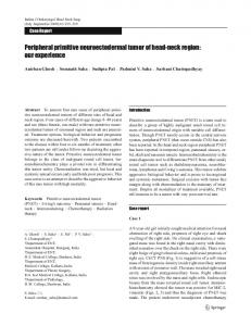
Fig. 1 Pancreatic peripheral primitive neuroectodermal tumor. Computed tomography in axial (left panel) and coronal (right panel) views showing a 4.5 × 4.0-cm well-delimited mass in the head of the pancreas (red arrows) with heterogeneous content, foci of calcification, and cystic/necrotic areas.
An 8-year-old girl presented with abdominal pain and jaundice of 1 month’s duration. She had conjugated hyperbilirubinemia and negative hepatitis serology. Computed tomography showed a mass in the head of the pancreas, with foci of calcification and cystic/necrotic areas " Fig. 1). Pancreatoblastoma and Frantz (● tumor were suspected. The patient underwent a cholecystojejunal anastomosis, and intraoperative biopsy of the pancreatic mass yielded inconclusive results. She was referred for endoscopic ultrasound (EUS) to re-evaluate the pancreatic mass. EUS showed a solid–cystic lesion in the head of the pancreas without vascular in-
Fig. 2
" Fig. 2, " Fig. 3). The main volvement (● ● pancreatic duct and common bile duct were slightly dilated. EUS-guided fineneedle aspiration of the pancreatic mass was done with a 22-gauge needle (EchoTip; Cook Medical, Limerick, Ireland) " Fig. 4). Cytopathologic evaluation of (● cell block material revealed a small cell neoplasm, and immunohistochemical analysis confirmed the diagnosis of peripheral primitive neuroectodermal tu" Fig. 5, " Fig. 6). mor (PNET) (● ● PNET belongs to a rare group of tumors called the Ewing sarcoma family of tumors [1 – 3]. Few PNETs arise in solid organs, and pancreatic PNETs are extremely rare [4 –
8]. Pancreatic PNETs are highly aggressive. Metastasis and recurrence are common, so that the prognosis is very poor. With modern multidisciplinary treatment, longterm survival can be achieved in 70 % to 80 % of patients with disease that has not metastasized [9]. The correlation of clinical symptoms with imaging, cytopathologic, and immunohistochemical analysis is useful to establish the diagnosis [10, 11]. An atypical rosette array of the cells, cytoplasmic neuronal secretory granules and neurofilaments, and pyknotic nuclear granules are important diagnostic criteria [4 – 8, 12]. Most tumors of the Ewing sarcoma family express high levels of a cell surface glycoprotein, CD99 [13, 14]. According to a 2014 review article [15], 14 cases of pancreatic PNET have been reported. This is the first case of a pancreatic PNET diagnosed by EUS. Endoscopy_UCTN_Code_CCL_1AF_2AZ_3AB
Competing interests: None
Flávio Amaro, Rogério Colaiácovo, Augusto Carbonari, Mauro Saieg, Ana Claudia Baraldi, Lúcio Rossini Centro Franco Brasileiro de Ecoendoscopia (CFBEUS), Santa Casa de São Paulo, São Paulo, Brazil References 1 Askin FB, Rosai J, Sibley RK et al. Malignant small cell tumor of the thoracopulmonary region in childhood: a distinctive clinicopathologic entity of uncertain histogenesis. Cancer 1979; 43: 2438 – 2451 2 Llombart-Bosch A, Lacombe MJ, Contesso G et al. Small round blue cell sarcoma of bone mimicking atypical Ewing's sarcoma with neuroectodermal features. An analysis of
Endoscopic ultrasound (stomach views) showing a solid cystic heterogeneous lesion in the pancreatic head.
Amaro Flávio et al. Primitive neuroectodermal tumor diagnosed by endoscopic ultrasound … Endoscopy 2015; 47: E11–E13
Electronic reprint for personal use
Pancreatic peripheral primitive neuroectodermal tumor diagnosed by endoscopic ultrasound
E11
E12
Cases and Techniques Library (CTL)
Fig. 3 Endoscopic ultrasound (stomach view) showing no echographic signs of portal vein impairment.
3
4
5
6 Fig. 4 Endoscopic ultrasound (stomach view) showing endoscopic ultrasound-guided fine-needle aspiration (22-gauge needle) of the solid cystic mass.
7
8
9
Electronic reprint for personal use
10
CEA
Negative
D1 CYCLIN
Focal positive
SYNAPTOPHYSIN
Focal positive
CHROMOGRANIN
Negative
Alpha-fetoprotein
Negative
Beta-catenin
Negative
CK7
Negative
Ki-67
Positive in 30 % of neoplastic cells
Tdt
Negative
Alpha1-antitrypsin
Negative
VIMENTIN
Negative
CD99
Positive
FLY-1
Focal positive
Fig. 5 Immunohistochemical profile suggestive of primitive neuroectodermal tumor. CEA, carcinoembryonic antigen; CK, cytokeratin; Tdt, terminal deoxynucleotidyl transferase; CD, cluster of differentiation.
11
12
13
five cases with immunohistochemical and electron microscopic support. Cancer 1987; 60: 1570 – 1582 Grier HE. The Ewing family of tumors. Ewing’s sarcoma and primitive neuroectodermal tumors. Pediatr Clin North Am 1997; 44: 991 – 1004 Movahedi-Lankarani S, Hruban RH, Westra WH et al. Primitive neuroectodermal tumors of the pancreas: a report of seven cases of a rare neoplasm. Am J Surg Pathol 2002; 26: 1040 – 1047 Bülchmann G, Schuster T, Haas RJ et al. Primitive neuroectodermal tumor of the pancreas. An extremely rare tumor. Case report and review of the literature. Klin Padiatr 2000; 212: 185 – 188 Perek S, Perek A, Sarman K et al. Primitive neuroectodermal tumor of the pancreas. A case report of an extremely rare tumor. Pancreatology 2003; 3: 352 – 356 Danner DB, Hruban RH, Pitt HA et al. Primitive neuroectodermal tumor arising in the pancreas. Mod Pathol 1994; 7: 200 – 204 Lüttges J, Pierré E, Zamboni G et al. Malignant non-epithelial tumors of the pancreas. Pathologe 1997; 18: 233 – 237 Granowetter L, Womer R, Devidas M et al. Dose-intensified compared with standard chemotherapy for nonmetastatic Ewing sarcoma family of tumors: a Children’s Oncology Group Study. J Clin Oncol 2009; 27: 2536 – 2541 Panicek DM, Gatsonis C, Rosenthal DI et al. CT and MR imaging in the local staging of primary malignant musculoskeletal neoplasms: report of the Radiology Diagnostic Oncology Group. Radiology 1997; 202: 237 – 246 Fuccio L, Larghi A. Endoscopic ultrasoundguided fine needle aspiration: how to obtain a core biopsy? Endosc Ultrasound 2014; 3: 71 – 81 Papierz W, Alwasiak J, Kolasa P et al. Primitive neuroectodermal tumors: ultrastructural and immunohistochemical studies. Ultrastruct Pathol 1995; 19: 147 – 166 Ambros IM, Ambros PF, Strehl S et al. MIC2 is a specific marker for Ewing’s sarcoma and peripheral primitive neuroectodermal tumors. Evidence for a common histogenesis of Ewing’s sarcoma and peripheral primitive
Fig. 6 Pancreatic peripheral primitive neuroectodermal tumor. a Cell block section showing clusters of rather uniform neoplastic cells arranged in a lobular pattern (hematoxylin and eosin, original magnification × 10). b Details of the neoplastic cells, showing scant cytoplasm, mild atypia, and a trabecular architecture. c Immunohistochemical reaction showing strong diffuse positivity for CD99.
Amaro Flávio et al. Primitive neuroectodermal tumor diagnosed by endoscopic ultrasound … Endoscopy 2015; 47: E11–E13
Cases and Techniques Library (CTL)
Bibliography DOI http://dx.doi.org/ 10.1055/s-0034-1377982 Endoscopy 2015; 47: E11–E13 © Georg Thieme Verlag KG Stuttgart · New York ISSN 0013-726X
Corresponding author Flávio Amaro, MD Rua Haddock Lobo 807, Apt. 14 São Paulo, São Paulo 01414-001 Brazil
[email protected]
Electronic reprint for personal use
neuroectodermal tumors from MIC2 expression and specific chromosome aberration. Cancer 1991; 67: 1886 – 1893 14 Fellinger EJ, Garin-Chesa P, Triche TJ et al. Immunohistochemical analysis of Ewing’s sarcoma cell surface antigen p30/32MIC2. Am J Pathol 1991; 139: 317 – 325 15 Mao Y, Sang X, Liang NX et al. Peripheral primitive neuroectodermal tumors arising in the pancreas: the first case report in Asia and a review of the 14 total reported cases in the world. Hepatobiliary Surg Nutr 2013; 2: 51 – 60
E13
Amaro Flávio et al. Primitive neuroectodermal tumor diagnosed by endoscopic ultrasound … Endoscopy 2015; 47: E11–E13