pinealoblastoma) or infratentorial (e.g. medulloblastoma in the cerebellum), the latter being more common. pPNETs are classified as part of the Ewing family of ...
Case Report
Singapore Med J 2011; 52(6) : e138
Peripheral type of primitive neuroectodermal tumour arising from the left orbital floor Santra G, Sinha P K, De D, Phaujdar S
ABSTRACT Primitive neuroectodermal tumours (PNETs) are rare tumours that originate from primitive neural crest cells. They are usually found in children below ten years of age. Peripheral PNETs (pPNETs) occur in soft tissues of the body, but have the same genetic changes as Ewing’s sarcoma of the bone (now called soft tissue Ewing’s sarcoma). They commonly present in the thoracopulmonary region, abdomen, pelvis and the extremities. The head and neck regions may also be involved. Our case demonstrates a PNET in the peripheral tissue arising from the left orbital floor and spreading
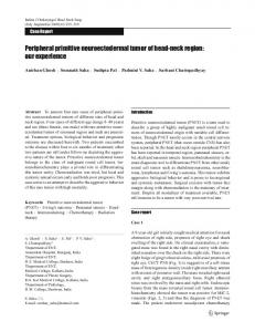
Fig. 1 Photograph shows a large tumour involving the left maxillofacial region and orbit in our patient.
locally to involve the left maxillofacial region, cheek and gum. The incidence of pPNETs is likely to be under-reported in the literature. Recent diagnostic advances, including cytogenetic and immunohistochemical analysis, have allowed these tumours to be distinguished from other small, poorly differentiated round cell tumours such as rhabdomyosarcoma, lymphoma and poorly differentiated synovial sarcoma.
below the lower margin of the left orbit as a bruise over
the area. Initially, the swelling was small, painless, firm and smooth, causing limited disfigurement to the face and no visual impairment. It gradually became enlarged,
resulting in distortion of the face and a loss of vision in the left eye (Fig. 1). There was no history of fever, nasal
discharge, bone pain, gum bleeding, patechial spots Keywords: immunohistochemical analysis, orbital floor, peripheral primitive neuroectodermal tumour, round cell tumour
or recurrent chest infections. The patient had a normal antenatal and developmental history, and his sibling was
also healthy. There was no history of consanguineous
Singapore Med J 2011; 52(6): e138-e140
marriage in the parents or any significant family history.
INTRODUCTION
examination revealed a regular pulse rate of 84/min and
Primitive neuroectodermal tumours (PNETs) are rare tumours originating from the cells of the primitive neural
crest. They are found mainly in children below ten years of age. Peripheral PNETs (pPNETs) commonly present
in the thoracopulmonary region, abdomen, pelvis and
the extremities but infrequently in the head and neck regions.(1-3)
CASE REPORT A 12-year-old Indian boy was admitted to our hospital
with complaints of a large painless swelling in the left cheek region for a period of four months. The swelling also involved the left orbit, and was incidentally noticed
The patient was conscious and well-oriented. Physical
blood pressure of 120/70 mmHg. A large, tense bosselated swelling measuring 13 cm × 11 cm was present in the
left half of his face, which involved the left orbit, cheek and gum. The overlying skin was shiny, with prominent
Department of Medicine, Medical College, 88 College Street, Kolkata 700073, West Bengal, India Santra G, MD Assistant Professor
superficial veins and ulcerations over it. The mass had
Sinha PK, MD, DM Associate Professor,
few teeth being uprooted. Other systemic examinations
De D, MBBS Postgraduate Trainee
Phaujdar S, MBBS Postgraduate Trainee
displaced the eye ball, nose and gum outwards, with a revealed no significant abnormalities.
Straight radiography and computed tomography
(CT) imaging of the posterior nasal sinus revealed a large
heterogeneously enhancing space-occupying lesion in the left inferolateral orbital region causing bony destruction and proptosis, with invasion of the extraocular muscles
Correspondence to: Dr Gouranga Santra Tel: (91) 94340 60591 Fax: (91) 33 2644 8773 Email: g.santra@ yahoo.com
Singapore Med J 2011; 52(6) : e139
2a
2c
2b
Figs. 2 Contrast-enhanced (a-b) coronal and (c) axial CT images show a large heterogeneously enhancing space-occupying lesion in the left inferolateral orbital region, causing bony destruction and proptosis with invasion of extraocular muscles and extension into the left maxillary sinus.
differential diagnoses being PNET, rhabdomyosarcoma and high-grade non-Hodgkin’s lymphoma (Fig. 3).
Immunohistochemical study was performed. The tissue was immunoreactive to MIC2/CD 99. Cytogenetic study revealed t(22,11) translocations. The combination of
morphology and immunochemistry was consistent with pPNET. (Figs. 2a–c). The lesion had extended into the left
DISCUSSION
channel. A neoplastic process was suggested. Fine-
from neuroectoderm, but as these tumour cells have not
maxillary sinus, causing a widening of the left osteomeatal
The majority of cells in PNET tumours are derived
needle aspiration cytology of the mass revealed sheets of
differentiated in a similar manner as a normal neuron, they
dissociated population of immature lymphoid cells, with
open, immature nuclear chromatin, prominent nucleoli and few mitotic activities. The possibility of a high-grade non-Hodgkin’s lymphoma (Burkitt) was suggested.
CT imaging of the chest was normal and bone
marrow biopsy revealed no significant abnormality.
appear primitive; this is how the term PNET is derived. Based on the tumour location, it is classified into two
types: (1) PNETs of the brain and central nervous system (CNS) and (2) pPNET outside the brain and nervous
system. A third category arising in the autonomic nervous
system, such as neuroblastoma, has also been suggested.(4)
CT imaging of the abdomen, however, revealed a
PNETs of the CNS are supratentorial (e.g.
mass showed infiltrating tumour composed of small
in the cerebellum), the latter being more common. pPNETs
few retroperitoneal lymph nodes. Biopsy from the
round cells in solid sheets and clusters. In some places, the cells were arranged around eosinophilic
fibrinous material in a rosette-like pattern. These cells showed scanty to moderate cytoplasm, with a high
nucleocytoplasmic ratio. The nuclei had open chromatin
and single-to-multiple distinct nucleoli. Occasional mitotic figures were noted. Classical lymphoglandular
bodies, splaying of chromatin material or a vacuolated background were not seen. The morphological features were those of a malignant round cell tumour, with the
pinealoblastoma) or infratentorial (e.g. medulloblastoma are classified as part of the Ewing family of tumours.(5) Ewing’s sarcoma family of tumours includes Ewing’s sarcoma, pPNET, neuroepithelioma, atypical Ewing’s sarcoma and Askin’s tumour (tumour of the chest wall).
pPNETs occur in soft tissues of the body, resembling soft tissue sarcoma (small, round blue-cell tumours) but have
identical chromosome translocation as Ewing’s sarcoma
of the bone (now called soft tissue Ewing’s sarcoma).
Immunohistochemical and cytogenetic studies suggest that these tumours have a common origin.
Singapore Med J 2011; 52(6) : e140
The incidence of pPNET is likely to be under-
reported in the literature. Recent diagnostic advances
like cytogenetic and immunohistochemical analysis have enabled these tumours to be distinguished from other small, poorly differentiated round cell tumours, including
rhabdomyosarcoma and lymphoma.(9,12) As pPNETs exhibit aggressive clinical behaviour and worse outcomes
compared to other small, round cell tumours, accurate
diagnosis of these tumours is of paramount importance. By using cytogenetic and immunohistochemical analysis, better management can be facilitated. Fig. 3 Photomicrograph shows malignant round cells arranged in a rosette pattern, with hyperchromatic nuclei and scanty cytoplasm (Haematoxylin & eosin, × 100).
pPNETs commonly present in the thoracopulmonary
region (Askin’s tumour), abdomen, pelvis and the
extremities. pPNETs that occur in the head and neck region are uncommon in most published case series.(1-3) However,
Jones and McGill reported 11 cases in the head and neck
region out of 26 patients with pPNETs.(6) The clinical
features of pPNETs depend on the site of presentation.
Symptoms commonly include pain and swelling of surrounding structures due to mass effect. pPNETs are
highly aggressive, and metastatic disease may be the first
presentation. The most common sites of pPNET metastases include the lung, bone and bone marrow.(7)
On histopathology, PNETs are highly cellular,
showing sheets of small, round cells with hyperchromatic nuclei. PNETs differ in their degree of neuroectodermal differentiation.
Tumours
demonstrating
neural
differentiation by light microscopy, immunohistochemistry or electron microscopy are traditionally called PNETs, and tumours undifferentiated by these analyses are known as Ewing’s sarcoma.(8) PNETs and Ewing’s sarcomas
commonly have t(11; 22) (q24; q12) translocation.(9) Expression of MIC2 gene produces a glycoprotein antigen
MIC2 (CD99), which consistently identifies pPNETs/ Ewing’s sarcoma. These sarcomas typically co-express
CD99 (MIC2) and vimentin,(10) but CNS PNETs (except
meningeal PNET) lack the expression of CD99.(11) The treatment for pPNET usually involves a combination of
surgery and chemotherapy with or without radiotherapy. The modality of treatment depends on the type, site and size of tumour, extent of metastasis as well as the age and general health status of the patient.
REFERENCES 1. Jürgens H, Bier V, Harms D, et al. Malignant peripheral neuroectodermal tumors. A retrospective analysis of 42 patients. Cancer 1988; 61:349-57. 2. Kushner BH, Hajdu SI, Gulati SC, et al. Extracranial primitive neuroectodermal tumors. The Memorial Sloan-Kettering Cancer Center experience. Cancer 1991; 67:1825-9. 3. Marina NM, Etcubanas E, Parham DM, Bowman LC, Green A. Peripheral primitive neuroectodermal tumor (peripheral neuroepithelioma) in children. A review of the St. Jude experience and controversies in diagnosis and management. Cancer 1989; 64:1952-60. 4. Batsakis JG, Mackay B, el-Naggar AK. Ewing’s sarcoma and peripheral primitive neuroectodermal tumor: an interim report. Ann Otol Rhinol Laryngol 1996; 105:838-43. 5. Khoury JD. Ewing sarcoma family of tumors. Adv Anat Pathol 2005; 12:212-20. 6. Jones JE, McGill T. Peripheral primitive neuroectodermal tumors of the head and neck. Arch Otolaryngol Head Neck Surg 1995; 121:1392-5. 7. Miser JS, Krailo MD, Tarbell NJ, et al. Treatment of metastatic Ewing’s sarcoma or primitive neuroectodermal tumor of bone: evaluation of combination ifosfamide and etoposide--a Children’s Cancer Group and Pediatric Oncology Group study. J Clin Oncol 2004; 22:2873-6. 8. Fletcher CDM, Unni K, Mertens F. World Health Organisation Classification of Tumours: Pathology and Genetics of Tumours of Soft Tissue and Bone. Lyon: IARC Press, 2002: 298-300. 9. Folpe AL, Goldblum JR, Rubin BP, et al. Morphologic and immunophenotypic diversity in Ewing family tumors: a study of 66 genetically confirmed cases. Am J Surg Pathol 2005; 29:1025-33. 10. Armbruster C, Huber M, Prosch H, Dworan N, Attems J. Ewing’s sarcoma and peripheral primitive neuroectodermal tumor in adults: different features of a rare neoplasm. Onkologie 2008; 31:179-84. 11. Ishii N, Hiraga H, Sawamura Y, Shinohe Y, Nagashima K. Alternative EWS-FLI1 fusion gene and MIC2 expression in peripheral and central primitive neuroectodermal tumors. Neuropathology 2001; 21:40-4. 12. Stafford EM, Honrado CP, Moscatello AL, Wilson YL, Weissbrod PA. Primitive neuroectodermal tumors [online]. Available at: emedicine.medscape.com/article/855644-overview. Accessed May 5, 2010.