Primary Primitive Peripheral Neuroectodermal Tumor of the Prostate Immunophenotypic and Molecular Study of a Case Maurizio Colecchia, MD; GianPaolo Dagrada, PhD; Pietro Luigi Poliani, MD; Antonella Messina, MD; Silvana Pilotti, MD
● A case of primitive peripheral neuroectodermal tumor arising in the prostate gland of a 31-year-old man and first diagnosed through a biopsy is reported. Microscopically, the tumor was made up of solid nests and sheets of small round cells, and it was difficult to distinguish the neoplasm from other small round cell tumors, such as small cell carcinoma, rhabdomyosarcoma, or malignant lymphoma. Immunohistochemically, the tumor cells showed immunoreactivity for CD99, vimentin, neuron-specific enolase, and synaptophysin. The neoplasm was excised by a radical surgical procedure preceded by chemotherapy and radiation therapy. The morphologic diagnosis of the prostatectomy specimen was complemented by molecular analysis performed on viable microdissected tissue obtained from formalin-fixed, paraffin-embedded tumor sections. Polymerase chain reaction and sequencing assessment showed the presence of EWS/FLI1 type 2 chimeric transcript, confirming the diagnosis of peripheral primitive neuroectodermal tumor. To our knowledge, this is the first description of a primary peripheral primitive neuroectodermal tumor in the prostate gland. (Arch Pathol Lab Med. 2003;127:e190–e193)
P
rimitive peripheral neuroectodermal tumors (pPNET) are malignant proliferations of small undifferentiated neuroectodermal cells occurring mainly in children. These tumors show a predilection for bones and soft tissues in the paraspinal region and lower extremities.1 Primitive peripheral neuroectodermal tumor is closely related to osseous and extraosseous Ewing sarcoma (EWS), in that it shares the same chromosomal translocation resulting in the fusion of EWS gene with a member of erythroblast transformation-specific domain family genes. The vast majority of pPNETs express a surface antigen encoded by the Mic-2 gene product and carry a nonrandom cytogenetic marker, which allows laboratorians to identify histologically or immunohistochemically equivocal cases. Although the molecular detection of transcripts by reverse transcrip-
Accepted for publication September 17, 2002. From the Dipartimento di Patologia (Drs Colecchia, Dagrada, Poliani, and Pilotti) and Unita` Operativa di Radiologia A (Dr Messina), Istituto Nazionale per lo Studio e la Cura dei Tumori, Milano, Italy. Corresponding author: Colecchia Maurizio, MD, Dipartimento di Patologia, Istituto Nazionale per lo Studio e la Cura dei Tumori, Via Venezian 1, 20133 Milano, Italy (e-mail:
[email protected]). e190 Arch Pathol Lab Med—Vol 127, April 2003
tion–polymerase chain reaction (RT-PCR) and sequencing is usually performed on cryopreserved material, protocols using archival paraffin-embedded tissues have been established recently. These procedures represent a useful tool in the diagnostic assessment of cases in which frozen material is not available.2 Primitive peripheral neuroectodermal tumor has been described in a variety of primary visceral sites,3–8 but to the best of our knowledge, it has never been reported in the prostate gland. We report a case of pPNET occurring in a 31-year-old man who presented with a mass within the prostate gland. The diagnosis was confirmed by molecular analysis performed on microdissected paraffinembedded material. REPORT OF A CASE A 31-year-old man with no previous history of malignancy was referred to another institution because a mass was identified by computed tomographic scan within the prostate with a suggestion of involvement of the left seminal vesicle. The patient’s serum prostate-specific antigen level was 2.06 ng/mL, and b-human chorionic gonadotropin and prostate-specific acid phosphatase values were within normal ranges. A prostate biopsy was performed, and a diagnosis of small round cell tumor consistent with pPNET was rendered. Two weeks later, the patient was admitted to the Istituto Nazionale per lo Studio e la Cura dei Tumori in Milan, Italy, for definitive therapy. Review of the biopsy confirmed the diagnosis of pPNET. Histologically, the proliferation consisted of small round tumor cells containing intracytoplasmic glycogen, accompanied by a scanty fibrovascular stroma (Figure 1). Immunocytochemically, the cells were immunoreactive for vimentin, CD99, and synaptophysin and were negative for all the other immunostains performed (cytokeratins, prostatespecific antigen, prostate-specific acid phosphatase, chromogranin, CD45, WT1, myogenin, a-smooth muscle actin, and neuronspecific enolase) (Table). Magnetic resonance imaging was performed to evaluate the extent of the lesion and to plan a possible surgical approach. Multiplanar imaging showed a 7 3 6 3 5.5-cm, plurinodular mass within the prostate with ill-defined borders, resulting in posterior displacement of the left seminal vesicle (Figure 2). A supplementary study consisting of total body computed tomographic scan and abdominal ultrasonography did not demonstrate any other site of tumor involvement. The mass molded the bladder without infiltrating it. The patient received adjuvant chemotherapy (3 cycles of ifosfamide, etoposide, vincristine, and adriamycin) and radiotherapy (4200 rad [42 Gy]) followed by radical surgery, which consisted of a combined excision of the prostate, seminal vesicles, and bladder. Peripheral Neuroectodermal Tumor of the Prostate—Colecchia et al
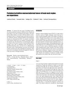
Figure 1. Prostate gland biopsy. The tumor is composed of round small cells with scanty cytoplasm and hyperchromatic nuclei (hematoxylineosin, original magnification 340). Figure 2. Magnetic resonance imaging of the pelvic region showing the prostate gland almost entirely replaced by the tumor mass and its relationship with the uninvolved surrounding structures. Figure 3. Histologic section from the prostatectomy specimen showing regressive changes (upper right) and closely associated residual tumor areas (bottom right). Sclerosis and uninvolved prostatic parenchyma are visible on the left side (hematoxylin-eosin, original magnification 315). Figure 4. High-power magnification of residual neoplastic cells with solid proliferation of tumor cells showing membrane-associated CD99 reactivity (immunoperoxidase staining, CD99 antibody, original magnification 3400). Figure 5. Detection of the EWS-FLI1 fusion transcript in the tumor by reverse transcription–polymerase chain reaction analysis. A 343-base pair (bp) product is detected in the tumor (lane 2). MW indicates molecular weight marker; lane 2, a 343-bp product detected in the tumor; lane 3, positive control (patient with a 277-bp product representing the type 1 fusion transcript EWS/FLI1); and lane 4, negative control. Arch Pathol Lab Med—Vol 127, April 2003
Peripheral Neuroectodermal Tumor of the Prostate—Colecchia et al e191
Dilutions and Sources of Antibodies Antibody
Host/Clone
Dilution
S100 Rabbit/polyclonal 1:4000 Synaptophysin Mouse/monoclonal CA 08 325 1:400 Desmin Mouse/monoclonal D 33 1:200 Myogenin Mouse/monoclonal F5D 1:50 a-Smooth muscle actin Mouse/monoclonal 5 C5 1:1000 CD45 Mouse/monoclonal B11 1 PD7/26 1:100 Cytokeratins Mouse/monoclonal AE1-AE3 1:50 Vimentin Mouse/monoclonal V9 1:200 Neuron-specific enolase Mouse/monoclonal MIG-N3 1:200 CD99 Mouse/monoclonal 013 1:100 WT1 Mouse/monoclonal C-19 1:400 * Dako indicates Dakopatts A/S, Glostrup, Denmark; Sigma, Sigma-Aldrich, St Louis, Mo; Sanbio, Sanbio BV, Uden, Signet Laboratories Inc, Dedham, Mass; and Santa Cruz, Santa Cruz Biotechnology Inc, Santa Cruz, Calif.
MATERIALS AND METHODS The surgical specimens were processed and fixed in 10% neutral buffered formalin and embedded in paraffin, and 5-mm-thick sections were stained with hematoxylin-eosin and periodic acid– Schiff. A fragment of tumor tissue was fresh frozen and stored at 2808C.
Immunohistochemical Staining
Source*
Dako Dako Dako Dako Sigma Dako Dako Dako Sanbio Signet Santa Cruz The Netherlands; Signet,
apeutic treatment, and corresponding to two thirds of the tumor mass. In the viable areas, the tumor presented as sheets and lobules of small round cells with dark round to oval nuclei (Figure 3). Occasional nuclei with prominent nucleoli were observed. The cytoplasm was scanty and was positive with periodic acid–Schiff. Abortive HomerWright rosettes were observed. Immunohistochemical Staining
A panel of immunohistochemical stains was performed using the streptavidin-biotin complex method (Vector Laboratories, Burlingame, Calif). Primary antibodies (vimentin, S100 protein, synaptophysin, neuron-specific enolase, CD99 [MIC2], CD45, chromogranin A, cytokeratins, smooth muscle actin, WT1, desmin, and myogenin) were used. Clones, dilutions, antigen-retrieval techniques, and manufacturers used are listed in the Table.
The viable neoplastic cells showed diffuse, intense cellsurface immunoreactivity for CD99 (Figure 4). The tumor cells were also positive for vimentin, neuron-specific enolase, and synaptophysin (the latter focally). All other immunostains were negative.
Reverse Transcription–Polymerase Chain Reaction
RT-PCR and Sequencing
The RNA extraction procedure from the fresh-frozen tumor tissue failed because the frozen stored fragment was entirely necrotic. Subsequently, total RNA was extracted from one 8-mmthick section obtained from a paraffin-embedded tumor sample. RNA extraction on RT-PCR was performed according to the methods of Hisaoka et al.2 A control PCR was done on a housekeeping gene, hypoxanthine guanine phosphoribosyl transferase (HPRT). Complementary DNA from 6 normal tissue samples (kidney, stomach, liver, lung, colon, and lymph node) derived from different patients surgically treated at our Institute were also analyzed. Subsequently, a nested PCR was carried out. The outer primers were EWS 22.3 and FLI1 11.3, and the inner primers were EWS 22.3N and FLI1 11.3N.2 The second round of PCR was performed using 1 mL of the first reaction product. The amplification profile for both PCRs was 40 cycles of denaturation at 948C for 40 minutes, annealing at 608C for 45 minutes, and extension at 728C for 1 hour.
A single, 343-base pair (bp) cDNA product was detected by RT-PCR for EWS/FLI1 mRNA (Figure 5). The band corresponded to the EWS/FLI1 fusion transcript, type 2 (EWS exon 7 and FLI1 exon 5), as confirmed by the obtained sequence (59AGCAGAACCCTT39). The chimeric transcript was not detected in the normal tissue samples tested.
DNA Sequencing Sequencing of PCR product was carried out using an automated sequencing system (377 DNA Sequencer, AB1 PRISM PE, Applied Biosystems, Foster City, Calif) following standard protocols.
RESULTS Pathologic Findings Grossly, the tumor mass measured 7 3 5 cm and was confined to the prostate, with involvement of the prostatic parenchyma located in the triangle between the urethra anteriorly and the plane of the ejaculatory ducts posteriorly. Infiltration of the base of the prostate gland and dislocation of the left seminal vesicle was grossly evident in sagittal sections. The tumor showed areas of extensive necrosis, probably caused by the previous radio-chemothere192 Arch Pathol Lab Med—Vol 127, April 2003
COMMENT Two features that render the diagnosis of pPNET difficult are its immunophenotypic profile, which often shows multidirectional differentiation along epithelial, neuroendocrine, and/or mesenchymal lines, and its not infrequent occurrence in unusual sites, that is, sites other than bone or soft tissues. Extraskeletal pPNET mainly occurs in the paravertebral region, the chest wall, and the lower extremities, and less commonly in the pelvis, retroperitoneum, or upper extremities. Exceptional sites of occurrence include several organs of the genitourinary system, such as kidney,3 ureter,4 bladder,5 testis,6 and seminal vesicles,7 as well as many other visceral sites (ovary, pancreas, uterus, parotid gland, and lungs).8 We describe a primary pPNET tumor arising in the wall of the prostate gland between the urethra, ejaculatory ducts, and seminal vesicles, in the absence of a lesion involving bone or soft tissues. The diagnosis of pPNET is mainly based on conventional histologic examination, but sometimes a confident categorization of the tumor may be difficult. The main differential diagnoses considered in our case, taking into account the age of the patient and the tumor site, were embryonal rhabdomyosarcoma, small cell carcinoma, maPeripheral Neuroectodermal Tumor of the Prostate—Colecchia et al
lignant lymphoma, and desmoplastic small round cell tumor. Rhabdomyosarcoma is the most common sarcoma of the bladder and prostate gland, but only a few cases have been reported in patients older than 20 years. The tumor cells are usually small and stellate, with hyperchromatic nuclei often showing atypical mitoses and exhibiting a greater variability in size and shape than those of pPNET. In our case, the morphologic findings and the negative immunostaining for desmin and myogenin ruled out this possibility. Small cell carcinoma of the prostate gland is a rare and aggressive neoplasm that generally shows a solid pattern of growth with occasional rosettes. The immunoprofile of this tumor shares several features with pPNET, such as immunoreactivity for chromogranin and synaptophysin, as well as cytokeratin decoration. However, CD99 positivity was not consistent with such a diagnosis in our case. Malignant lymphoma was easily ruled out by negativity for CD45. Finally, a possible secondary involvement of the prostate gland by desmoplastic small round cell tumor was excluded by the lack of expression of cytokeratins, desmin, and the WT1 gene product. CD99, a glycoprotein product of the MIC2 gene, is present on the cell surface of the vast majority of PNET cases (90%), and therefore represents a useful marker for the diagnosis of pPNET, but it is not entirely specific. Indeed, CD99 positivity has been observed in lymphoblastic lymphoma (90%), rhabdomyosarcoma (20%–25%), poorly differentiated sarcoma (75%), and approximately 50% of mesenchymal chondrosarcomas.9 A greater degree of diagnostic reliability is offered by molecular analysis, since PNETs are characterized by a nonrandom translocation, which results in the fusion of the EWS gene on chromosome 22q12 to FLI1 in 11q24, ERG in 21q22, and, rarely, ETV1 in 7p22, E1AF in 17q12, and FEV in 2q33.10 Both EWS-FLI1 and EWS-ERG rearrangements show different combinations of exons encoding different fusion transcripts and producing different chimeric proteins. In the present case, PCR sequence analysis of RNA obtained from viable paraffin-embedded tissue showed fragments corresponding to the in-frame junctions between EWS exon 7 and FLI1 exon 5, also called type 2 according to Delattre et al,11 which accounts for the 25% of EWS-FLI1 fusion transcripts and seems to be associated with a more aggressive clinical behavior.10
Arch Pathol Lab Med—Vol 127, April 2003
The advantage provided by the application of molecular analysis on paraffin-embedded tissue in cases such as ours, which occurred in an unusual site and in which chemotherapy induced extensive regressive changes, is obvious. Recently, cytogenetic analysis showed that visceral PNETs share the same translocation characteristic of EWS/PNET of soft tissue and bone, demonstrating the presence of chimeric EWS-FLI1 fusion transcripts in 18 visceral PNETs.8 Our case confirms the genetic link between visceral EWS/PNET and EWS/PNET of soft tissue and bone and, although several cases of pPNET arising in the wall of the ureter, bladder, kidney, and seminal vesicle have been described in the urinary tract, to the best of our knowledge, this is the first report of a case arising within the substance of the prostate gland. We gratefully acknowledge Juan Rosai, MD, of the Department of Pathology, Istituto Nazionale dei Tumori, Milan, Italy, for the revision of this manuscript and Gianni Roncato, Istituto Nazionale dei Tumori, Milan, Italy, for his skillful technical assistance. References 1. Dehner LP. Peripheral and central primitive neuroectodermal tumors: a nosologic concept seeking a consensus. Arch Pathol Lab Med. 1986;110:997– 10051. 2. Hisaoka M, Tsuji S, Morimitsu Y, et al. Molecular detection of EWS-FLI1 chimeric transcripts in Ewing family tumors by nested reverse transcription-polymerase chain reaction: application to archival paraffin-embedded tumor tissues. APMIS. 1999;107:577–584. 3. Quezado M, Benjamin DR, Tsokos M. EWS/FLI-1 fusion transcripts in three peripheral primitive neuroectodermal tumors of the kidney. Hum Pathol. 1997; 28:767–771. 4. Charny CK, Glick RD, Genega EM, Meyers PA, Reuter VE, La Quaglia MP. Ewing’s sarcoma/primitive neuroectodermal tumor of the ureter: a case report and review of the literature. J Pediatr Surg. 2000;35:1356–1358. 5. Banerjee SS, Eyden BP, McVey RJ, Bryden AA, Clarke NW. Primary peripheral primitive neuroectodermal tumour of urinary bladder. Histopathology. 1997; 30:486–490. 6. Aguirre P, Scully RE. Primitive neuroectodermal tumor of the testis: report of a case. Arch Pathol Lab Med. 1983;107:643–645. 7. Bjerklund Johansen TE, Huseby A, Stenwig JT. Extraskeletal Ewing’s sarcoma contiguous with the seminal vesicle. Scand J Urol Nephrol. 1988;22:237–239. 8. O’Sullivan MJ, Perlman EJ, Furman J, Humphrey PA, Dehner LP, Pfeifer JD. Visceral primitive peripheral neuroectodermal tumors: a clinicopathologic and molecular study. Hum Pathol. 2001;32:1109–1115. 9. Folpe AL, Chand EM, Goldblum JR, Weiss SW. Expression of Fli-1, a nuclear transcription factor, distinguishes vascular neoplasms from potential mimics. Am J Surg Pathol. 2001;25:1061–1066. 10. de Alava E, Gerald WL. Molecular biology of the Ewing’s sarcoma/primitive neuroectodermal tumor family. J Clin Oncol. 2000;18:204–213. 11. Delattre O, Zucman J, Melot T, et al. The Ewing family of tumors: a subgroup of small-round-cell tumors defined by specific chimeric transcripts. N Engl J Med 1994;331:294–299.
Peripheral Neuroectodermal Tumor of the Prostate—Colecchia et al e193