PARTITIONING OF THE MORPHOMETRIC DIFFUSION CAPACITY OF RESPIRATORY ORGANS IN SOUTH AMERICAN DIPNOI, Lepidosiren paradoxa Marisa N. Fernandes Federal University of São Carlos Department of Physiological Sciences C. Postal 676, Phone: 55 16 260-8314 email:
[email protected] Marcos Flávio P. G. Moraes Dept. Morphology/University of Alagoas Oscar Tadeu F. Costa Dept. Morphology/University of Amazonas Federal University of São Carlos Post-Graduate Program in Ecology and Natural Resources C. Postal 676, Phone: 55 16 260-8314 Sabine Holler Steven F. Perry University of Bonn Zoology Institute Phone: +49-228-733807 e-mail:
[email protected] Abstract The transition from water to air breathing marked a significant transition in the evolution of vertebrates and many aspects of the fractionating of the respiratory organs’ diffusion capacity are still unknown. This study, which is based on stereologic principles, quantifies the respiratory surface area of the gills, lungs and skin of the “piramboia”, Lepidosiren paradoxa (Dipnoi). Based on morphometric data of the respiratory surface area, the water- and/or air-blood thickness of the diffusion barrier and the values calculated for the diffusion capacity of O2 through the respiratory epithelium, the lungs are the main
105
respiratory structure for O2 uptake, followed by the skin and the gills, the latter being insignificant as an O2 uptake organ. Introduction The respiratory transition from water to air was a significant event in the evolution of vertebrates (Johansen, 1968) and the living lungfish genera represent the link between water- and air-breathing species. There are several functional and morphological data on the gills and lungs of Protopetrus and Neoceratodus (Klika & Lelek, 1967; Hughes, 1973; Laurent et al., 1978; Maina & Maloy, 1985; Fritsche et al., 1993; Olson 1994). However, only a few brief descriptions or comparisons with the pattern of the two aforementioned species exist for the structurally more advanced South American lungfish, L. paradoxa (Johansen & Lenfant, 1967; Graham, 1997; Laurent et al., 1978; Burggren & Johansen, 1986; Axelson et al., 1989; Abe & Steffensen, 1996a,b; Harder et al., 1999). Morphometric data of the respiratory structure provide a morphological reference point for the interpretation of physiological studies as well as a starting point for further studies on adaptation (Perry et al., 1994). A detailed descriptive and morphometric study of the respiratory surface and the air-blood thickness of the lungs of L. paradoxa was carried out by Hughes and Weibel (1976). However, no data on the gills and skin are available. Therefore, the main purpose of this study was to determine the morphometric partitioning of the respiratory surface and the diffusion capacity of O2 in the respiratory structures, i.e., the gills, skin and lungs, of this South American lungfish. Materials and Methods Adult specimens of South American lungfish Lepidosiren paradoxa weighing 864 ± 196 g, were obtained in Cuiabá, MT, Brazil. Prior to manipulation, the lungfish were anesthetized by 30 minutes’ immersion in a 0.05% solution of ethyl p-aminobenzoate (Benzocaine, Sigma). Their hearts were exposed and their blood vessels were perfusated with a saline solution, followed by 2.5% glutaraldehyde buffered to pH 7.4 with 0.1 M phosphate buffer (300 mOsmol). The gills and lung were removed and skin samples were taken. The gills and skin were processed by light microscopy and embedded in historesin (Leica). The lungs were processed by light and transmission electron microscopy and embedded, respectively, in historesin (Leica) and Araldite 6005 (Ladd). The
106
skin samples were decalcified with a solution of 10% EDTA buffered with phosphate buffer at pH 7.4) before being processed. Morphometric measurements were done following the stereological method. The reference volume of gills, lungs and skin was determined based on the Cavalieri principle (Gundersen & Jensen, 1987). The respiratory surface areas and the barrier thickness of gills and skin (water-blood distance) and lungs (airblood distance) were calculated using the vertical section method (Gundersen et al., 1988). The barrier thickness of gills and skin was measured at the light microscopic level and the barrier thickness of the lung was defined at the electron microscopic level using a half-logarithmic ruler in randomly chosen directions (Perry, 1981). The harmonic average of each structure was calculated according to Weibel and Knight (1964). The latter calculations were used to calculate the anatomic diffusion factor of each respiratory structure (ADF = surface area/barrier thickness, Perry, 1978) The O2diffusing capacities were calculated based on the product of ADF and Krogh’s diffusion coefficient of tissue layers. Results and Discussion L. paradoxa’s gill morphology differs from those of other teleosts. Five gill arches present hemibranches with lobe-like filaments irregularly arranged in the posterior region (opercular side) (Fig 1A). The connective tissue and blood vessels evolve from the basement membrane and from a multilayered epithelium consisting of at least four cell layers (Fig. 1B). The skin consists of an epidermis and a dermis. The epidermis has 6 to 10 cell layers. The dermis consists of dense connective tissue, pigment cells, bony ridge scales, blood vessels and capillaries (Fig 2).
107
A
B F F A A
Figure 1. L. paradoxa. A. Lobe-like filament (arrows) bearing the gill arch. B. Light micrograph of several lobe-like filaments (F) showing the distribution of blood vessels and capillaries (arrows) in the connective tissue. Toluidine blue/acid fucsin. Scale bars are 100 µm.
Ep
C
M
D
Figure 2. Light micrograph of a transverse section of L. paradoxa skin. Structure of the epidermis (Ep) and dermis (D), showing the capillaries in the connective tissue below the epithelial cell layers. C: capillary; M: Melanophore; MC: mucous cells; S: scale. Toluidine blue/acid fucsin. Scale bars are 50 µm.
108
Paired lungs are located in the median line of the body, dorsally to the digestive tube and ventrally to the vertebral column. They have a common anterior region (cranial region) that opens to the glottis. A large air duct in each lung, which is surrounded by septa and trabecullae, consists of asymmetric faveoli. The septa consist of connective tissue, smooth muscles and blood vessels (Fig. 3a, b). The epithelium consists of pavement cells with short microvilli and sparsely distributed ciliated cells. The faveolate epithelium presents pneumocytes resembling types I and II on the basement membrane. Innumerous capillaries are close to the basement membrane (Fig. 3C).
A
B
C F
D
P
T
C Pn
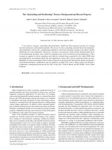
D Figure 3. Transversal section of the lung’s middle region, showing the parenchyma (P) and air duct (D). Scale bar: 5 mm. B. Light micrograph of parenchyma of lung showing the faveolus (F) and trabecula (T). Scale bar: µm C. TEM micrograph of parenchyma showing the lung’s respiratory epithelium. Pn: pneumocyte; C: capillary. Scale bar: µm The surface percentage of gills, skin and lungs and the respective respiratory and non-respiratory percentage of each structure are shown in Figure 4. The morphometric values of the surface area, barrier thickness, anatomic diffusion factor and O2 diffusion capacity of the gills, skin and lungs of L. paradoxa are shown in Table 1. The lungs of L. paradoxa have a more extensive respiratory surface, lower airblood diffusion distance and higher anatomic diffusion factor than the gills and skin. ADF is a morphological indicator of gas exchange capability and is directly proportional to the diffusion capacity (Perry, 1978). The high ADF of lungs is congruent with the hypothesis that the lungs alone are capable of sustaining the metabolic requirements of species, an assumption that is corroborated by the DO2 value for lungs, gills and skin.
109
A
B Lungs 61 %
Surface area (% )
Gills 0.1 %
S kin 39 %
100 80 60 40 20 0
SNR
SNR
SNR
SR
SR
SR
G ills
Skin
Lungs
Figure 4. L. paradoxa. A. Partitioning of the respiratory surface (gills, skin and lungs). B. Respiratory (SR) and non-respiratory (SNR) surfaces of the gills, skin and lungs. The values given here refer to a hypothetical lungfish weighing 0,87 kg. Table 1. Absolute surface area (S), harmonic average (τh) of the barrier diffusion distance between air-blood and or/water/blood, anatomic diffusion factor (ADF) and morphometric O2 diffusion capacity of gills, skin and lungs of a 0.87 kg South American lungfish, L. paradoxa
Structure Gills Skin Lungs
S (cm2) 0.6 404 614
τh (µm) 110 138 1.3
ADF cm2µm-1 kg-1 5.52 10-3 3.46 466.9
DO2 cm3min-1mmHg-1kg-1 1.0.10-6 6.9.10-4 1.1.10-1
Acknowledgments This research work was supported by grants awarded to Drs. M.N. Fernandes and S.F. Perry by CAPES (Brazil) and DAAD (Germany), respectively. M.F.P.G. Moraes and O.T.F. Costa acknowledge CAPES for the award of a scholarship.
References
110
Abe, AS and Steffensen, JF 1996a Bimodal respiration and cutaneous oxygen loss in the lungfish, Lepidosiren paradoxa.. Rev. Bras. Biol. 56: 211216. Abe, AS. and Steffensen, JF 1996b Lung and cutaneous respiration in awake and estivating South American lung fish Lepidosiren paradoxa. Rev. Bras. Biol. 56: 485-489. Axelson, M, Abe, AS, Bicudo, JEPW 1989 On the cardiac control in the South American lung fish, Lepidosiren paradoxa. Comp. Biochem. Physiol. 93A: 561-566 Burggren, WW, Johansen, K 1986 Circulation and respiration in lungfish (Dipnoi). J. Morphol. (Supp) 1: 217-236 Costa, OTF, Perry, SF, Schmitz, A, Fernandes, MN 2001 Stereological analysis of fish gills: Method. 6th International congress of Vertebrate Morphology Proceedings, Jena, Germany, July 21-26,2001. J Morphol 248:219 Fritsche, R, Axelsson, M, Franflin, CE 1993 Respiratory and cardiovascular responses to hypoxia in the australian lungfish. Respir. Physiol. 94: 173-178. Graham, JB 1997 Air-breathing fishes. Evolution, diversity and adaptations. Academic Press, San Diego, 299p. Gundersen, HJG, Bendtsen, TF, Korbo, L, Marcussen, N, Moller, A, Nielsen, K, Nyengaard, JR, Pakkenberg, B, Sorensen, FB, Vesterby, A and West, MJ 1988 Some new, simple and efficient stereological methods and their use in pathological research and diagnosis. APMIS 96: 379-394. Gundersen, HJG and Jensen, EB 1987 The efficiency os sistematic sampling in stereology and its prediction. J. Microsc. 147: 229-263 Harder, V, Souza, RHS, Severi, W, Rantin, FT and Bridges, CR 1999 The South American lung fish – adaptations to an extreme habitat. In: The Biology of Fishes (Val, AL and Almeida-Val, VMF, eds) INPA, Manaus, p. 87-98.
111
Hughes, GM 1973 Ultrastructure of the lung of Neoceratodus and Lepidosiren in relation to the lung of other vertebrates. Folia Morphol. 21: 155-161. Hughes, GM and Weibel, ER 1976 Morphometry of fish lungs. In: Respiration of Amphibious Vertebrates (Hughes, GM, ed) Academic Press, London. p. 213-232. Johansen, K 1968 Air breathing fishes. Sc. Am. 219: 102-111. Johansen, K and Lenfant, C 1967 Respiration function in the South American lungfish, Lepidosiren paradoxa (Fitz.) J. Exp. Biol. 46: 205-218. Klika, E and Lelek, A 1967 A contribution to the study of the lungs of Protopterus annectens and Polypterus senegalensis. Folia Morphol. 15, 168-174. Laurent, P, Delaney, RG. and Fishman, AP 1978 The vasculature of the gills in the aquatic and aestivating lungfish (Protopterus aethiopicus). J. Morphol. 156: 173-208. Maina, JN and Maloiy, GMO 1985 The morphometry of the lung of the african lungfish Protopterus aethiopicus: Its structural-functional correlations. Proc. Royal Soc. London B Biol. Sci. 224 : 399-420. Moraes, MFPG, Costa, OT, Perry, SF and Fernandes, MN 2001 Morphology and stereological analysis of pulmonary surfaces of Lepidosiren paradoxa (Dipnoi). XVIII Brazilian Congress of Microscopy and Microanalysis Society Proceedings, Águas de Lindóia, Brazil, October 28 - 31, 2001, Acta Microscopica (in press). Olson, KR 1994 Circulatory anatomy in bimodally breathing fish. Am. Zool. 34: 280-288 Perry, SF 1978 Quantitative anatomy of the lungs of the red-eared turtle Pseudemys scripta elegans. Resp Phyisiol. 35: 245-265. Perry, SF 1981 Morphometric analysis of pulmonary structure: methods for evaluation and comparison of unicameral lungs. Mikroskopie 38: 278293.
112
Perry, SF, Hein, J and van Dieken, E 1994 Gas exchange morphometry of the lungs of the tokay, Gekko gecko L. (Reptilia: Squamata: Gekkonidae). J. Comp. Physiol. B, 164: 206-214. Perry, SF, Höller, S, Schmitz, A, Costa, OTF, Moraes, MFPG and Fernandes, MN A new stereological procedure for gills of radically different structure, demonstrated on two obligatory air breathers, Arapaima and Lepidosiren. 94th Annual meeting of the Deutsche Zoologische Gesellschaft, Osnabrück Proceedings, Germany, June 4-8, 2001. Zool (Supp IV) 104: 42 Weibel, ER and Knight, BW 1964 A morphometric study on the thickness of the pulmonary air-blood barrier. J.Cell Biol. 21: 367-384.
113
114