... Vol 90 Drug-DNA Interaction Protocols. Edited by K R Fox Humana Press Inc , Totowa, NJ .... 3rd Hound PCR |. Anneal and extend end- labelled primer 3 3.
11 PCR-Based Methods for Detecting DNA Damage and Its Repair at the Subgene and Single Nucleotide Levels in Cells Keith A. Grimaldi and John A. Hartley 1. Introduction A large category of anticancer drugs owe their cytotoxicity to their ability to interact with, and damage, DNA. Many agents form bulky adducts that block RNA and DNA polymerases, inhibiting transcription, and DNA replication. It is well-known that cancer chemotherapy is far from satisfactory. The many problems include unpleasant, sometimes life threatening side effects, tumor resistance to drugs, and mutagenic effects of the drugs themselves that can cause secondary cancers and can increase the mutation rates of existing tumors leading to the emergence of more invasive and aggressive disease. DNA damage and repair is central to many of these problems and therefore studying this process at fine levels of resolution in mammalian cells, both transformed and normal, will be important. Also, with the aim of improving the specificity of cancer therapy, novel sequence specific cytotoxic agents are being developed that may allow some degree of gene targeting. Clearly, for the development and rational design of such agents, it is important to have methods that will allow the sequence selectivity of binding to be studied in cells to see if the intended target sequence is being hit and to what extent individual lesions are repaired. Since its first description in 1985 (1), the polymerase chain reaction (PCR) has been adapted for use in just about every branch of molecular biology and beyond. It is no surprise then that it should find a place in the study of DNA damage and repair resulting from drug—DNA interactions. This chapter will focus on uses of PCR developed in the authors' laboratory that allow drugDNA interactions to be studied at various levels of resolution from gene regions From Methods in Molecular Biology, Vol 90 Drug-DNA Interaction Protocols Edited by K R Fox Humana Press Inc , Totowa, NJ
157
Grimaldi and Hartley
158 Template DNA (genomic) Primer NRAS A
_»
«
..*—
Primer NflAS B
EXPONENTIAL AMPLIFICATION
> •
_ ^
9
> • -
UUANIIIAIIVfc
-
—
p=
PCR
>-*
4 ? h
EXPONENTIAL AMPLIFICATION OF ONE STRAND ONLY
* " ~r~U
> •
i
A
NO EXPONENTIAL AMPLIFICATION
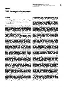
Fig 1 QPCR method
(300-3000 bp) (2,3) right down to the ultimate level of detection—individual nucleotides in single-copy genes in mammalian cells (4) The overall PCR method to be described in this chapter exploits the fact that covalent drug-DNA adducts can block taq polymerase. It can be separated into three parts and used according to the level of resolution desired by the investigator Quantitative PCR (QPCR) will measure the aggregate damage on both strands in a gene region of choice. It is sensitive enough to be used to look at subgene functional regions such as introns, exon, promoters, and so forth. Currently, a convenient size would be between 300—3000 bp, however, with new reagents allowing "long-PCR" becoming available the upper limit may be extended up to 20-30 kbp allowing QPCR to be used to study entire genes. Strand-specific QPCR (ss-QPCR) incorporates adaptations that allow damage to be measured in the same region as QPCR, but in a strand specific way This is particularly important in the light of recent discoveries showing heterogeneity of repair among gene regions and that by means of transcription coupled repair the transcribed strand of an expressed gene can be repaired more efficiently than the nontranscnbed strand (5). Single-strand ligation PCR (sslig-PCR) extends further the method to allow the detection of adduct formation at the level of single nucleotides, on individual strands, in a single copy gene in mammalian cells.
1.1. Overview of the Protocols 1.1.1. QPCR (see Fig. 1) A pair of oligonucleotide primers is used that defines the region of the gene selected for study. In the PCR each strand of genomic DNA serves as a potential template for exponential amplification and the presence of one or more
DNA Damage Detected by PCR-Based Methods
159
adducts will block the amplification of that strand. Therefore, when drugtreated DNA is amplified, and the reaction is stopped in the exponential phase, the amount of product will be reduced compared to untreated DNA. Furthermore, the extent of the reduction will be proportional to the amount of damage caused by the drug treatment and by including a radioactive nucleotide in the PCR the extent of damage caused by particular treatments (and subsequent repair) can be accurately quantified. 1.1.2. SS-QPCR The method is outlined in Fig. 2 (as set up to measure lesions on the transcribed strand). DNA extracted from drug-treated and untreated cells is subjected to a first round "linear" PCR using a single biotinylated primer (5 ItB), complementary to the transcribed strand. This PCR generates a family of single-stranded molecules, some of which will be truncated because of the presence of a blocking lesion on the transcribed strand of the template DNA. All are captured on streptavidin-coated paramagnetic beads and washed with NaOH to remove genomic DNA including any hybridized to the PCR products. After neutralization, the single-stranded molecules, while still attached to the beads, serve as templates in a second, exponential, PCR In the exponential amplification the downstream primer (primer 2) is complementary to the transcribed strand and is nested with respect to primer 1 The upstream primer (primer 3) is complementary to the nontranscnbed strand and its binding site determines the length of the gene region in which damage is to be measured. In this PCR only those DNA molecules that were extended past the site of primer 3, i.e., those that were not blocked by lesions on the genomic DNA, will be exponentially amplified. Thus, provided the PCR remains in the exponential phase when stopped, the amount of product will be directly proportional to the amount of undamaged template present in the region under study of the original genomic DNA. 1.1.3. Sslig-PCR The method of sshg-PCR is outlined in Fig. 3. As with ss-QPCR it involves a first round PCR using a single 5'-biotinylated primer, which defines the area of the gene to be investigated. Thirty cycles of linear amplification by PCR generates a family of single-stranded molecules of varying length for which the 5' end is defined by the primer and for which the 3' ends are defined by the positions of the DNA-drug adducts. In order to exponentially amplify these molecules, which are captured and isolated by binding to streptavidin coated magnetic beads, a single stranded, 5'-phosphorylated, oligonucleotide is hgated to their 3'-OH ends using T4 RNA ligase. This oligonucleotide also bears a 3'-terminal amine group to block self-ligation. With both ends of the DNA molecules defined, they can then be exponentially amplified and detected. The sequence
Grimaldi and Hartley
160
•
DNA isolated from drug treated cells
TS UIIIIIIHHimiHIIIIIIIIIIIHI
iiiiiiiiiiiiiiiiiiHijiiiiiiitiiimiii
NTS Denature and anneal biotlnylated primer 1-tB
TS ¥=1-tB PCR1 Extend pnmer 1-1 1-tB 20 Cycles
Linear Amplilication
Full length product
_ „ _ 1>tB Truncated product
*—1-tB - Capture on beads, - NaOH wash to remove hybridized DNA - TE wash
3
>
--
•
3»—2
1st Cycle Extension of pnmer 3 only on full-length template PCR2 Exponential Amplification
.
,r
No Product
••—1-tl
Denature and anneal primers
•—2 '==*
_ i .
t B
^
Exponential Amplification and Quantification of PCR product
Fig 2.
DNA Damage Detected by PCR-Based Methods
161
positions of the adducts are determined by electrophoresing the sshg-PCR products on a sequencing gel.
2. Materials The following lists equipment and reagents required to detect damage at all levels, i.e., from gene region down to single bases. Individual needs will determine what is necessary. For example, if damage and repair is to be studied at the level of a gene region (300-2000 bp) and without any strand specificity then only those items necessary for QPCR will be needed.
2.1. Cell and DNA Treatment 1. Cells in suspension or monolayer culture (see Note 1) 2 DNA-binding drug, e.g., Cisplatin, mechlorethamine, and so on 3 10X Teoa (store at 4°C; for treatment of naked DNA)' 250 mMTriethanolamine, pH7.2, lOmMEDTA 4 Drug stop solution. 0.6M Sodium acetate, pH 5.2 5. Tissue culture plates (6-well, 24-well, and/or Petri dishes)
2.2. DNA Isolation 1 Cell lysis buffer (store at room temp): 400 mMTns-HCl, pH 8.0, 60 mM EDTA, 150 mMNaCl, 1% (w/v) sodium dodecyl sulfate (SDS) 2 5 M Sodium perchlorate (store at room temperature) 3. Chloroform. 4 37 and 65°C water bath 5 Rotary mixer 6 Microfuge 7. Vacuum dryer
2.3. Oligonucleotides Oligonucleotides were obtained from Genosys UK, or Pharmacia. Store all oligonucleotides at-20°C. The oligonucleotide sequences will obviously depend on the region of the gene to be studied. Those described here were used to study damage and repair in a region comprising intron 1 of the human N-ras gene (see Table 1 for sequence).
2.3.1. QPCR 1. NRAS-A 5'-CCT AAA TCT GTC CAA AGC AGA GGC from the coding strand. 2. NRAS-B: 5'-CAG CAA GAA CCT GTT GGA AAC CAG from the noncoding strand This primer pair defines a 523-bp region of the N-ras gene intron 1 Fig 2 (opposite page) Ss-QPCR method (as set up to measure lesions on the transcribed strand)
Grimaldi and Hartley
162 /!
DNA Isolated Irom treated cells
HMMIIIHIIIIIIIIIilllllllllllllll
Denature and anneal biotlnylated primer 31nB
Linear amplification with faa polymerase (30 cycles)
HI Hound PCB I
u M
2N
"*• *&
Wash to remove unbound genomic DNA etc
Ugate •ligation oligonucleotide" YwithT4RNAIIgase
1
Wash to remove excess oligonucleotide
Amplify by PCR 2nd Round PCR I
Anneal ligation pnmer"
1st Cycle
Denature and anneal primers "ligation pnmer* + pnmer 3 2
Cycles 2-X
Exponential amplification, X cycles
Amplified top strand
| 3rd Hound PCR |
Anneal and extend endlabelled primer 3 3 (4 cycles)
Denature and run on sequencing gel
Fig. 3.
DNA Damage Detected by PCR-Based Methods
163
2.3.2. Strand-Specific QPCR (ss-QPCR) The following primers will measure damage in a 350-bp region of the N-ras gene intron 1. 2.3.2 1.STEP1
1 To measure damage on the nontranscribed strand of N-ras: 3 1 nB (5'-Biotinylated): 5'-CAG CAA GAA CCT GTT GGA AAC CAG. 2 To measure damage on the transcribed strand of N-ras. 5 ItB (5' Biotinylated): 5'-GGT CCT TCC ATT TGG TGC CTA CG. (These primers were obtained synthesized with biotin incorporated at the 5'-end) 2.3.2 2. STEP 2
3 Oligo 3.2- 5'-CCA GTA ATC AGG GTT AAT TGC GAG C 4 Oligo 5 2 5'-ACG TGG GGA GAT CTT GGA GA
2.3.3. Single-Strand Ligation PCR (sslig-PCR, to measure damage on individual nucleotides) The following primers are required in addition to the primers described above for ss-QPCR (3 InB, 5.ItB, Oligo 3.2, Oligo 5.2): 1 To measure damage on the nontranscribed strand- Oligo 3 3' 5'-GCG AGC CAC ATC TAC AGT AC 2 To measure damage on the transcribed strand Oligo 5 3 5'-TGG AGA CAG AAG GGA GAA TG 3 "Ligation Oligonucleotide " 5'-p-ATC GTA GAT CAT GCA TAG TCA TA-n This oligonucleotide should be supplied gel or HPLC purified. It must also be 5'-phosphorylated (p) and bear a 3'-terminal amine group (n) to block self-hgation (these modifications should be incorporated at synthesis) 4. "Ligation Primer"- 5'-TAT GAC TAT GCA TGA TCT ACG AT This oligonucleotide, which is complementary to the "ligation oligonucleotide," must be gel or HPLC purified.
2.4. End Labeling Oligonucleotides (at 5' end) Oligonucleotides were end-labeled with T4 polynucleotide kinase using Gibco-BRL (Gaithersburg, MD) kits with forward reaction buffer 1. [y-32P]-ATP 10 UCI/|LIL (Amersham). 2. Forward buffer- 300 mMTns-HCl, pH 7.8, 75 mM 2-mercaptoethanol, 50 mM MgCl2, 1.65 uMATP
Fig. 3. (opposite page) Sslig-PCR method (as set up to measure lesions on the nontranscribed strand).
164
DNA Damage Detected by PCR-Based Methods
165
2.5. PCR 2.5.1. All Reactions 1. Taq Polymerase (Perkin-Elmer, Promega, Advanced Biotechnologies, UK, and so on). 2 10X PCR buffer (see Note 5)- 200 mM (NH4)2S04, 750 mM Tris-HCI, pH 9.0, 0.1%(w/v)Tween 3. 25 mM MgCl2 (store at 4°C). 4 10X dNTP's (Pharmacia): make a mixture containing 2 mM concentration each of dATP, dGTP, dCTP, and dTTP; store at-20°C 5 Thermal cycler (e g , MJ PTC-100 with heated lid see Note 6) 6. Mineral oil (if thermal cycler is without heated lid facility) 7 PCR tubes—0 5 or 0.2 mL
2.5.2. QPCR and ss-QPCR [a-32P]-ATP 10 uO/uL (Amersham). Quantitation of the PCR product may be performed by one of two methods (see Note 13). One method (item 1) involves TCA precipitation of the PCR product and scintillation counting. The alternative (item 2) is quantitation by densitometric scanning of autoradiographs or phosphor image analysis after agarose gel electrophoresis. 1. TCA precipitation. a. Whatman GFC filters (24-mm diameter) b. Multiple filtration manifold (Milhpore) c. 5% TCA. 5% (w/v) trichloroacetic acid, 20 mM tetrasodium pyrophosphate (store at 4°C) d. Scintillation fluid (Ecoscint, National diagnostics) e. Scintillation counter (e g., Beckman LS1800) 2. Agarose gel electrophoresis and densitometnc scanning or phosphor image analysis. a Equipment for horizontal agarose gel electrophoresis. b. 50X TAE: 2 M Tns-acetate; 0 05 MEDTA (per L 242 g Tns base; 57 1 mL glacial acetic acid; 100 mL, 0 5 MEDTA, pH 8 0) c Agarose. d 6X Agarose gel loading buffer- 0.25% bromophenol blue, 4% (w/v) sucrose in water. e. Gel dryer (suitable for agarose and acrylamide gels, e g , Hoeffer). f. Autoradiography cassette or phosphor image cassette. g. Autoradiography film h. Standard equipment for X-ray film development l. Gel Scanner or Phosphor Image analyzer.
2.5.3. ss-QPCR Only 1. Freshly prepared 0.4 M NaOH.
166
Grimaldi and Hartley
2.5.4. ss-QPCR and sslig-PCR 1 Streptavidin M-280 Dynabeads (Dynal, UK) 2 Magnet to capture beads—capacity at least six Eppendorf tubes, e g , MPC-E6 (Dynal, UK) 3 5X Washing and binding buffer (WBB, store at 4°C) 25 mM Tris-HCl, pH 7 6, 5 mAfEDTA, 5MNaCl. 4 TE (pH 7 6) (store at 4°C) 10 mM Tris-HCl, pH 7 6, 1 mM EDTA
2.5.5. sslig-PCR Only 1 PEG (store at 4°C) 50% (w/v) PEG 8000 2 1 OX Ligation Buffer (store at -70°C): 0.5 MTns-Cl,pH 8.0,100mMMgCl2, lOmM hexammine (III) cobalt chloride, 100 ug/mL bovine serum albumin, 200 \iM ATP. To make 1 mL 500 |iL 1 MTns-HCl, pH 8 0,100 uL 1 MMgCl 2 , 10 ^L 10 mg/mL BSA, 2.68 mg hexammine (III) cobalt chloride, 2 uL 100 mM ATP, 388 |^L H 2 0. 3 T4 RNA Ligase (New England Biolabs, activity = 20 U/^L, store at -20°C) 4. Sequencing gel loading buffer. 96% (v/v) formamide (deionized), 20 mMEDTA, 0.03% (w/v) xylene cyanol, 0.03% (w/v) bromophenol blue. 5. Sequencing gel' 6% sequencing gels were prepared with Sequagel (National Diagnostics) Composition , 5 7% acrylamide, 0.3% b«-acrylamide, 8 3 MUrea, 0 1 Mtns-borate, pH 8 3, 2 mMEDTA 6. TEMED. 7 50% (w/v) ammonium persulfate (store at 4°C) 8. Sequencing gel apparatus: at least 60 cm long, 20 cm wide, and 0 4 mm thick Suppliers include Kodak-IBI and Life Technologies 9. 10X TBE: 0.9 M Tns-borate; 0.02 M EDTA (per L- 108 g tns base; 55 g boric acid, 7.44 g Na-EDTA) 10 Equipment for horizontal agarose gel electrophoresis. 11 Gel dryer (suitable for acrylamide gels; e.g., Hoeffer) 12 Autoradiography cassettes (43 x 35 cm) 13 Autoradiography film 14. Standard equipment for X-ray film development
3. Methods 3.1. Treatment of Isolated DNA 1 Use 0.5 ug DNA for QPCR and ss-QPCR and 3 ng DNA for sslig-PCR per reaction. 2. Incubate DNA with drug for 1 h at 37°C in Teoa in a total volume of 50 (iL m 1,5-mL microfuge tubes 3. Add 50 uL 0.6 M sodium acetate "drug stop" solution and precipitate DNA with 3 vol 95% ethanol 4. Wash DNA pellet with 2 x 1 mL 75% ethanol (room temperature) and dry under vacuum 5 Resuspend DNA in deionized 10 uL H 2 0 ready for PCR.
DNA Damage Detected by PCR-Based Methods
167
3.2. Treatment of Cells 3.2.1. Suspension Cultures 1. Count cells and resuspend at a density of 2 x 10⁶ cells/mL m tissue culture medium with or without fetal calf serum as required (see Note 2). 2 Add required amount of drug (dissolved in tissue culture medium or isotonic solution) to the wells of 24-well tissue culture plates. 3. Add tissue culture medium to make the volume up to 0.5 mL 4 Add 0.5 mL of cell suspension (1 x 10⁶ cells) and incubate at 3 7°C for appropriate time 5 Transfer cells to 1.5-mL microfuge tubes, wash out wells with 0 4-mL tissue culture medium and add to tubes Spin for 5 mm at 270g, 4°C 6 Remove supernatant and wash cells—by resuspending and spinning*—with 3 x 1 mL tissue culture supernatant 7 After washings remove supernatant At this point the cell pellet may be stored at -20°C until DNA isolation For repair experiments the cells are resuspended in 1 mL tissue culture medium, with fetal calf serum, transferred to a fresh 24-well plate and incubated at 37°C for appropriate times before harvesting the cells.
3.2.2. Adherent Cells 1 Grow cells to almost confluence in 2-cm diameter wells 2 Treat with drug as for suspension cells except the drug is added together in 1 -mL tissue culture medium to avoid adding concentrated drug directly to the cells. 3. Incubate as for suspension cells 4. Remove drug medium and gently wash cells three times with 1 mL fresh tissue culture medium (see Note 3). 5 If repair experiments are to be earned out, add tissue culture medium with serum and incubate for appropriate times. 6 Harvest the cells by trypsinization and spin as for suspension cells These cells may be stored at -20°C
3.3. DNA Isolation 1. Resuspend cell pellet in 340 |iL cell lysis buffer 2 Add 100 uL 5 M sodium perchlorate. 3. Incubate at 37°C for 20 min, mixing occasionally 4. Transfer to a 65°C water bath and incubate for 20 min with occasional mixing by inversion 5. Add 580 uL chloroform precooled to -20°C. 6. Mix by rotation for 20 min at room temperature. 7. Spin in microfuge at 1 l,600g for 10 min. 8 Remove half (220 uL; equivalent to 5 x 10⁵ cell from suspension cultures) upper aqueous layer, transfer to fresh 1.5-mL microfuge tube and add 440 uL absolute ethanol (kept at -20°C) to precipitate DNA (see Note 4). 9. Spin at top speed in a microfuge for 20 min and wash DNA pellet with 2 x 1 mL 75% ethanol (kept at room temperature).
168
Grimaldi and Hartley
10 Dry the DNA pellet under vacuum 11 Resuspend pellet in H 2 0. The amount of H20 to use depends on which experiment is to be carried out—QPCR' resuspend in 250 uL H 2 0, use 50 uL per PCR; ssQPCR and sshg-PCR. resuspend in 50 uL H20, use 10 uL per PCR
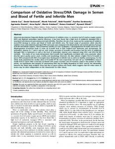
3.4. QPCR The efficiency and specificity of PCR often depends on the MgCl2 concentration in addition to the annealing temperature. These parameters should, therefore, be established by titration before proceeding with damage experiments. Initial optimization of PCR can be performed without radioactivity, the performance being assessed by ethidium staining of agarose gels. With QPCR (and ss-QPCR, see Subheading 3.5.) DNA damage m the region of the gene under study leads to a reduction in the amount of PCR product, (see Subheading 1.). It is essential to ensure that the only limiting component of the PCR is the template DNA and that the reaction remains in the exponential phase when terminated so that any damage to DNA will cause a directly proportional reduction in the amount of radioactive product. The important factors are cycle number and quantity of DNA, so preliminary experiments must be performed to determine the conditions required. The first experiment to do is to keep the amount of DNA constant at, for example, 0.5 ug and vary the number of cycles between 20 and 30 cycles. After quantitation of the radioactive product, the results should show an exponential increase in the amount of amplified product with increasing cycle number (see Fig. 4A) A cycle number is then chosen, which is well within the exponential range but which generates sufficient amplified DNA to be easily measured. In the N-ras example 26 cycles were chosen. Next a DNA titration is performed using this fixed number of cycles, the amount of DNA is varied between 0.1 and 1.0 p.g and the amount of amplified product should increase linearly in direct proportion to the amount of starting DNA (see Fig. 4B). These experiments thus establish the conditions under which QPCR and ss-QPCR will give a quantitative measurement of the amount of DNA template available for amplification (i.e., free of damage) and DNA damage experiments can be performed. 1. Reaction components and template DNA are mixed in a volume of 100 ju.L containing 50 pmol of each primer NRAS-A and NRAS-B; 2 U taq polymerase; 2 jnCi [a-32P]-dATP; 200 uMeach dATP, dGTP, dCTP, dTTP; 1 5 mMMgCl2 (see Note 7), 10 uL 10X PCR buffer. If necessary, add 40 uL mineral oil overlay. 2. Place tubes in thermal cycler and carry out cycling as follows- 3 min at 94°C initial denaturation then 26 cycles of: 1 mm at 94°C, 1 min at 60°C (annealing temp—see Note 8), 1 min at 72°C This is followed by a final incubation of 4 min at 72°C 3. For quantitative results each PCR should be carried out in triplicate and the following controls are essential: a. No DNA m PCR.
169
DNA Damage Detected by PCR-Based Methods 40000
30000 -
5 Q. O
20000 -
10000 -
Cycles 30000 •
B / n
20000 -
2 o.
o
•
/^
1
1
10000 -
0