Sambrook, J. (1989) Nature (London) 339, 721-723. 4. Wiman, B., Chimeluska, J. & Ranby, M. (1984) J. Biol. Chem. 259, 3644-3647. 5. van Mourik, J. A., ...
Proc. Natl. Acad. Sci. USA Vol. 89, pp. 7422-7426, August 1992 Cell Biology
Complexes of tissue-type plasminogen activator and its serpin inhibitor plasminogen-activator inhibitor type 1 are internalized by means of the low density lipoprotein receptor-related protein/!a2-macroglobulin receptor KIM ORTH*, EDWIN L. MADISON*, MARY-JANE GETHING*t, JOSEPH F. SAMBROOK*,
AND
JOACHIM HERZ4§
Departments of *Biochemistry and *Molecular Genetics and tHoward Hughes Medical Institute, University of Texas Southwestern Medical Center, Dallas, TX 75235
Communicated by Michael S. Brown, May 12, 1992
The requirement for complex formation of a protease with its inhibitor for efficient receptor-mediated endocytosis is not unprecedented. The protease inhibitor a2-macroglobulin forms a complex with a variety of endoproteinases that then cleave a specific region of the inhibitor, termed "the bait region," causing a conformational change in the inhibitor that leads to formation of a covalent bond between the attacking protease and the inhibitor. The conformational change also exposes a receptor-binding site on the a2-macroglobulin molecule. The endocytic receptor responsible for clearance of the a2-macroglobulin-protease complex has recently been shown to be the low density lipoprotein receptor-related protein (LRP) (11, 12). LRP is a large plasma-membrane protein of 4525 amino acids (13). The extracellular portion of 4400 amino acids undergoes a proteolytic cleavage during its passage through a post-Golgi compartment. In the resulting heterodimeric protein, the large subunit, LRP-515, remains noncovalently attached to the smaller LRP-85, which is anchored in the plasma membrane by a single membranespanning segment (13). Like the low density lipoprotein receptor, LRP can bind and endocytose lipoproteins containing apolipoprotein E (apoE) and has, therefore, been postulated to participate in the clearance of chylomicron remnants (13, 14). Although LRP is expressed in many different tissues, specific ligands for this receptor, when injected into the circulation, are almost exclusively taken up by the liver (15). During purification of LRP/a2-macroglobulin receptor, a small soluble protein of Mr ==39,000 was found to copurify with the receptor (11). A cDNA encoding this 39-kDa protein has been cloned and expressed in Escherichia coli as a fusion protein that consists of glutathioneS-transferase (GST) sequences at its N terminus and 39-kDa protein sequences at its C terminus. This fusion protein (GST-39-kDa protein) inhibits LRP-mediated uptake of apoE-enriched lipoproteins as well as a2-macroglobulin by cultured cells (16, 17). The parallels between the mechanism of inactivation of endoproteinases by a2-macroglobulin and the interaction between t-PA and PAM-i have led us to investigate the possibility that LRP might also function as a receptor for the t-PA-PAI-1 complex. MATERIALS AND METHODS t-PA and tyrosylprolylarginyl chloromethyl ketone (YPACK) were provided by W. F. Bennett and B. A. Keyt, Genentech. lodo-Beads and Iodo-Gen were purchased from Pierce. PAM-i was isolated and activated as described (5, 18). Protein
ABSTRACT Tissue-type plasmingen activator and urokinase are serine proteases secreted by many cell types that participate in biological processes, such as tissue restructuring, cell migration, and tumor metalst . Clinically, these proteases are used to dissolve coronary fibrin clots that are the proximal causes of acute myocardial infarction. In vivo, the activity of these enzymes is controlled by plasminn-activator inhibitors, members of the serpin family of protease inhibitors. This study shows that tissue-type plsminogen activator-inhibitor complexes bind in solution to low density lipoprotein receptorrelated protein (LRP), a large heterodimeric ubiquitous membrane receptor. In cultured cells, endocytosis and degradation of these complexes is reduced by polyclonal antibodies directed against LRP and inhibited by a Mr 39,000 protein that binds to LRP and inhibits its interaction with previously known ligands, including apolipoprotein E and a2-macroglobulin. We propose a role for LRP in the clearance of plasminogen activatorinhibitor complexes that is analogous to its function in the endocytosis of a2-macroglobulin-protease complexes.
Tissue-type plasminogen activator (t-PA) is a blood-borne serine protease of Mr -66,000 that converts the zymogen plasminogen to the highly active fibrinolytic protease plasmin. This activity is exploited clinically to dissolve thrombi that form in coronary arteries and cause acute myocardial infarction. t-PA is secreted as an enzymatically active protein of 527 amino acids. The N-terminal portion of the molecule is organized into four highly conserved structural motifs, whereas the C-terminal portion is a typical serine protease and contains a characteristic catalytic triad (1). Under normal physiological conditions, the concentration of t-PA in plasma is low (:0.1 nM), and much of the endogenous protein circulates as an enzymatically inactive complex composed of t-PA and a powerful inhibitor, the serpin plasminogen-activator inhibitor type 1 (PAI-1) (1, 2). PAl-i reacts rapidly with a series of positively charged residues located near the catalytic site of t-PA (3), forming a heterodimeric complex that is stable to treatment with SDS (4, 5). PAM- acts as a suicide substrate for both t-PA and urokinase, a plasminogen activator with many structural similarities to t-PA (1, 2). These plasminogen activator-inhibitor complexes are cleared from the circulation by the liver (1, 6, 7). In fact, in a perfused liver system t-PA-PAI-1 complex is cleared considerably faster than free t-PA (8). The hepatic receptor(s) responsible for this efficient removal of t-PA-PAI-1 complexes have not been identified, although studies with the human hepatic cell line HepG2 have shown that interaction between t-PA and PAT-i associated with the extracellular matrix is required for recognition and endocytosis (9, 10).
Abbreviations: apoE, apolipoprotein E; DMEM, Dulbecco's modified Eagle's medium; GST, glutathione-S-transferase; LRP, low density lipoprotein receptor-related protein; PAMI, plasminogenactivator inhibitor type 1; t-PA, tissue-type plasminogen activator; u-PA, urokinase plasminogen activator; YPACK, tyrosylprolylarginyl chloromethyl ketone. §To whom reprint requests should be addressed.
The publication costs of this article were defrayed in part by page charge payment. This article must therefore be hereby marked "advertisement" in accordance with 18 U.S.C. §1734 solely to indicate this fact.
7422
Cell
Biology: Orth et al.
A-Sepharose was purchased from Pharmacia. Computer software (ENZFITTER) was from Sigma; molecular weight markers and Affi-Gel Hz hydrazide were from Bio-Rad. Tissue culture media and plasticware were obtained from GIBCO/BRL. Iodination of Proteins and YPACK. Iodination was done by using Iodo-Beads, according to the manufacturer's instructions. YPACK (1.8 nmol) was iodinated with 100 j.Ci of Na125I (1 Ci = 37 GBq) by using the Iodo-Gen method. The YPACK/Na'25I mixture was transferred from the Iodo-Gencontaining vial to a new tube, and L-tyrosine was added to a final concentration of 5 mM. After incubation for 15 min at 220C, 1.8 nmol of t-PA was added, and incubation was continued at 220C for 2 hr. Ninety nanomoles of unlabeled YPACK was then added, and incubation was continued for an additional 30 min. Free 125I was removed by gel filtration. t-PA bound to 125I-labeled YPACK was unable to form complexes with PAM-i, as assayed by SDS/polyacrylamide gel analysis. Ligand Blotting. Protease-inhibitor complexes were prepared as follows: 0.05-1 pg of 1251-labeled t-PA (750-1794 cpm/ng) or urokinase plasminogen activator (u-PA) (3669 cpm/ng) was incubated in 180 ,ul of buffer (20 mM sodium phosphate, pH 7.0/0.1% Tween 20) in the absence or presence of 0.1 ,ug of YPACK or in the presence of 4-16 ,ug of PAI-i for 15 min at 22°C. Complex formation was assessed by SDS/PAGE under reducing conditions. Fractions of rabbit or rat liver membranes enriched for LRP by DEAE-cellulose chromatography (14) were separated by electrophoresis through nonreducing SDS/polyacrylamide gradient gels (310%) and transferred to nitrocellulose. Strips (containing =30 pg of protein) were processed for ligand blotting in the presence of 2 mM CaCl2 as described (14), except that 0.1% (vol/vol) Triton X-100 was added to the blotting buffer. Immunoblotting. Immunoblotting was done with monoclonal antibody at 5 pg/ml directed against rabbit or rat LRP515. Bound IgG was detected with 125I-labeled goat antimouse antibody (106 cpm/ml), as described (15, 16). Cell Culture. Stock cultures of COS-1 cells were grown in Dulbecco's modified Eagle's medium (DMEM)/10% fetal calf serum in an atmosphere of 5% CO2. After trypsinization 0.5 x 10⁶ cells were seeded into 12-well plates. Twelve hours later the medium was removed, and the cells were incubated in DMEM without fetal calf serum. After 1 hr the medium was replaced with fresh DMEM lacking glutamine but containing the indicated amounts of 175I-labeled reagents. Cellular degradation of radiolabeled ligands was determined by measuring the appearance of trichloroacetic acid-soluble radioactivity in the medium. Nonspecific degradation was measured in parallel dishes that did not contain cells. Experiments were done at subsaturating ligand concentrations to obtain the maximal ratio of signal-to-background (ratio -10:1 at 1 nM t-PA). Typically, -10% of the added ligand was converted to an acid-soluble form during the experiment. In experiments not shown here, cellular degradation was found proportional to the observed binding at all substrate concentrations. 125I-labeled t-PA (1293-1911 cpm/ng) was bound to COS cells by incubating 0.6 x 10⁶ cells at 4°C in 0.5 ml of Hepes-buffered DMEM, pH 7.4/5% bovine serum albumin and the indicated amounts of labeled ligand. After 90 min, cells were rinsed three times with phosphate-buffered saline/ 0.2% bovine serum albumin followed by two washes for 10 min in the same buffer. Cell-associated radioactivity was measured by adding 0.2 ml of 1 M NaOH per well and counting of the lysate. Apparent Kd and number of binding sites were calculated by using the approximation program ENZFIrrER.
The rat hepatoma cell line H4-II-E-C3 was obtained from L. Cottam (University of Texas Southwestern Medical Cen-
Proc. Natl. Acad. Sci. USA 89 (1992)
7423
ter). Tissue culture and assay conditions were the same as described for COS cells. Isolation of LRP by Immunoaffinity Chromatography. Monoclonal anti-LRP 515 (2.5 mg) was treated with Affi-Gel Hz hydrazide gel (1.5-ml bead vol). Approximately 1 ml of anti-LRP resin was then incubated overnight at 40C with a crude membrane fraction prepared from rabbit liver (14). Control anti-LRP resin was incubated with blotting buffer. Resins were then washed six times with blotting buffer and resuspended in a final volume of 3 ml and 1.5 ml, respectively. 1251-labeled t-PA or 1251-labeled t-PA-PAI-1 complexes were incubated for 1 hr at 220C with 100 ul of 1:1 (beads/blotting buffer) slurry of LRP resin or control resin with or without GST or GST-39-kDa fusion protein at 10 pg/ml. Samples were washed three times with blotting buffer, boiled for 3 min in sample buffer containing 1% SDS and 100 mM dithiothreitol, and analyzed by electrophoresis through an SDS/8% polyacrylamide gel.
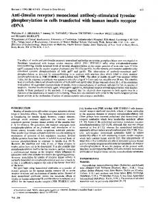
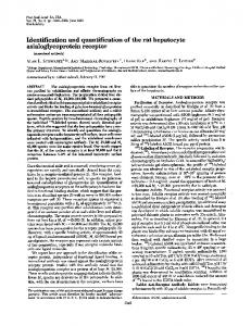
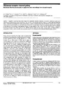
RESULTS Complexes of t-PA-PAI-1 Bind Specifically to LRP in Vitro. To investigate the possibility that LRP might function as a receptor for clearance of t-PA-PAI-1 complexes (1, 2, 7), we tested whether such complexes could specifically interact with LRP in vitro. Fig. la shows the results of an experiment in which 12-I-labeled t-PA, 125I-labeled t-PA that had been inactivated by the tetrapeptide inhibitor YPACK (19), and t-PA-PAI-1 complexes radiolabeled in the t-PA moiety were tested for their ability to bind to a partially purified fraction of rabbit liver membranes containing LRP. t-PA-PAI-1 complex (lane 3) but not uncomplexed t-PA (lane 1) or t-PAYPACK, which cannot form a complex with PAI-1 (lane 2), bound to a protein that comigrates with LRP, as identified by probing the nitrocellulose filter with a monoclonal antibody specific to LRP (lane 4) (15). When a similar experiment was done by using 125I-labeled u-PA, u-PA-PAI-1 complexes (lane 7), but not free u-PA (lane 5) or u-PA-YPACK (lane 6), bound to a protein the molecular mass of which is indistinguishable from that of LRP. Binding of both t-PA-PAI-1 and u-PA-PAI-1 complexes depended on Ca2+ (Fig. lb, lane 4) as is so for apoE (14) and for a2-macroglobulin-protease complexes (20). We next examined the effect of the 39-kDa protein on the binding of t-PA-PAI-1 complex to immobilized LRP. As shown in Fig. lb, GST-39-kDa protein (lane 6), but not GST alone (lane 5), completely inhibited binding of the t-PA-PAI-1 complex to LRP. Human t-PA-PAI-1 complex bound to both rabbit (Fig. la) and rat (Fig. lb) LRP. To confirm that plasminogen activator-PAI-1 complexes bind to LRP, we tested whether LRP immobilized on an immunoaffinity matrix can bind 125I-labeled t-PA-PAI-1 complex (Fig. 2). By contrast to 1251-labeled t-PA (lane 1), 125I-labeled t-PA-PAI-1 bound to the immobilized receptor (lane 2). In the presence of GST-39-kDa protein (lane 4), but not of GST alone (lane 3), binding of 125I-labeled t-PA-PAI-1 complexes to the immunoaffinity matrix was abolished. Furthermore, no binding was seen when an equivalent amount of immunoaffinity matrix containing no LRP was incubated with 1251-labeled t-PA (lane 5) or 125I-labeled t-PA-PAI-1 complex (lane 6). Binding of the enzyme-inhibitor complexes is, therefore, mediated by LRP and is not from nonspecific interaction with the matrix. Binding and Degradation of t-PA-PAI-l Complexes by COS Cells. To corroborate these in vitro findings, we assayed the ability of simian COS cells, which express both LRP and PAI-1 (K.O., unpublished observations), to specifically bind and endocytose 125I-labeled t-PA. The binding of t-PA to COS cells is not affected by amino acid substitutions in the epidermal growth factor domain (K.O., unpublished observations) that affect the ability of t-PA to interact with a
7424
Cell Biology: Orth et al.
Proc. Natl. Acad. Sci. USA 89 (1992)
a
b
_11251
1
+
+
+
PAl-i , + YPAC K __ + + _V2511 t-PA ±. ±+ i - + antl-LRP Lane 1 42 l 3 4 5 67 .
GST-39 GST EDTA _ PAl-1 _ [12511 t-PA __
l
l+
+
+
+ + .i. + + + anti-LRP + - - - - Lane 1 2 3 4 5 6
+
+ +
kDs
kDa *.
4
LRP-515>-
e
V
LRP-515>'
*
0
200-
116
-
116 -
92 -
92 -
68
-
66-
FIG. 1. (a) Binding of t-PA-PAI-1 and u-PA-PAI-1 complexes to LRP. 125I-labeled t-PA (lane 1), t-PA-YPACK (lane 2), or t-PA-PAI-1 (lane 3) were incubated with nitrocellulose strips containing rabbit liver membrane fraction and processed for ligand blotting. An adjacent strip (lane 4) was immunoblotted with anti-LRP-515 monoclonal antibody (15), and bound IgG was detected with 1251-labeled goat anti-mouse antibody. Parallel experiments were done by using 125I-labeled u-PA (lanes 5-7). Autoradiographs were exposed overnight at -70TC (t-PA blot), overnight at 22TC (u-PA blot), or 30 min at -70TC (anti-LRP blot) with an intensifying screen. (b) Binding of t-PA-PAI-1 complex to LRP requires Ca2+ and is inhibited by GST-39-kDa protein. 1251-labeled t-PA-PAI-1 complex was formed as described above. Nitrocellulose strips containing rat liver-membrane fraction were preincubated for 15 min at 22TC in blotting buffer with or without 5 mM EDTA, GST at 25 pg/ml or GST-39-kDa protein (GST-39) at 25 pg/ml before adding 1251-labeled t-PA or 125I-labeled t-PA-PAI-1 complex. An adjacent strip was processed for immunoblotting with monoclonal anti-rat LRP-515 IgG (16). Strips were exposed at -70TC with an intensifying screen for 20 hr (t-PA) and 2.5 hr (immunoblot), respectively.
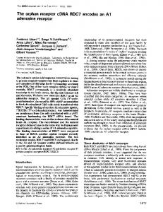
specific receptor on the rat hepatoma cell line H4 (21). To control for the possibility that COS cells clear t-PA from the medium also when it is not complexed to PAT-i, we measured the ability of the cells to degrade YPACK-t-PA complexes that had been radiolabeled in the YPACK moiety (Fig. 3). Although t-PA that had been directly labeled with 1251 was efficiently degraded by the cells, no acid-soluble radioactivity t-PA
-+I
~
tl t-PA/ 'Al-I l+ GST 39 I ] GST-39+
Lane
I
.2 I3
4
was recovered from the medium when the label resided on the YPACK moiety. t-PA degradation by COS cells, therefore, requires a functionally active site on the t-PA molecule. This situation is consistent with degradation occurring only by a
complex-dependent pathway. Fig. 4a shows that COS cells express a single class of binding sites for 1251-labeled t-PA with an apparent Kd of 8 x 10⁻⁹ M and -50,000 sites per cell. When a fixed concentration of 125I-labeled t-PA (100 ng/ml = 1.52 nM) was incubated with COS cells for various periods of time, increased amounts of trichloroacetic acid-soluble radioactivity were
5 6
Complex _ ~0 0.)
* _o-
FIG. 2. Recombinant 39-kDa protein inhibits binding of t-PAPAT-i complex to immunoaffinity-purified LRP. Resin containing
anti-LRP monoclonal antibody with or without LRP bound was incubated with 125T-labeled t-PA or t-PA-PAI-1 complex in the absence or presence of GST or GST-39-kDa protein (GST-39) at 10 Ag/ml. Bound ligand was detected by SDS/8% PAGE under reducing conditions. The gel was calibrated by using high-molecular-mass markers. Positions corresponding to the M, of t-PA-PAI-1 complex (arrowhead at 110 kDa) and two-chain t-PA (asterisk at -35 kDa) are indicated. The presence of two-chain t-PA in lanes 2 and 3 is due to partial lability of t-PA-PAI-1 complex to boiling in SDS under reducing conditions (9). The autoradiograph was exposed for 4 days at -70"C with an intensifying screen.
(U
*t-PA
*YPACK/t-PA
FIG. 3. Endocytosis of 1251-labeled-t-PA or 125I-labeled YPACKt-PA in COS cells. Degradation of 125I-labeled t-PA (50 ng/ml, 0.76 nM, 1073 cpm/ng) or YPACK-t-PA complex labeled on the YPACK moiety (50 ng/ml, 0.76 nM, 1779 cpm/ng) incubated at 37"C for 20 hr with 0.5 x 10⁶ cells in the presence of either GST at 10 pg/ml or recombinant GST-39-kDa protein at 10 pg/ml (GST-39) was determined by measuring acid-soluble radioactivity released into the medium. Means of duplicate experiments are shown. Absolute amounts of degraded 1251-labeled t-PA are indicated and normalized to 106 cells. *, 125I label.
Proc. Natl. Acad. Sci. USA 89 (1992)
Cell Biology: Orth et al.
b
a - .80 0
A 0
0
*/
10
,i0
60
at
PC A"~ ~
C
*
0
.I
7425
~
~
-v
100
A.
~
40
ND
20
0
F10
0 0
60 80
20 40
100
120
0
C
1.0Norwm
400
@
Do &^3x
d
2to0
0
ao
6
3
t-PA, uM
9
12
Time, hr .o GST
*t-PA
Is
P0 449
PC LOOD
200D
I
04
%
I
z 1
~Anti-I"P
d
Lb1 0
150 300 450 600
IgG, ,ug/ml
0
o
x
I*
0
10
GST-39
20
GST or GST-39-kDa protein, ;ug/ml
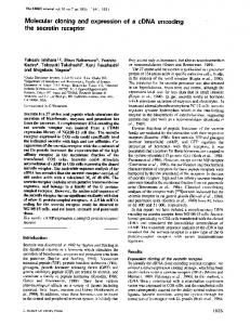
FIG. 4. Binding and degradation of 1251-labeled t-PA by COS cells. Absolute amounts of degraded 125I-labeled t-PA are indicated and normalized to 106 cells. (a) Binding of 125I-labeled t-PA (1293 cpm/ng) to COS cells (0.6 x 10⁶ cells per well) was done in the absence (A) or presence (A) of 1 AM unlabeled t-PA; *, specific cellular binding. One experiment representative of three is shown. (b) Degradation of 125I-labeled t-PA (100 ng/ml, 1.52 nM, 755 cpm/ng) incubated at 370C for various periods of time with 0.5 x 10⁶ cells per well. (c) Degradation of 125I-labeled t-PA (50 ng/ml, 0.76 nM, 518 cpm/ng) incubated at 370C for 20 hr with 0.5 x 10⁶ cells per well in the presence of the indicated concentrations of nonimmune IgG or polyclonal anti-LRP IgG. Cells were preincubated with IgG for 2 hr before adding 125I-labeled t-PA. One experiment representative of three is shown. (d) Degradation of 1251-labeled t-PA (50 ng/ml, 0.76 nM, 800 cpm/ng) incubated at 37TC for 20 hr with 0.8 x 10⁶ cells per well with the indicated concentrations of either GST or recombinant 39-kDa fusion protein (GST-39). GST or GST-39-kDa protein were added to the cells 15 min before adding 125I-labeled t-PA. One experiment representative of three is shown.
recovered in the medium (Fig. 4b), indicating that 1251-labeled t-PA was actively taken up and degraded. Based on the measured Kd and the number of binding sites per cell a receptor-recycling time of -12 min can be calculated. This value agrees well with those determined for other constitutively recycling receptors (22), demonstrating a physiologically relevant uptake mechanism. When the cells were incubated with increased concentrations of polyclonal anti-LRP IgG (16), degradation of 125I-labeled t-PA could be reduced by -90% (Fig. 4c). Nonimmune control IgG had no effect on the ability of the cells to degrade 1251-labeled t-PA. The IgG concentrations required to reduce 125I-labeled t-PA degradation by 50%o ('150 ug/ml) closely resemble those required for reduction of apoE-mediated uptake of lipoproteins and uptake and degradation of a2-macroglobulin by human fibroblasts (16). Degradation of t-PA by COS cells is also inhibited by GST-39-kDa fusion protein but not by GST alone (Fig. 4d). The amount of GST-39-kDa fusion protein required to reduce the degradation of t-PA by 50% (-0.5 pg/ml) (i) is very similar to the amount needed to inhibit apoE-dependent uptake of lipoproteins and a2-macroglobulin by human fibroblasts (16) and (ii) correlates closely with the Kd determined for the binding of 39-kDa protein to LRP/a2-macroglobulin receptor (11). Degradation of t-PA by H4 Cells. To investigate a possible relationship between the complex-dependent pathway seen in COS cells and previously described mechanisms (21) for
*YPACKC/t-PA
FIG. 5. Endocytosis of 1251-labeled-t-PA or 125I-labeled YPACKt-PA in H4 cells. Degradation of 125I-labeled t-PA (50 ng/ml, 0.76 nM, 1073 cpm/ng) or YPACK-t-PA complex labeled in the YPACK moiety (50 ng/ml, 0.76 nM, 1779 cpm/ng) incubated at 370C for 20 hr with 1.0 x 10⁶ cells with either GST at 10 pZg/ml (n) or recombinant 39-kDa protein at 10 pLg/ml (GST-39, o) was determined by measuring acid-soluble radioactivity released into the medium. Means of duplicate experiments are shown. Absolute amounts of degraded '25I-labeled t-PA are indicated and normalized to 106 cells. *, mlI label.
endocytosis of free t-PA in a liver-derived cell line, we performed the experiment of Fig. 5. Both COS cells and H4 cells synthesize comparable amounts of LRP, as demonstrated by metabolic labeling and immunoprecipitation (data not shown). Figs. 3 and 5 show that both cell lines degrade t-PA with approximately equal efficiency and that degradation of t-PA in both cases is inhibited by GST-39-kDa protein. These results demonstrate that LRP is involved in endocytosis oft-PA by COS cells and H4 cells. By contrast with COS cells, however, a fraction of cellular t-PA degradation is independent of complex formation of the protease with its inhibitor PAI-1, as shown by the ability of the H4 cells to degrade YPACK-t-PA complex labeled in the YPACK moiety. This PAI-i-independent degradation of t-PA is also inhibited by GST-39-kDa protein, suggesting that LRP is an essential component in both complex-dependent and independent pathways. DISCUSSION In this paper we have shown that complexes of t-PA and u-PA with the inhibitor PAM- can specifically bind in vitro to LRP. This interaction depends upon Ca2+ and is disrupted by 39-kDa protein, which interferes with the binding of all known ligands to LRP. Degradation of 125I-labeled t-PA by COS cells is greatly reduced in the presence of the 39-kDa protein and by antibodies directed against LRP. These independent lines of evidence show that LRP is involved in clearance of the plasminogen activator t-PA and possibly of u-PA as well. Both our in vitro data (Figs. 1 and 2) and the finding that COS cells are unable to degrade uncomplexed t-PA (Fig. 3) suggest that complex formation of t-PA with PAIMl is essential for binding to LRP. When taken together with previous results (11, 12), our experiments indicate that LRP has an important and general role in the constitutive clearance of proteases that have been inactivated by complex formation with their respective inhibitors. By analogy, other members of the serpin family (e.g., antithrombin III, a2antiplasmin, Cl-inhibitor, etc.) may be recognized and internalized by the same receptor system (LRP) after complex formation with their respective target proteases. Specific ligands of LRP when injected into the circulation are predominantly cleared by the liver (15), which is also the predominant site that clears t-PA from the circulation (1, 6, 7). However, because LRP is ubiquitously expressed, our
7426
Cell Biology: Orth et al.
data suggest that it might also play an important role in the uptake and degradation of extracellular protease-inhibitor complexes, thus preventing their accumulation in the intercellular space. Our observation that u-PA in conjunction with PAM-i can also bind to LRP on ligand blots agrees with previous studies that found that u-PA-PAI-1 and t-PA-PAI-1 complexes compete for the same binding site (10). Although both t-PA and u-PA are secreted proteins and active in solution, u-PA is thought to express its regulated biological activity while bound to a glycosylphosphatidylinositol-linked cell-surface receptor (23, 24). The mode of u-PA clearance from the plasma membrane after inactivation by PAl-i (24) or PAI-2 (25) is unclear; however, this process could involve the participation of another receptor (26). Because LRP is a constitutively recycling cell-surface receptor (15) capable of binding u-PA-PAI-i complexes in solution, as shown here, conceivably LRP also interacts with these complexes when they are bound to the u-PA receptor and, therefore, have been restricted in their mobility to the two dimensions of the lipid bilayer. This hypothesis raises interesting questions about the role of LRP in the regulation oftissue restructuring, cell migration, tumor invasion, and implantation of developing embryos in the uterus (2, 25, 27-29). Our findings strongly indicate that PAM-i mediates binding of two plasminogen activators, t-PA and u-PA, to LRP. Bu et al. (30) have shown that free t-PA binds to LRP on the surface of rat MH1C1 hepatoma cells in a manner apparently independent of PAI-1. This binding may correspond to the fraction of t-PA that, after inhibition by YPACK, cannot complex with PAM-i but is, nevertheless, degraded by rat H4 hepatoma cells (Fig. 5). Using purified reagents in our in vitro experiments (Figs. 1 and 2), we were unable to detect a direct interaction of free t-PA with LRP, suggesting that another component is essential for the binding of uncomplexed t-PA to LRP in hepatic cells. Because uptake and degradation of YPACK-t-PA is inhibited by GST-39-kDa protein, LRP is probably involved in this clearance mechanism. We propose that t-PA binds to a specific receptor on the surface of hepatic cells via the t-PA epidermal growth factor domain (21). This t-PA-receptor complex could be stabilized by LRP, which then mediates the clearance of the protease in a trimeric complex in which PAM-i plays no role. In the absence of such a specific receptor, as in COS cells, t-PA clearance would depend on complex formation with PAI-1. While the question as to how PAI-i-dependent and -independent clearance pathways of t-PA are related to each other clearly requires further investigation, the studies presented here and in the accompanying report by Bu et al. (30) provide strong evidence that LRP is essential for the clearance of plasminogen activators from the circulation and from the extracellular space, in general. We thank Wen-Ling Niu for excellent technical assistance, Y. K. Ho for helpful discussions, and Michael Brown, Joseph Goldstein, and David Russell for invaluable suggestions and critical reading of the manuscript. Ralph Shohet generously provided PAI-1. This work was supported by research grants from the National Institutes of Health. J.H. is supported by a Lucille P. Markey Scholar Award and by the Syntex Scholar Program.
Proc. Natl. Acad. Scl; USA 89 (1992) 1. Lijnen, H. R. & Collen, D. (1991) Thromb. Haemostasis 66, 88-110. 2. Dano, K., Andreasen, P. A., Grondahl-Hansen, J., Kristensen, P., Nielsen, L. S. & Skriver, L. (1985) Adv. Cancer Res. 44, 139-266. 3. Madison, E., Goldsmith, E., Gerard, R., Gething, M. J. & Sambrook, J. (1989) Nature (London) 339, 721-723. 4. Wiman, B., Chimeluska, J. & Ranby, M. (1984) J. Biol. Chem. 259, 3644-3647. 5. van Mourik, J. A., Lawrence, D. A. & Loskutoff, D. J. (1984) J. Biol. Chem. 259, 14914-14921. 6. Korninger, C., Stassen, J. M. & Collen, D. (1981) Thromb. Haemostasis 46, 658-661. 7. Rijken, D. C., Otter, M., Kuiper, J. & van Berkel, T. J. C.
(1990) Thromb. Res. Suppl. X, 63-71. 8. Wing, L. R., Hawksworth, G. M., Bennett, B. & Booth, N. A.
(1991) J. Lab. Clin. Med. 117, 109-114. 9. Morton, P. A., Owensby, D. A., Sobel, B. E. & Schwartz, A. L. (1989) J. Biol. Chem. 264, 7228-7235. 10. Morton, P. A., Owensby, D. A., Wun, T. C., Billadello, J. J. & Schwartz, A. L. (1990) J. Biol. Chem. 265, 14093-14099. 11. Strickland, D. K., Ashcom, J. D., Williams, S., Burgess,
W. H., Migliorini, M. & Argraves, W. S. (1990) J. Biol. Chem. 265, 17401-17404. 12. Kristensen, T., Moestrup, S. K., Gliemann, J., Bendtsen, L.,
Sand, 0. & Sottrup-Jensen, L. (1990) FEBS Lett. 276, 151-155.
13. Brown, M. S., Herz, J., Kowal, R. C. & Goldstein, J. L. (1991) Curr. Opin. Lipidol. 2, 65-72. 14. Kowal, R. C., Herz, J., Weisgraber, K. H., Mahley, W. H., Brown, M. S. & Goldstein, J. L. (1990) J. Biol. Chem. 265, 10771-10779. 15. Herz, J., Kowal, R. C., Ho, Y. K., Brown, M. S. & Goldstein, J. L. (1990) J. Biol. Chem. 265, 21355-21362. 16. Herz, J., Goldstein, J. L., Strickland, D. K., Ho, Y. K. & Brown, M. S. (1991) J. Biol. Chem. 266, 21232-21238. 17. Moestrup, S. K. & Gliemann, J. (1991) J. Biol. Chem. 266, 14011-14017. 18. Hekman, C. M. & Loskutoff, D. J. (1985) J. Biol. Chem. 260, 11581-11587. 19. Bennett, W. F., Paoni, N. F., Keyt, B. A., Botstein, D., Jones, A. J. S., Presta, L., Wurm, F. M. & Zoller, M. J. (1991) J. Biol. Chem. 266, 5191-5201. 20. Moestrup, S. K., Kaltoft, K., Sottrup-Jensen, K. & Gliemann, J. (1990) J. Biol. Chem. 265, 12623-12628. 21. Bassel-Duby, R., Jiang, N. Y., Bittick, T., Madison, E., McGookey, D., Orth, K., Shohet, R., Sambrook, J. & Gething, M. J. (1992) J. Biol. Chem. 267, 9668-9677. 22. Brown, M. S. & Goldstein, J. L. (1986) Science 232, 34-47. 23. Ploug, M., Ronne, E., Behrendt, N., Jensen, A. L., Blasi, F. & Dano, K. (1991) J. Biol. Chem. 266, 1926-1933. 24. Cubellis, M. V., Wun, T. C. & Blasi, F. (1990) EMBO J. 9, 1079-1085. 25. Estreicher, A., Muhlhauser, J., Carpentier, J. L., Orci, L. & Vassalli, J. D. (1990) J. Cell Biol. 111, 783-792. 26. Ploug, M., Behrendt, N., Lober, D. & Dano, K. (1991) Semin. Thromb. Hemostasis 17, 183-193. 27. Saksela, 0. & Rifikin, D. B. (1988) Annu. Rev. Cell Biol. 4, 93-126. 28. Sappino, A. P., Huarte, J., Belin, D. & Vassalli, J. D. (1989)J. Cell Biol. 109, 2471-2479. 29. Stephens, R. W., Pollanen, J., Tapiovaara, H., Leung, K. C., Sim, P. S., Salonen, E. M., Ronne, E., Behrendt, N., Dano, K. & Vaheri, A. (1989) J. Cell Biol. 108, 1987-1995. 30. Bu, G., Williams, S., Strickland, D. K. & Schwartz, A. L. (1992) Proc. Natl. Acad. Sci. USA 89, 7427-7431.