Exp Brain Res (1999) 126:315–335
© Springer-Verlag 1999
R E S E A R C H A RT I C L E
Ilsun M. White · Steven P. Wise
Rule-dependent neuronal activity in the prefrontal cortex
Received: 15 July 1998 / Accepted: 7 January 1999
Abstract We studied single-neuron activity in the prefrontal cortex (PF) while a monkey performed a task according to two different rules, termed conditional and spatial. The monkey viewed a video screen, and its task required a hand movement in response to the dimming of a light spot. There were four light spots on the screen: right, left, up, and down from the center. Only one of the four spots dimmed, and the degree of dimming was slight. Accordingly, the monkey needed to foveate the “correct” light spot to detect the dimming. A visual cue indicated which of the four light spots would be deemed correct and, thus, would dim on each trial. The sequence of events was as follows: a fixation spot appeared at the center of the screen; then, a cue appeared twice at one of the four potential target locations; then, the four target spots appeared; and, finally, one of them dimmed. Except for the color of an initial fixation point, the cues, their locations, and other events were identical for the conditional and spatial rules. The rules differed in one essential way. For the conditional rule, nonspatial attributes of the visual cue indicated which of the four light spots would dim, and the cue’s location was irrelevant. For the spatial rule, the cue’s location determined the correct target on that trial. The light spot at the location of the cue always dimmed, regardless of which cue appeared there. Our sample included 221 PF neurons showing significant task-related activity modulation, distributed among dorsal, dorsolateral, and ventral PF regions. Between one-third and one-half of the I.M. White · S.P. Wise (✉) Laboratory of Systems Neuroscience, National Institute of Mental Health, P. O. Box 608, Poolesville, MD 20837, USA e-mail:
[email protected] Tel.: +1-301-496-1201, Fax: +1-301-402-0236 Express mail service: S.P. Wise NIH Animal Center, Elmer School Road, Building 110, Room 119, Poolesville, MD 20837, USA Current address: I.M. White Behavioural Biology Laboratory, Institute of Toxicology, Swiss Federal Institute of Technology (ETH Zürich), Schorenstrasse 16, CH-8603 Schwerzenbach, Switzerland
sample in each of those regions showed statistically significant activity differences that could be attributed to the rule. Selectivity for cues and/or their locations was common. However, there was no significant regional segregation of such selectivity. These data support the hypothesis that PF plays a role in the guidance of behavior according to previously learned rules. Key Words Frontal cortex · Executive functions · Rules · Arbitrary mapping · Working memory
Introduction The prefrontal cortex (PF) was among the first subjects of behavioral neurophysiology (Fuster and Alexander 1971; Kubota and Niki 1971), and an extensive series of studies over the subsequent quarter century sought insight into its functional organization. Those studies, along with the neuroanatomical and neuropsychological literature of the period, have been reviewed previously (Wise et al. 1996). Several recent studies have extended the pioneering work by examining the role of PF in processing information about stimuli, responses, rewards, errors, and other behaviorally significant events (Hasegawa et al. 1998; Lecas 1995; Miller et al. 1996; Rao et al. 1997; Rolls and Baylis 1994; Rolls et al. 1996a, 1996b; Sakagami and Niki 1994b; Watanabe 1996; Wilson et al. 1993). Studies of stimulus-related information processing have emphasized the storage of stimulus information in short-term memory (e.g., Wilson et al. 1993) and attentional selection among stimuli (e.g., Miller et al. 1996). The present study emphasized a more abstract aspect of information processing in PF, one involving behaviorguiding rules rather than objects, places, or events such as rewards. Developing the ideas of Passingham (1993) and others, we hypothesized that a principal function of PF involves the application of behavior-guiding rules (Wise et al. 1996). In the present study, we tested that hypothesis by comparing the activity of PF cells during the performance of a task according to two well-learned
316
rules. Our strategy in searching for the predicted rule effects was analogous to that generally used in neuroimaging studies in which two “activity” maps are compared: we contrasted the activity of each PF neuron during task performance according to one rule with that of the same neuron during performance according to the other rule. These data have been previously reported in abstract form (White and Wise 1997).
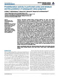
Material and methods Subject One male rhesus monkey (Macaca mulatta), 6–7 kg, was used in this study. All procedures conformed with the Guide for the Care and Use of Laboratory Animals (revised 1996, ISBN 0–309–05377–3) and complied with an institutionally approved animal study proposal. The monkey sat, head fixed, in a primate chair facing a video screen 43 cm away with a switch, termed a bar, attached to the front of the chair and within easy reach. Behavioral design We trained the monkey to perform a task with two rules, conditional and spatial, alternating in blocks of 40–70 trials. ConditionalFig. 1 Events in the behavioral tasks, showing one example trial for both rules. The example involves presentation of cue A in the up location. Events and task periods are described in the Methods section. Inset at lower right shows monochrome versions of the cues, placed in the locations with which they are uniquely associated for the conditional rule. Q1 First presentation of the cue, Q2 second cue presentation, Q cue
rule trials began when the monkey fixated a blue spot (1° visual angle) at the center of the video screen. Later, the fixation spot disappeared and, simultaneously, one of four complex (2°) visual cues appeared for 0.25 s at one of four locations: (7° up, down, left, or right from the center). The four visual cues were distinctively different in color and shape, each being a combination of two shapes (circles, squares, and rectangles) of different color. Monochrome versions of the cues, designated A, B, C, and D, are shown in Fig. 1 (lower right). The locations of each cue and the order of their appearance were randomized to produce 16 cue-location combinations. After either a variable 0.45- to 0.75-s delay or a fixed 0.5-s delay, the cue reappeared at the same location for 0.25 s. The first presentation of the cue was termed Q1, the second Q2. [We presented the cue twice so that it would appear both without (Q1) and with (Q2) a high level of predictability of that cue. However, that aspect of the experimental design yielded little of interest and will not be pursued in this report.] Following Q2, another variable 0.45to 0.75-s delay or a fixed 0.5-s delay ensued, after which four white target spots simultaneously appeared 7° up, down, left, and right from center. These were the four possible targets of eye movement on every trial. For conditional-rule trials, the cue’s shapes and colors indicated which of the four target spots would dim, and each cue was uniquely associated with one of the four potential targets. A correct eye movement consisted of a saccade to the location instructed by the cue. [After the first fifth of the recording sessions, a saccade to the correct target was reinforced by a drop of liquid diet (Ensure, Abbott Laboratory) and incorrect eye movements aborted the trial. During the early recording sessions, no reward was given for the eye movement and the trial continued if the initial saccade was to an incorrect target]. If a saccade was made to the correct target spot, the monkey needed to continue to fixate it in order to detect a subtle dimming. Dimming occurred 0.5, 1.0, or 1.5 s after target onset (pseudorandomly selected), at which time the monkey was required to release the bar within 0.55 s to receive reinforcement. Thus, during conditional-rule trials, the nonspatial attributes of each cue (mainly shape and color) indicated the correct target location for that trial, and the cue’s location was irrelevant.
317 During spatial-rule trials, all events and cues were identical to those for conditional-rule trials, except that the fixation spot was yellow. Fixation-spot color was intended to help the monkey readily distinguish the rule at the beginning of each trial – notwithstanding the fact that, within blocks of trials, the rule was the same – and to assist in the transition from one rule to the other. For the spatial rule, the location of the cue indicated where a target would dim on that trial, which was at the same location as the cue had previously appeared. The nonspatial attributes of the cue were irrelevant. One example each of a conditional-rule trial (left) and a spatial-rule trial (right) is shown in Fig. 1. For both kinds of trials, cue A appeared in the “up” location. As noted by the window surrounding the target (which was not visible to the monkey), this cue signaled that the right target would dim on conditional-rule trials, whereas the target above the fixation point would dim on spatialrule trials. Surgical and recording procedure A stainless steel recording chamber (27×36 mm) was implanted over the left frontal cortex, and a head-restraint device was implanted in the same procedure. The monkey received the analgesic banamine (0.5 mg/kg, IM) for 3 days postoperatively, as well as antibiotics. Postoperative training resumed one week after surgery. Tungsten microelectrodes (Haer Instruments, Brunswick, Maine, 1–3 MΩ measured at 1 kHz) were used to record neuronal activity in the monkey’s frontal cortex. Single-unit potentials were filtered with a bandpass of 600 Hz to 6 kHz, amplified and discriminated using a Multispike Detector (Alpha-Omega Engineering, Nazareth, Israel) or a time-amplitude waveform discriminator (BAK Electronics, Rockville, Maryland, USA). Single-neuron activity was isolated as the monkey performed the task according to either the conditional or spatial rule, in roughly equal numbers. Cell activity was recorded during the first rule for a block of 40–70 trials, then the alternative rule was enforced for a similar number of trials. For 62% of the neuronal sample, we could gather at least one subsequent block of data for the first rule (to make at least three blocks overall), a percentage that did not differ significantly among the regions of cortex studied. The gaze angle of the right eye was sampled at 250 Hz and recorded with an infrared oculometer (Bouis Instruments, Karlsruhe, Germany). Oculomotor behavior was recorded during each trial for every neuron. Electromyographic (EMG) activity of shoulder, neck, and trunk muscles was recorded using surface cup electrodes at the end of the recording sessions. EMG signals were filtered and discriminated in the same manner as the neuronal signal to create a pulse replica. Muscles monitored on the right (performing) side included deltoid, triceps, biceps, extensor carpi ulnaris, latissimus dorsi, infraspinatus, pectoral and temporal muscles. Trapezius was examined bilaterally. Intracortical microstimulation was employed during the early phases of neural recording. Trains of 0.2-ms cathodal constantcurrent pulses (10–80 µA), delivered at 350 pulses/s for a duration of 100–350 ms (PSIU-6, Grass Instruments, Quincy, Massachusetts, USA), enabled the identification of the frontal eye field. Data analysis PF activity was analyzed during time windows in eight task periods: five stimulus periods and three delay periods. The five stimulus periods consisted of fixation spot onset (during the pre-cue interval), the first presentation of the cue (Q1), the second presentation of the cue (Q2), target onset, and target dimming (see Fig. 1). Each of these time windows extended from 50 ms until 250 ms after stimulus onset. The three delay periods consisted of time windows prior to Q2 (the inter-Q delay), target presentation (the pretarget delay), and target dimming (the pre-dim delay). Activity was measured from 200 ms until 100 ms before the relevant event.
Fig. 2 Penetrations sites at which task-related neurons were found. Circle diameter is proportional to the number of task-related neurons at each coordinate. Filled squares indicate the location of pins inserted at known coordinates. PFd Dorsal prefrontal cortex, PFdl dorsolateral prefrontal cortex, PFv ventral prefrontal cortex, F2/F7 supplementary eye field and dorsal premotor areas, Vedge the ventral extreme of the lateral convexity, Medge the medial limit of the hemisphere, p principal sulcus, a arcuate sulcus, s superior precentral sulcus, c central sulcus, i intraparietal sulcus A reference period was defined during the pre-Q interval. It lasted from 300 ms until 100 ms before Q1. While this epoch was not ideal as a reference period due to anticipatory activity during the pre-Q interval in some neurons, it was the most useful choice among the alternatives available and was appropriate for the bulk of the population. Both task relatedness and rule effects were detected with a two-factor ANOVA (α=0.05). A neuron was considered task-related if it had activity that significantly differed from that in the reference period for one or more of the eight task periods examined. Cells showing rule effects were identified by comparing activity for the conditional and spatial rules. A neuron was considered rule dependent if, in addition to being task related, it showed a significant rule-to-rule activity difference for a given task period. For each cell, a Bonferroni correction was applied to compensate for testing up to eight task periods (P0.05). Saccadic eye-movement latencies were measured from the onset of Q1, trial-by-trial, for a typical data set (taken while recording the activity of the cell illustrated in Fig. 14). The mean saccadic latency was 118±32 ms (n=205), with a range from 55 ms to 210 ms. The distribution was bimodal, with one mode at 75 ms and the other at 135 ms. The faster mode appears to correspond to express saccades, which were first reported when
319 Fig. 4 A Dorsolateral prefrontal cortex cell showing a rule effect, with a preference for conditional-rule trials. All data aligned on the onset of Q1. Data for all cues and cue locations are combined for each rule. Averages based on 154 conditional-rule trials and 114 spatial-rule trials. B Ventral prefrontal cortex cell showing a preference for spatial-rule trials, in the format of A. Averages based on 69 conditional-rule trials and 51 spatial-rule trials. Q1 First presentation of the cue, Q2 second cue presentation
there was a temporal “gap” between a fixation-point offset and the onset of a saccade target (Fischer and Boch 1983). In the present task, there was no gap between the fixation-point offset and Q1 onset. However, the monkey was highly practiced in making oculomotor responses to one of the four (and only four) possible cue locations. Such overtraining to saccade-target location has been shown to be the most important factor in promoting express saccades (Paré and Munoz 1996). The task was designed primarily as a study of rule-dependent oculomotor behavior. Except for the first onefifth of the recording sessions, the monkey was explicitly rewarded for making a saccade to the correct target. Even in the first fifth of the recording sessions, the monkey was implicitly reinforced for a saccade to the correct target, because only by fixating that light spot could the subtle dimming be detected. Notwithstanding the focus on oculomotor behavior, the monkey began each trial by depressing a bar and, when the target light dimmed, earned a reward by releasing the bar. This task requirement ensured that the monkey maintained active engagement in the task throughout each trial. Accordingly, we examined aspects of this skeletomotor behavior. Figure 3 shows the reaction time for bar release during the conditional-rule trials and the spatial-rule trials, divided by cue and location. Overall, the mean bar-release reaction time was 346±63 ms (SD, n=471) for the conditionalrule trials and 351±55 ms (n=379) for the spatial-rule trials. The differences in the median values among the groups were not statistically significant (Kruskal-Wallis one-way ANOVA on ranks, H=12.8, df=15, P>0.6). We examined the EMG activity throughout the trial, with special emphasis on the activity immediately after target dimming. There was no systematic variation of the EMG
activity in relation to any of the task events prior to target dimming, except for a small increase in deltoid activity during the period immediately after the fixation spot appeared. We found no evidence in the EMG records to indicate that the monkey made different movements during spatial- versus conditional-rule trials at times near bar release or at other times during a trial. Neuronal data base Neurons showed a mean reference-period discharge rate of 7.3±13.1 impulses/s over the entire task-related population (n=221 neurons). There was no significant difference in reference activity between the conditional-rule (7.6±13.6 impulses/s) and spatial-rule (7.0±12.5 impulses/s) trials (Wilcoxon signed rank test, P>0.05). As noted above, the choice of a reference period was problematic; we did not record activity during the intertrial interval. Therefore, we chose a reference period before Q1. This led to some false-negative tests for task relatedness, but did not materially affect the main results. Of 311 PF neurons adequately tested, 221 showed significant taskrelated activity. We place no emphasis on the proportion of task-related neurons in our sample (221/311) because a great deal of selection occurred during the isolation of neurons for recording. In addition to those 221 task-related PF neurons, a small additional sample of 54 taskrelated neurons was obtained in the cortex immediately caudal to PFd (termed F2/F7), but this was not considered to be part of PF for the purpose of the present analysis. In that analysis, we concentrated on the comparison of neuronal activity for blocks of trials for which different rules prevailed. We did not observe any dramatic ac-
320
Fig. 5A–E Dorsolateral prefrontal-cortex neuron with rule effect: repeatability of rule-to-rule activity differences and lack of dependence on oculomotor behavior or fixation-point color. For each part of the figure, horizontal (Eh) and vertical (Ev) eye-position traces are shown above the raster and reciprocal interval plot (rip) displays. All four records (Eh, Ev, rip, and raster) are aligned on the onset of Q1. The vertical tick marks on each line of the raster indicate the time of occurrence of action potentials for that neuron. The plus signs to the left of the alignment show the onset of the fixation spot, those to the right indicate the offset of Q1 in A–D. The unfilled squares show Q2 onset time, and the two plus signs to the right of the squares indicate target onset and target dimming, respectively. All data are from trials in which cue C was presented in the left location. A Activity for the first block of trials, which involved the spatial rule. B Activity for the third block of trials, which also used the spatial rule. C Activity for the fourth block of trials, which used the spatial rule and a fixation point with a neutral gray color rather than a yellow one. D Activity during the second block of trials, which involved the conditional rule. E Trials for the two rules matched for saccadic eye movement, with emphasis on saccades to the left. The unfilled square marks the offset of Q1. Time scale differs from A–D. Note that the rule effect, as reflected in the lower activity during conditional-rule trials, does not depend on the difference in saccades. Q1 First presentation of the cue, Q2 second cue presentation
tivity effects of the transition from one rule to the next. That is, activity in the first few successful trials for a given rule did not differ in any obvious way from a cells’ activity for the remainder of the block in which the same rule applied.
Rule effects The principal result of the present study was that between one-third and one-half of the task-related PF neurons, depending on the region and task period, showed statistically significant activity differences, which could be attributed to the rule. We will consider the population data below, but we will first describe some specific examples of rule effects. Examples of rule effects are shown in Fig. 4 for two PF cells, one with a preference for conditional-rule trials, the other for spatial-rule trials. Figure 4A shows a PFdl cell with greater activity for conditional-rule trials. This neuron had a phasic increase in activity during the Q1 period, which began 80–90 ms after Q1 onset, regardless of
321
Fig. 6A–F Dorsolateral prefrontal cortex cell with rule effect: lack of dependence on cue and location. The vertical tic marks indicate the times of action potentials. All data are aligned an Q1 onset, and the two plus signs to the right of that line indicate Q1 offset (off) and Q2 onset, respectively. The third plus sign indicates the time of target onset and the unfilled square indicates the time of dimming. The rightmost plus signs indicate the time of bar release. A Activity for the first block, which required the conditional rule. B Activity for the second, spatial block matched with A and B for the cue presented on those trials (cue B). C Activity for the third block, also for the conditional rule. D Activity in the second block of trials, the same block as in B, but matched with A and B for the location of the target on each trial. E Comparison of histograms for the illustrated spatial-rule trials (solid line) versus conditional-rule trials (dotted line). F Average of B and D (spatialrule trials) minus average of A and C (conditional-rule trials). Q1 First presentation of the cue, Q2 second cue presentation
the cue or its location. This activity decayed rapidly during spatial rule trials. However, for conditional-rule trials, the early increase in activity decayed much more slowly than in spatial-rule trials and persisted for the remainder of the Q1 period and for 100–200 ms after Q1 offset. Figure 4B shows the activity of a PFv cell with greater activity for spatial-rule trials, especially during the Q1 period
and again during the inter-Q delay. A detailed trial-by-trial examination of the eye-position records (not illustrated) ruled out an oculomotor basis for the rule effect. Figure 5 illustrates three important features of the data and its analysis: (1) the reliability of the rule effect, as observed in a repeated-block design; (2) the lack of influence of fixation-spot color; and (3) the elimination of an eye-movement correlate on a trial-by-trial basis. The PFdl neuron illustrated in Fig. 5 had a preference for spatial-rule trials. For those trials, the cell shows a significant increase in modulation at approximately the offset of Q1, continuing at roughly that intensity through the inter-Q delay, and decreasing gradually through the remainder of the trial (Fig. 5A). For conditional-rule trials, the cell was much less modulated (Fig. 5D). Figure 5B confirms that the activity of the cell returned to the level and pattern seen in the first block of spatial-rule trials (Fig. 5A) in a subsequent block of spatial-rule trials, recorded after the conditional-rule block (Fig. 5D). We can, therefore, confirm that rule effects were not a reflection of an overall change in cell excitability or other nonspecific factors. For the cell illustrated in Fig. 5, as with seven others, we also tested the effect of fixa-
322 Table 1 Rule effects by task period, task-related cases in prefrontal cortex (dorsal + dorsolateral + ventral prefrontal cortex) only, excluding the data from premotor areas. A single neuron contributes for all taskrelated task periods
Cases with rule effects Task related cases Percentage with rule effects
Pre-Q interval
Q1
47
51
99 47%
95 106 54% 45%
Table 2 Rule effects by area. Left: case-by-case analysis, task-related cases only. A single neuron contributes for all task-related task periods. Right: cell-by-cell analysis, cells with task-related cases only. A single neuron contributes only once (with Bonfer-
Inter-Q delay 48
Rule effect Task related Percentage with rule effect
59
Pretarget
Target on
57
59 116 51%
114 118 52% 48%
Pre-dim dim
Target delay
Total
73
72
466
215 34%
215 33%
1078 43%
roni correction for multiple comparisons). PFd Dorsal prefrontal cortex, PFdl dorsolateral prefrontal cortex, PFv ventral prefrontal cortex, F2/F7 supplementary eye field and dorsal premotor areas
Cases PFd
Q2
Cells PFdl
156 159 380 451 41% 35%
PFv
F2/F7
151 132 247 253 61% 52%
tion-point color on the differences between rules. The pattern of activity and its magnitude were not affected when spatial-rule trials began with the fixation of a neutral gray fixation point, in alternation with blocks of conditional-rule trials using the same neutral fixation point (compare Fig. 5C with A and B in contrast to D). Although it is clear that the oculomotor behavior differed between the two rules (e.g., Fig. 5A vs. D), trials with comparable leftward saccades in conditional-rule trials were compared with spatial-rule trials (Fig. 5E). In accord with the conditional-rule data shown in Fig. 5D, trials with matched eye movements also showed little or no modulation (Fig. 5E, left) compared to spatial-rule trials (Fig. 5E, right). Figure 6 emphasizes another important aspect of the experimental design, the ability to match both the cues and their locations in order to eliminate the possibility that those factors underlie the rule effects reported here. This PFdl cell showed a spatial-rule preference. Figure 6A and C show trials in which cue B was presented when the non-preferred, conditional rule was in effect. According to the conditional rule, the target of eye movement would be the upward choice (depicted by the arrow in the upper, left inset) regardless of the location of cue B. When the rule was switched to spatial, this activity was significantly greater than in conditional-rule trials (Fig. 6B and D). The cell showed an anticipatory build up of activity prior to Q1 onset and a phasic increase after Q1 onset. Figure 6B shows trials matched for the cue used in Fig. 6A and C. Figure 6D shows trials matched for target location. Figure 6E compares all four histograms, with the activity for the spatial rule indicated by the solid lines and that for the conditional rule depicted by the dotted lines. The difference between rules was clear and significant (Fig. 6F). The comparisons in Fig. 6 show that neither the cues per se, the location of the cues, nor the location of the target account for the rule effect.
Total
PFd
PFdl
PFv
total PF
F2/F7
Total
598 1331 45%
31 78 40%
31 84 37%
27 59 46%
89 221 40%
27 54 50%
116 275 42%
Distribution of rule effects across task periods and areas Rule effects were observed in a substantial minority of PF neurons, as shown in Tables 1 and 2. The data in Table 1 are limited to the PF population. In Table 2, taskrelated neurons from the region immediately caudal to PF (F2/F7) are included. Rule-effects were observed in each of the eight task periods examined (Table 1). For 79% of cells showing a rule effect, the effect was observed in more than one task period (a mean of 3.6±2.2 task periods per cell). We term the activity of a each cell in a given task period a case, and we performed both a case-by-case and cell-by-cell analysis. Each neuron contributed to Table 1 for each task period in which it had significant modulation and, for the top row, also a significant rule effect. The number of tested cases varies because most cells show significant task-related activity in only a subset of task periods. There was relatively little variation in the proportion of cases showing rule effects, at least among the first six task periods. There were no dramatic variations among the various PF areas. Table 2 gives a breakdown of rule effects by cortical region. The left half of the table shows a case-by-case analysis, in which a given cell could contribute several times, the right half gives the cell-by-cell analysis, based on a strict Bonferroni correction for multiple tests. The proportions of neurons showing rule effects are lower for the cell-by-cell analysis because of this correction. Nevertheless, in all of the frontal regions examined, a substantial minority of the cells, between one-third and onehalf of the sample, showed a significant rule effect. Note particularly the values for the “total PF”, that is, PFd, PFdl, and PFv combined. There was no difference, by region, in the proportion of cases or cells with rule effects (Pearson’s χ2, P>>0.1). Figure 7 shows the cortical penetration sites for both the cells tested for rule effects (Fig. 7A) and the cells
323 Fig. 7A–C Locations of penetrations having cells with rule effects. A Circle diameter is proportional to the number of task-related neurons at each coordinate. B Circle diameter proportional to the proportion of cases showing a significant rule effect at each coordinate. C Locations of penetrations with cells showing spatial-rule preferences (plus signs) and/or conditional-rule preferences (unfilled circles). Dashed line, division for χ2 test showing a rostral concentration of conditional-rule preferences. Sulci as in Fig. 2. Coordinate scale in mm
Fig. 8A, B Comparison of activity levels for the two rules. A Scatter plot of activity levels for the two rules. Each cell is represented once, with the point showing the largest absolute value of the rule-effect index (see Methods section). B The distribution of rule-effect indices for the dorsolateral prefrontal cortex (PFdl), dorsal prefrontal cortex (PFd), and ventral prefrontal cortex (PFv)
showing rule effects (Fig. 7B). Cells with rule effects were distributed across the entire sampled area, with no apparent clustering or, as emphasized in Table 2, statistically significant differences among the regions demarcated in Fig. 2 (Pearson’s χ2, P>>0.1).
Table 3 Number and percentage of cells with activity in conditional-rule trails greater than that in the spatial-rule trails and vice versa, by area. Each cell contributes once to the table, according to the stimulus object and task period with the largest rule-effect index value. PFd Dorsal prefrontal cortex, PFdl dorsolateral prefrontal cortex, PFv ventral prefrontal cortex, F2/F7 supplementary eye field and dorsal premotor areas PFd
Rule preferences If a significant rule effect was observed, we then examined each case’s rule preference. This analysis was done on a case-by-case basis, rather than a cell-by-cell basis,
Conditional > spatial Spatial > conditional
PFdl
PFv
F2/F7
23 (55%) 24 (47%) 20 (50%) 14 (39%) 19 (45%) 27 (53%) 20 (50%) 22 (61%)
Total number of cells 42
51
40
36
324
Fig. 9 Dorsolateral prefrontal cortex neuron with location selectivity, as well as a preference for spatial-rule trials. Each raster and histogram pair shows the neuron’s discharges for the spatial-rule trials at the top and for conditional-rule trials at the bottom. In each plot, the vertical tics indicate the time that an action potential occurred relative to the onset of Q1 (solid vertical line in each raster). The plus sign to the right of the vertical line indicates the time of Q2 onset, and the unfilled square indicated the time of target onset. Event markers and axes are equivalent in every plot. The pairs of plots to the left, up, down, and right show activity on trials for which the cues were presented in the corresponding locations (see Fig. 1). Data for all four cues are combined. Note that this neuron showed location selectivity for the left, and, when cues were presented at that location, the rule effect showed a spatial-rule preference. Q1 First presentation of the cue, Q2 second cue presentation
because we found that a given cell could show a spatialrule preference in one task period and a conditional-rule preference in another (not illustrated). As shown in Table 3, PF cells in each region had preferences for either the spatial and conditional rules in approximately equal proportions. There was no significant difference in rule preference by the cortical regions, as defined pre hoc (Pearson’s χ2=1.35, df=3, P>>0.1). Note, however, that the pre hoc demarcations of cortical regions primarily
focused on a distinction between dorsal and ventral PF regions. Figure 7C shows that there was a significant preponderance of cells with conditional-rule preferences in the rostral parts of the sampled region. This difference was statistically significant (χ2=15.2, df=1, P>0.1). These distributions are plotted in Fig. 13. There was no obvious clustering or regional preference for spatial selectivity, cue selectivity, or the combination of both within the sampled region. Note that cells with cue and/or location selectivity, but without rule effects, were not included in the analysis above. All the cells included in Table 4 or illustrated in Fig. 13 showed significant rule effects. However, we did examine cue and location selectivity for cells lacking rule effects. For those neurons as well, we could not observe any segregation by cortical region according to cue or location selectivity (not illustrated). In a further examination, a cell-by-cell analysis was performed for all
327 Table 4 Number and percentage of cases with significant cue selectivity, location selectivity, or both by cortical region (ANOVA, α=0.05). PFd Dorsal prefrontal cortex, PFdl dorsolateral prefron-
Cue selective Location selective Both Total selective/cases
tal cortex, PFv ventral prefrontal cortex, F2/F7 supplementary eye field and dorsal premotor areas
PFd
PFdl
PFv
F2/F7
20 (11%) 21 (11%) 15 (8%) 56/188 (30%)
33 (13%) 50 (20%) 33 (13%) 116/247 (47%)
19 (15%) 19 (15%) 7 (6%) 45/124 (36%)
20 (17%) 12 (10%) 10 (8%) 42/119 (35%)
task-related neurons in the sample, including cells both with and without rule effects. Because the properties of the cells differed by task period, a cell with cue selectivity in one task period, but location selectivity in a different task period was classed as having “both” kinds of selectivity. Averaged over the entire PF sample, 15% of the cells showed only cue selectivity or only location selectivity (which accounted for 30% of the sample). An additional 33% showed both kinds of selectivity. By area, 16%, 9%, and 24% of the cells recorded had only cue selectivity in PFd, PFdl, and PFv, respectively. Similarly, 14%, 9%, and 24% showed only location selectivity. Of course, a cell-by-cell analysis increases the number of cells showing combined location and cue selectivity, due to collapsing data across task periods. The percentages of PF cells showing both kinds of selectivity were 24%, 48%, and 24%, in PFd, PFdl, and PFv, respectively. Cells lacking rule effects Many cells showed task-related activity, but lacked a rule effect (see Table 2). Particularly common was the pattern shown in Fig. 14. Although showing no main effect of the rule, the cell had activity during the inter-Q delay, continuing until the four potential targets appeared. That discharge pattern was highly specific for trials in which the dimming target was to the right of the fixation point. This was true for conditional-rule trials whenever cue A was presented, whether it was in the right, up, left, or down location (Fig. 14A). It was equally true for the spatial-rule trials, regardless of which cue was presented in the right location (Fig. 14B). Hence, the cell’s activity appeared to be selective for the location of the target (to the right), regardless of the rule or cues that indicated that target.
Discussion Rule effects A substantial minority of the PF neurons in the present study showed activity differences that could be attributed to the behavior-guiding rule. Some neurons showed highly selective activity for the spatial task, some showed selectivity for the conditional task, and some showed a more complex selectivity, involving preferenc-
es for different rules during different task periods. These data support the hypothesis that PF plays a role in guidance of behavior according to previously learned rules (Passingham 1993; Wise et al. 1996) and that much of the special information processed by PF is managerial in nature (Grafman 1995). As noted in the Introduction, the experimental design reported here has many similarities to those previously used to analyze PF activity. However, most previous studies have attempted to construct, as independent variables, either sensory information processing (Fuster 1973; Rolls and Baylis 1994; Rolls et al. 1996b; Rosenkilde et al. 1981; Tanila et al. 1991; Yamatani et al. 1990), stimulus localization and attention (Lecas 1995; Mikami et al. 1982; Vaadia et al. 1986), response guidance (Barone and Joseph 1989; Boch and Goldberg 1989; Komatsu 1982; Kubota and Niki 1971; Kubota et al. 1974; Niki 1974; Watanabe 1986a, 1986b), short-term memory (Abeles et al. 1993; Barone and Joseph 1989; Funahashi et al. 1993a, 1993b; Kojima and GoldmanRakic 1982; Miller et al. 1996; Quintana and Fuster 1993; Watanabe 1990, 1992), reward-related information processing (Critchley and Rolls 1996; Kubota and Komatsu 1985; Rosenkilde et al. 1981; Thorpe et al. 1983; Watanabe 1996), timing (Niki and Watanabe 1979), or various combinations of those factors. The present experiment, by contrast, was designed to identify rule effects per se by attempting to exclude the variables listed above. That is, we tested the hypothesis that PF activity should exhibit signals reflecting behavior-guiding rules (Wise et al. 1996) in addition to signals that reflect stimuli, events, responses, rewards, attention, etc. Our strategy was to identify these more abstract signals by comparing activity during the same stimuli and responses, while attempting to control other factors to the extent possible. We emphasize that nothing here contradicts previous findings about stimulus, response, reward, or attention effects on PF activity. Rather, our data support the view that, in addition to those specific factors, PF activity reflects more general information processing, such as that reflecting rules. The cells lacking rule effects in the present study may be solely involved in processing information about specific cues, actions or knowledge of results, or they may be sensitive to rules other than the two tested here. The present results can be contrasted with those of Kurata and Wise (1988), who studied activity in PMd while monkeys performed according to a nonspatial (col-
328 Fig. 12A, B Dorsolateral prefrontal-cortex cell with complex combination of location and cue selectivity, as well as a preference for conditional-rule trials. A Conditional-rule trials. Each set of raster and rip (reciprocal interval plot) displays is presented in a location analogous to their location on the screen, separately for each of the four cues. The arrows above the right side of each display show the location, relative to center, of the target on those trials. All data are aligned on the onset of Q1. The plus sign to the right of alignment indicates Q1 offset, and the unfilled square shows the time of Q2 onset. All axes are of the same scale in all plots. B Spatial-rule trials, format as in A. Q1 First presentation of the cue, Q2 second cue presentation
A
or) versus a spatial mapping rule. They found that the rule rarely affected delay-period activity. The present findings, then, might then be taken as evidence of a difference between PMd and PF. However, we do not adopt such a view for several reasons: the simplicity of a twochoice task (Kurata and Wise 1988) versus the complexity of the present four-choice task that had various additional delays and a bar release requirement; the fact that, in the study of Kurata and Wise, nonspatial cues always appeared in the center of the display rather than to the left and right; and the present findings for premotor cortex (F2/F7 in Table 2), which do not suggest a major difference in the proportion of cells with rule effects.
There are results in the neurophysiological literature that are consistent with those presented here, although they have not been interpreted as reflections of behaviorguiding rules. Rather, relevant neurophysiological findings have been attributed to sensorimotor transforms or as pertaining to decision making and response selection. For example, using a matching-to-sample design, Hasegawa et al. (1998) found that, after an identical pair of choice stimuli (one of which matched the sample), 73% of PFdl neurons (with a few cells probably in PFv) showed different activity, depending on the direction of eye movement, which was to be made to the matching stimulus regardless of where it appeared. They interpret-
329 Fig. 12B
B
ed the activity difference as a reflection of the response decision. However, this signal may have reflected a rule guiding eye movements to the left when the matching stimulus was in that location and vice versa. Other studies have led to similar results with similar interpretations (Komatsu 1982; Quintana et al. 1988; Watanabe 1986a; Yajeya et al. 1988). Watanabe (1986a, 1986b, 1990, 1992) has reported a series of results on paired associate tasks, i.e., arbitrarily mapped visual stimuli, which could be interpreted as rule effects. However, in those studies, either the movement was constant (Watanabe 1990, 1992) or was a simple go/no-go choice (Watanabe 1986a, 1986b). In none of those experiments could the
selection of action among alternatives be analyzed, as in the present study, which is a critical feature of behavior according to abstract rules. In a particularly intriguing study, Sakagami and Niki (1994a) found that 28% of PFv cells had an activity that differed when fixationpoint color indicated whether position, color, or shape of a subsequent stimulus was relevant to instructing a rapid versus slow bar release. Their finding compares to the present observation that 46% of PFv cells have rule effects.
330 Fig. 13A–C Distribution of location and cue selectivity for cases with significant rule effects. A Locations of cases with cue selectivity. B Penetrations with cases showing both cue and location selectivity. C Penetrations with location selectivity. Format as in Fig. 7
Functional localization We found no evidence for a dominance of nonspatial information processing in PFv or of spatial information in PFdl, as suggested by previous studies (O’Scalaidhe et al. 1997; Wilson et al. 1993). Cells with cue selectivity were not segregated from those with location selectivity. Indeed, both kinds of selectivity were often observed in the same cell (see Fig. 13C). Similarly, neurons with a preference for the conditional (nonspatial) rule were not concentrated in PFv, nor were cells with a spatial-rule preference concentrated in PFdl. Our results thus agree generally with those of Rao et al. (1997), who studied PF activity during the short-term storage of spatial and nonspatial information in a recognition memory task. They too found little evidence for segregation of spatial versus nonspatial information processing in PF. And, as in the present results, Rao et al. (1997) often observed the combination of spatial and nonspatial selectivity in individual neurons. Our results extend those of Rao et al. (1997), whose task required the use of information for stimulus recognition, to a task with requiring the mapping of stimulus information onto an action or goal. There are several possible explanations for the apparent discrepancy of the present results with those of Wilson et al. (1993). The tasks were very different, both in design and motivation. Wilson et al. (1993) aimed at confirming the working-memory theory of PF function. Thus, their nonspatial task included an imposed delay period to enhance mnemonic demands. In the present task, the stimulus working-memory demands were small.
The monkey could and did fixate the instructed target soon after the relevant information was received. While it is conceivable that differing mnemonic demands may explain the discrepancy between our results and those of Wilson et al. (1993), this perspective does not contribute to understanding the conflict between their results and those of Rao et al. (1997; Rushworth and Owen 1998). Further, other work has shown that a memory requirement is not necessary for observing nonspatial selectivity in PFv neurons (O’Scalaidhe et al 1997). Accordingly, other factors need to be considered. As we acknowledge in more detail below, our rules were neither purely spatial nor purely nonspatial. The monkey was required to map nonspatial information onto a response goal, sometimes at an arbitrarily associated location. Mapping nonspatial onto spatial information could have obscured an underlying regional specialization. Nevertheless, if nonspatial information processing were predominant in PFv, as has been suggested, we would predict greater activation there for conditional-rule trials than for spatial-rule trials. Nonspatial information was crucial to task performance for the conditional rule, but was irrelevant for the spatial rule. The present results lend no support to that idea. Another potentially important factor involves the reorientation of selective spatial attention. Wilson et al. (1993) presented nonspatial instruction stimuli only centrally, in contrast to the spatial instructions, which were presented only paracentrally (Funahashi et al. 1989). In the present study, this factor was eliminated: cues were always presented in paracentral locations. At least some of the
331
neurophysiological data that has been interpreted in terms of spatial versus nonspatial information processing may instead reflect central versus paracentral instructional information, and the shifts in spatial attention triggered by the latter. Finally, we need to consider the recording sites designated as PFdl and PFv in more detail. As far a we can judge from the published surface maps, the recording sites designated as PFdl here correspond reasonably closely to many of those considered to be PFdl (area 46) by Funahashi et al. (1989) and in subsequent studies from the same laboratory. As for the area designated PFv by Wilson et al. (1993), which comprised the caudal half of the ventral convexity adjacent to the principal sulcus, the present sample of PFv neurons includes most of this region, with a concentration of task-related cells in its more caudal aspects. Although we did not observe a segregation of spatial and nonspatial rule preferences along the dorsal-to-ventral dimension, we did observe a significant preponderance of cells with conditional rule preferences rostrally (see Fig. 7C). This finding agrees with Rushworth et al. (1997), who examined neuroimaging data from a wide range of studies contrasting spatial tasks (involving spatial memory and motion perception) with nonspatial tasks (emphasizing the perception or memory of colors, forms, or faces). Especially in the ventral portions of PF, their meta-analysis indicated that the nonspatial tasks tended to produce activation foci that were more rostral than spatial tasks. Interpretational limitations We have used the terms conditional and spatial for the two rules studied here, but, as we acknowledge above, neither rule was purely nonspatial. The conditional rule, indeed, demanded a spatially directed response. It may, thus, be argued that both rules were spatial in that they both required spatial information processing. Conversely, it may also be argued that both rules were conditional. In this view, the monkey may approach the so-called spatial rule as an arbitrary mapping exercise, which, in any event, can be construed as formally similar to a conditional rule (Passingham 1993). Further, despite the wellknown and dramatic effects of attention, in which prefrontal cells may be rendered virtually unresponsive to unattended stimuli (see di Pellegrino and Wise 1993), the importance of the nonspatial attributes of the cues may have taken on such overall importance in the task that they affected cell activity for both rules, even when behaviorally irrelevant. Accordingly, we do not argue that the rule effects are specific to conditional versus nonconditional rules, although we cannot rule out that possibility (see Parker and Gaffan 1998). The current experimental design also has the flaw that the monkeys could have ignored either the spatial information in the cue (in the conditional task) or its nonspatial information (in the spatial task). Thus, we cannot rule out the possibility that differ-
ences in neuronal activity may have reflected selective attention for either spatial or nonspatial information. The monkey failed to learn a more complex version of the task, with distractors and constant central fixation, that eliminated this flaw in the behavioral design. The use of a colored fixation spot must be acknowledged as a weakness in the experimental design. When we used the neutral (gray) fixation point, fixation-point color was not a significant factor in the activity of any of the eight cells tested or for the animal’s behavior, once the transition between rules had occurred. However, we acknowledge that this was a small neuronal sample. As noted in the Results, presumably because this manipulation caused a slight decrease in reward rate, the monkey’s behavior often led to the loss of cell isolation. Accordingly, we performed the neutral fixation-spot test only when the monkey’s behavior was extraordinarily reliable and not as often as originally planned. It is therefore difficult to completely rule out an effect of fixation-point color, and we acknowledge that some of the rule effects may reflect color (or light intensity) effects. However, based on the relative paucity of cells with color selectivity in PF (Quintana et al. 1988; Tanila et al. 1991; Yajeya et al. 1988) and the fact that the monkey could perform the task without fixation-point color, this appears to us to be an unlikely explanation for the rule effects. Further, the rule effects long outlasted the presentation of the fixation point during each trial. As shown in Table 1, substantial proportions of PF neurons showed rule effects in all of the task periods examined. That is, the fixation spot was absent during seven of the eight task periods, some occurring many seconds after the fixation spot had disappeared. We compensated for the fact that oculomotor behavior differed between the two rules, at least for parts of the trial, by performing an exhaustive trial-by-trial analysis for each of the cells showing a rule effect. We ruled out eye movement with up to three approaches, depending on the type of rule effect and activity pattern of each cell: 1. Some cells did not modulate activity at all immediately before, after, or during movement. 2. For the others, we first compared activity for spatialrule trials with the same eye movements made for those conditional-rule trials (25%) in which the cue was at the target location. In those conditional-rule trials, the monkey’s oculomotor behavior was very similar to the spatial-rule trials. For many cases with rule effects, this comparison was sufficient to rule out eye movement. 3. For those cases that still appeared to have rule effects that could possibly be attributed to eye movements, we made plots and examined the issue further. We chose examples of the same eye movement performed during the two rules, as illustrated in Fig. 5, to rule out a strictly oculomotor account of the rule effect. Cells with oculomotor relationships were not accepted as showing rule effects. Further, we observed approximately the same proportion of rule effects
332 Fig. 14A, B Dorsal prefrontalcortex cell without a rule effect, but showing target selectivity. Format as in Fig. 12. A Conditional-rule trials. B Spatial-rule trials. Q1 First presentation of the cue, Q2 second cue presentation
A
throughout the pre-Q interval, during which the monkey was fixating the central fixation spot, as for the other time periods sampled. This finding provides strong additional grounds to reject an oculomotor interpretation of the rule effect. Sampling problems have always been of concern in single-neuron studies. For the data presented here, we isolated neurons as the monkey performed the task according to both rules, in approximately equal proportions. Nevertheless, neurons that had little modulation during one rule (but might be highly task-related for the other rule) were likely to be under-represented in our sample.
The fact that this study is based on data from a single monkey is also noteworthy. Although there is ample precedent for one-monkey studies in behavioral neurophysiology, the absence of independent sampling needs to be acknowledged. However, we are not aware of any statistical justification for requiring a second subject in neurophysiological studies. We hypothesized that, under the experimental conditions we created, we would observe a neuronal signal reflecting the behavior-guiding rules. Such a signal was observed clearly in the first subject studied. The examination of further subjects would not subtract the signal from the first and, accordingly, we terminated the study after collecting data from one sub-
333 Fig. 14B
B
ject. We do not dispute the possibility that other monkeys might adopt different strategies, which might yield a different neurophysiological result. Finally, while we conclude that the activity of many PF neurons reflects the rule that guides current behavior, we do not mean to imply by this statement that cells showing rule effects are necessarily involved in rule implementation. Instead, cells with rule effects could be affected by other neurons, either nearby or at distant locations, that implement the rule. The rule effects reported here may simply reflect rules implemented by networks elsewhere. However, the substantial proportion of cells reflecting the behavior-guiding rule and their broad dis-
tribution through the prefrontal cortex suggests that this factor constitutes an important aspect of PF function. Conclusion Our data support the hypothesis that one function of PF involves the application of behavior-guiding rules to the information inherent in specific objects, places, and events. We have argued elsewhere that PF supports this function through the learning of new rules and the rejection of outdated rules as the basis for current action (Wise et al. 1996). We make no claim for a special role
334
of PF in rule guidance on the basis of the present results. We did not test any neurons outside the frontal lobe, and, therefore, we make no claim about the specificity of rule effects to PF. Wise et al. (1996) outline the case for such a role of PF based on a variety of studies, to which the present results can be added. We can also not make any statement about a role for PF in rule acquisition on the basis of the present study. We make here only the weaker claim that our results are consistent with the hypothesis that PF activity reflects the guidance of behavior according to well-learned rules. Our study aimed at testing an “executive function” hypothesis for PF, so termed to distinguish these related ideas from a view of PF function that emphasizes storage and manipulation of sensory information in working memory (Goldman-Rakic 1984, 1987, 1994; O’Scalaidhe et al. 1997). The underlying theme of the executive function hypothesis is that PF generates, evaluates, and guides appropriate actions and strategies (Grafman 1995; Owen et al. 1996; Parker and Gaffan 1998; Passingham 1993) and selects information relevant to current behavior (Miller et al. 1996; Petrides 1996; Rushworth et al. 1997). The present experiment was devoted to testing one prediction of these ideas, specifically that neuronal activity in PF should reflect behavior-guiding rules when stimuli, responses, reward information, and other aspects of behavior common to both rules are held constant. Our results support that hypothesis. Acknowledgments The authors thank Mr. Robert Gelhard for preparing the histological material. Experimental design, data collection, data analysis, and manuscript preparation were performed by both authors.
References Abeles M, Bergman H, Margalit E, Vaadia E (1993) Spatiotemporal firing patterns in the frontal cortex of behaving monkeys. J Neurophysiol 70:1629–1638 Barone P, Joseph J-P (1989) Prefrontal cortex and spatial sequencing in macaque monkey. Exp Brain Res 78:447–464 Boch RA, Goldberg ME (1989) Participation of prefrontal neurons in the preparation of visually guided eye movements in the rhesus monkey. J Neurophysiol 61:1064–1084 Critchley HD, Rolls ET (1996) Hunger and satiety modify the responses of olfactory and visual neurons in the primate orbitofrontal cortex. J Neurophysiol 75:1673–1686 Fischer B, Boch R (1983) Saccadic eye movements after extremely short reaction times in the monkey. Brain Res 260:21–26 Funahashi S, Bruce CJ, Goldman-Rakic PS (1989) Mnemonic coding of visual space in the monkey’s dorsolateral prefrontal cortex. J Neurophysiol 61:331–349 Funahashi S, Chafee MV, Goldman-Rakic PS (1993a) Prefrontal neuronal activity in rhesus monkeys performing a delayed anti-saccade task. Nature 365:753–756 Funahashi S, Inoue M, Kubota K (1993b) Delay-related activity in the primate prefrontal cortex during sequential reaching tasks with delay. Neurosci Res 18:171–175 Fuster JM (1973) Unit activity in prefrontal cortex during delayedresponse performance: neuronal correlated of transient memory. J Neurophysiol 36:61–78 Fuster JM, Alexander GE (1971) Neuron activity related to shortterm memory. Science 173:652–654
Goldman-Rakic PS (1984) Modular organization of prefrontal cortex. Trends Neurosci 7:419–429 Goldman-Rakic PS (1987) Circuitry of primate prefrontal cortex and regulation of behavior by representational memory. In: Plum F, Mountcastle VB (eds) Handbook of physiology: the nervous system, vol. 5. American Physiological Society, Bethesda, pp 373–417 Goldman-Rakic PS (1994) The issue of memory in the study of prefrontal function. In: Thierry A-M, Glowinski J, GoldmanRakic PS, Christen Y (eds) Motor and cognitive functions of the prefrontal cortex. Springer, Berlin Heidelberg New York, pp 112–121 Grafman J (1995) Similarities and distinctions among models of prefrontal cortical functions. Ann NY Acad Sci 769:337–368 Hasegawa R, Sawaguchi T, Kubota K (1998) Monkey prefrontal neuronal activity coding the forthcoming saccade in an oculomotor delayed matching-to-sample task. J Neurophysiol 79:322–333 Kojima S, Goldman-Rakic PS (1982) Delay-related activity of prefrontal neurons in rhesus monkeys performing delayed response. Brain Res 248:43–49 Komatsu H (1982) Prefrontal unit activity during a color discrimination task with go and no-go responses in the monkey. Brain Res 244:269–277 Kubota K, Komatsu H (1985) Neuron activities of monkey prefrontal cortex during the learning of visual discrimination tasks with go/no-go performances. Neurosci Res 3:106–129 Kubota K, Niki H (1971) Prefrontal cortical unit activity and delayed alternation performance in monkeys. J Neurophysiol 34:337–347 Kubota K, Iwamoto T, Suzuki H (1974) Visuokinetic activities of primate prefrontal neurons during delayed-response performance. J Neurophysiol 36:1197–1212 Kurata K, Wise SP (1988) Premotor cortex of rhesus monkeys: set-related activity during two conditional motor tasks. Exp Brain Res 69:327–343 Lecas J-C (1995) Prefrontal neurones sensitive to increased visual attention in the monkey. Neuroreport 7:305–309 Matelli M, Luppino G, Rizzolatti G (1991) Architecture of superior and mesial area 6 and the adjacent cingulate cortex in the macaque monkey. J Comp Neurol 311:445–462 Mikami A, Ito S-I, Kubota K (1982) Modification of neuron activity of the dorsolateral prefrontal cortex during extrafoveal attention. Behav Brain Res 5:219–223 Miller EK, Erickson CA, Desimone R (1996) Neural mechanisms of visual working memory in prefrontal cortex of the macaque. J Neurosci 16:5154–5167 Niki H (1974) Prefrontal unit activity during delayed alternation in the monkey. I. Relation to direction of response. Brain Res 68:185–196 Niki H, Watanabe M (1979) Prefrontal and cingulate unit activity during timing behavior in the monkey. Brain Res 171:213–224 O’Scalaidhe S, Wilson FAW, Goldman-Rakic PS (1997) Areal segregation of face-processing neurons in prefrontal cortex. Science 278:1135–1138 Owen AM, Morris RG, Sahakian BJ, Polkey CE, Robbins TW (1996) Double dissociations of memory and executive functions in working memory tasks following frontal lobe excisions, temporal lobe excisions or amygdalo-hippocampectomy in man. Brain 119:1597–1616 Paré M, Munoz DP (1996) Saccadic reaction time in the monkey: advanced preparation of oculomotor programs is primarily responsible for express saccade occurrence. J Neurophysiol 76:3666–3681 Parker A, Gaffan D (1998) Memory after frontal-temporal disconnection in monkeys: conditional and nonconditional tasks, unilateral and bilateral frontal lesions. Neuropsychologia 36: 259–271 Passingham RE (1993) The frontal lobes and voluntary action. Oxford University Press, Oxford Pellegrino G di, Wise SP (1993) Effects of attention on visuomotor activity in the premotor and prefrontal cortex of a primate. Somatosens Mot Res 10:245–262
335 Petrides M (1996) Lateral frontal cortical contribution to memory. Semin Neurosci 8:57–63 Quintana J, Fuster JM (1993) Spatial and temporal factors in the role of prefrontal and parietal cortex in visuomotor integration. Cereb Cortex 3:122–132 Quintana J, Yajeya J, Fuster JM (1988) Prefrontal representation of stimulus attributes during delay tasks. I. Unit activity in cross-temporal integration of sensory and sensory-motor information. Brain Res 474:211–221 Rao SC, Rainer G, Miller EK (1997) Integration of what and where in the primate prefrontal cortex. Science 276:821–824 Rolls ET, Baylis LL (1994) Gustatory, olfactory, and visual convergence within the primate orbitofrontal cortex. J Neurosci 14:5437–5452 Rolls ET, Critchley HD, Mason R, Wakeman EA (1996a) Orbitofrontal cortex neurons: role in olfactory and visual association learning. J Neurophysiol 75:1970–1981 Rolls ET, Critchley HD, Treves A (1996b) Representation of olfactory information in the primate orbitofrontal cortex. J Neurophysiol 75:1982–1996 Rosenkilde CE, Bauer RH, Fuster JM (1981) Single cell activity in ventral prefrontal cortex of behaving monkeys. Brain Res 209:376–394 Rushworth MFS, Owen AM (1998) The functional organization of the lateral frontal cortex: conjecture or conjuncture in the electrophysiology literature. Trends Cogn Sci 2:46–53 Rushworth MFS, Nixon PD, Eacott MJ, Passingham RE (1997) Ventral prefrontal cortex is not essential for working memory. J Neurosci 17:4829–4838 Sakagami M, Niki H (1994a) Encoding of behavioral significance of visual stimuli by primate prefrontal neurons: relation to relevant task conditions. Exp Brain Res 97:423–436 Sakagami M, Niki H (1994b) Spatial selectivity of go/no-go neurons in monkey prefrontal cortex. Exp Brain Res 100:165– 169
Tanila H, Carlson S, Linnankoski I, Lindroos F, Kahila H (1991) Functional properties of dorsolateral prefrontal cortical neurons in awake monkey. Behav Brain Res 47:169–180 Thorpe SJ, Rolls ET, Maddison S (1983) The orbitofrontal cortex: neuronal activity in the behaving monkey. Exp Brain Res 49:93–115 Vaadia E, Benson DA, Hienz RD, Goldstein MH (1986) Unit study of monkey frontal cortex: active localization of auditory and of visual stimuli. J Neurophysiol 56:934–952 Watanabe M (1986a) Prefrontal unit activity during delayed conditional go/no-go discrimination in the monkey. I. Relation to the stimulus. Brain Res 382:1–14 Watanabe M (1986b) Prefrontal unit activity during delayed conditional go/no-go discrimination in the monkey. II. Relation to go and no-go responses. Brain Res 382:15–27 Watanabe M (1990) Prefrontal unit activity during associative learning in the monkey. Exp Brain Res 80:296–309 Watanabe M (1992) Frontal units of the monkey coding the associative significance of visual and auditory stimuli. Exp Brain Res 89:233–247 Watanabe M (1996) Reward expectancy in primate prefrontal neurons. Nature 382:629–632 White IM, Wise SP (1997) Rule-dependent neuronal activity in the prefrontal cortex. Soc Neurosci Abstr 23:1303 Wilson FAW, O’Scalaidhe P, Goldman-Rakic PS (1993) Dissociation of object and spatial processing domains in primate prefrontal cortex. Science 260:1955–1958 Wise SP, Murray EA, Gerfen CR (1996) The frontal cortex – basal ganglia system in primates. Crit Rev Neurobiol 10:317–356 Yajeya J, Quintana J, Fuster JM (1988) Prefrontal representation of stimulus attributes during delay tasks. II. The role of behavioral significance. Brain Res 474:222–230 Yamatani K, Ono T, Nishijo H, Takaku A (1990) Activity and distribution of learning-related neurons in monkey (Macaca fuscata) prefrontal cortex. Behav Neurosci 104:503–531