Jan 13, 1981 - ABSTRACT Protein S, a recently described vitamin K-de- pendent plasma protein, is shown to exist in two forms in plasma- free protein andlin ...
Proc. Natl. Acad. Sci. USA.
Vol. 78, No. 4, pp. 2512-2516, April 1981 Immunology
High molecular weight complex in human plasma between vitamin K-dependent protein- S and complement component C4b-binding protein BJORN DAHLBACK AND JOHAN STENFLO Department of Clinical Chemistry, University of Lund, Malmo General Hospital, S-214 01 Malmo, Sweden
Communicated by Jan G. Waldenstrom, January 13, 1981
ABSTRACT Protein S, a recently described vitamin K-dependent plasma protein, is shown to exist in two forms in plasmafree protein andlin complex with C4b-binding protein. C4b-binding protein is involved in the regulation of the rate of complement activation. A major proportion of C4b-binding protein in plasma is in complexwith protein S. The complex is a major and previously unrecognized component of the group of plasma proteins that adsorbs to barium citrate. The complex dissociates in the presence of NaDodSO4, indicating that C4b-binding protein and protein S are held together by noncovalent bonds. Uncomplexed C4b-binding protein waspurified from the supernatant after barium citrate adsorption. On NaDodSO4/polyacrylamide gels without reduction, it appeared to have a slightly faster migration rate than the C4b-binding protein dissociated from the complex with protein S. After reduction, the subunits-of-the two forms of C4b-binding protein appeared to have identical molecular weights. Furthermore, there is an equilibrium between free and bound protein S in plasma. The role of protein S in the complex is unknown.
adsorption, and chromatography on DEAE-Sephacel and on Sepharose CL-4B (to be published). Uncomplexed C4bp was purified from the supernatant after barium citrate adsorption essentially as described (5). The purified proteins were labeled with, "2I by the lactoperoxidase method (10). Electrophoretic and Immunological Techniques. Antibodies against protein S and C4bp were raised in rabbits. Two other antisera against C4bp were also available, one kindly provided by Anders Sjoholm (Inst. of Medical Bacteriology, University of Lund, Lund, Sweden) and the other by A. R. Bradwell (Dept. of Immunology, University of Birmingham, Birmingham, England). NaDodSOpolyacrylamide disc gel electrophoresis, agarose gel electrophoresis, electroimmunoassay, crossed immunoelectrophoresis, and double immunodiffusion were performed by standard methods (for references see ref. 11). Human plasma protein S was analyzed by radioimmunoassay. Fifty jud of "II-labeled protein S (125I-protein S; approximately 25 ng), 50 uld of rabbit anti-protein S diluted 1:3200 in assay buffer (10 mM Tris/10 mM EDTA/0. 15 M NaCl, pH 8.0, containing S mg ofbovine serum albumin per ml), 50 Al of sample, and 350 tJ of assay buffer also containing normal rabbit serum diluted 1:320 were added to plastic tubes (11 x 75 mm). The tests were run in triplicate. The reaction mixtures were incubated overnight at 40C, and then 500 ,1 of goat anti-rabbit IgG antiserum diluted 1:40 in the assay buffer containing 5% (wt/ vol) polyethylene glycol 6000 were added. The tubes were incubated for 1 hr at room temperature and -then centrifuged at 2500 X g for 15 min. The supernatants were decanted, and the radioactivity in the precipitates was measured in a gamma counter. Standard curves were prepared with dilutions of purified protein S. The same procedure with anti-C4bp antiserum was used for precipitation of C4bp-125I-protein S complexes in column effluents. Aliquots ofeach fraction were incubated with undiluted rabbit anti-C4bp antiserum at 40C overnight and then mixed with goat anti-rabbit IgG antiserum. Nonspecific precipitation was determined in parallel incubation tubes with rabbit nonimmune serum. It was constantly found to be approximately 10% of the total radioactivity. Other Methods. y-Carboxyglutamic acid was determined after alkaline hydrolysis as described (12). Amino, acid analyses were obtained after hydrolysis in 6 M HC1 by standard methods (13). Sequence analyses were performed with a Beckman sequencer (14).
Protein S is a vitamin K-dependent protein recently purified from both human and bovine plasma (1-3). It is a single-chain protein with an apparent molecular weight of approximately 70,000. Like the vitamin K-dependent coagulation factors, it contains approximately 10 'y-carboxyglumatic acid residues positioned in the amino-terminal part of the molecule. This part of the molecule shows a striking homology to corresponding sequences of the vitamin K-dependent coagulation factors (4). Although efforts have been made, it has not yet been possible to demonstrate that protein S is a zymogen of a serine protease, and the function of the protein is still unknown. The C4b-binding protein (C4bp) is a recently characterized plasma protein presumably involved in the regulation ofthe rate of complement activation (5-9). C4bp forms a complex with the activated form ofthe fourth complement component (C4b) both when it is in fluid phase and when C4b is part of the surfacebound C4 complex. When C4b is bound to C4bp, it is sensitive to proteolytic degradation by the enzyme C3b inactivator (5-9). In this report we demonstrate that protein S in plasma is present in two forms-i.e., as free protein and in complex with another plasma protein, the C4b-binding protein. The C4bbinding protein-protein S complex (C4bp-S) constitutes a major and previously unobserved component of the group of plasma proteins that is adsorbed to barium citrate.
METHODS AND MATERULS Protein S was isolated from 4 liters of freshly frozen normal human plasma by using a modification of existing techniques '(3). C4bp-S was purified from human plasma by barium citrate
RESULTS On crossed immunoelectrophoresis ofhuman plasma and serum with an antiserum raised against human protein S, two im-
The publication costs ofthis article were defrayed in part by page charge payment. This article must therefore be hereby marked "advertisement" in accordance with 18 U. S. C. §1734 solely to indicate this. fact.
Abbreviations: C4b,. activated form of the fourth complement component; C4bp, C4b-binding protein; C4bp-S, complex between C4bp and protein S.
2512
Immunology: DANA and Stenflo 'T
12
'- -
Proc. Natl. Acad. Sci. USA 78 (1981)
T
A
10
8K C) oo eq
6
-
2 0
180
140
220 Fraction
260
B BAAAAAA AA A
i
189
199
223 Fraction
239
a
195
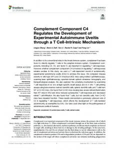
225 235 251 215 231 241 Fraction FIG. 1. Chromatography of barium citrate eluate on a DEAE-E3ephacel column. The barium citrate eluate from 4 liters of freshly froz4,en plasma was applied to a column (2.5 x 35 cm) with DEAE-Sephaccel equilibrated in 0.1 M sodium phosphate (pH 6.0) containing 1 mM besnzamidine and was eluted with a linear gradient of NaCl (0.1-0.6 M NaCl; 600 ml in each vessel). The flow rate was 13.5 ml/hr, and 3 fri actions per hr were collected. (A) Chromatogram; the start ofthe gradie,nt is indicated by an arrow. (B) Electroimmunoassay with anti-C4bp anItiserum (upper) and anti-protein S antiserum (lower); (C) Agarose ggel electrophoresis of fractions ofthe DEAE-Sephacel column in 0.075 M barbital buffer (pH 8.6) containing 2 mM EDTA. a, C4bp-S; b, proteein S. Prothrombin is the main component ofthe peak eluting in fractions 177
187
209
201
2513
to both immunoprecipitates were found to be adsorbed to barium citrate. After elution of the adsorbed proteins with 0.2 M EDTA (pH 7.4) and dialysis, the protein solution was applied to a DEAE-Sephacel column. The fractions were analyzed with electroimmunoassay by using antisera against protein S. Material reacting with anti-protein S antibodies appeared in two peaks, the second one just in front of prothrombin, which was the major component of fractions 240-260 (Fig. 1). The morphological characteristics ofthe immunoprecipitates in the first peak suggested that the material contained the complexed form of protein S, whereas the morphological shape of the immunoprecipitates in the second peak was identical to that obtained with purified protein S. Agarose gel electrophoresis offractions from the first peak showed a dominating protein band just anodal to the application slit. After testing these fractions with several different antisera against plasma proteins, this protein was identified as the C4bp by immunodiffusion (see below) and electroimmunoassay. The C4bp rockets coincided with the first peak of protein S immunoreactivity (Fig. 1). The complex between C4bp and protein S did not dissociate during agarose gel electrophoresis and was obtained in pure form after gel filtration on a column of Sepharose CL-4B (2.5 X 85 cm) in 0.1 M Tris.HCV1 M NaCV10 mM EDTA, pH 8.0. The C4bp-S was eluted after the void volume of the Sepharose CL-4B column as a symmetrical peak. As judged by electroimmunoassay, the distributions of C4bp and protein S were identical to that of C4bp-S and coincided in absorbance, indicating that the complex was homogeneous and did not dissociate during these conditions. The supernatant after barium citrate adsorption contained less than 20% of the original plasma C4bp as judged by electroimmunoassay. This form of C4bp was not in complex with protein S and accordingly did not react with the antibody against protein S (Fig. 2). An antiserum against C4bp appeared to react equally well with complexed and uncomplexed C4bp as judged by immunodiffusion. The same results were obtained by using two different antisera against C4bp raised in two different laboratories. Furthermore, apicture of immunological identity was obtained between our antiserum and the two other antisera against C4bp when tested with C4bp-S. Purified C4bp and C4bp-S were analyzed by 4% (wt/vol) and 10% NaDodSOJpolyacrylamide gel electrophoresis (Fig. 3). Before reduction they migrated as high molecular weight proteins. However, uncomplexed C4bp had a slightly faster electrophoretic migration rate than the C4bp dissociated from the complex with protein S. Protein S appeared to dissociate from the C4bp-S complex on NaDodSOJpolyacrylamide gels, and the reason for the small difference in migration rates between the two forms of C4bp is unknown. When approximately 10 Ag A-C4bp
C4bp
C4bp-S
I
240-260.
munoprecipitates were seen that differed both in position and in precipitation pattern. This suggests that two molecular species of protein S are present in plasma. The antigens giving rise
A-pS
FIG. 2. Double radial immunodiffusion of C4bp-S and C4bp. Purified C4bp-S and C4bp were analyzed by using an antiserum against C4bp (A-C4bp) and another against protein S (A-pS).
Immunology: Dahlback and Stenflo
2514
1
2
3
4
5
6
Proc. Natl. Acad. Sci. USA 78 (1981)
8
FIG. 3. NaDodSO4/polyacrylamide gel electrophoresis of C4bp-S, C4bp, and protein S. Samples applied to gels 4,5,6, and 8 (marked with an asterisk) were treated with 5% (vol/vol) 2-mercaptoethanol at 100°C for 2 min before application. Gels 1-3 and 4-8 were 4% and 10%, respectively, in acrylamide. C4bp-S was applied to gels 1 and 4; C4bp was applied to gels 2 and 5. A mixture of C4bp-S and C4bp was applied to gels 3 and 6, and protein S was applied to gels 7 and 8 (10-20 jig of protein was applied to each gel).
of C4bp-S was applied to 10% NaDodSOjpolyacrylamide gels without reduction, a barely visible doublet appeared with an approximate molecular weight of 75,000 (not shown). In gels heavily overloaded with C4bp-S (50-100 jig) this doublet (which had the same mobility as protein S) was distinct, whereas it was absent when the same amount of C4bp was analyzed. After reduction of C4bp and C4bp-S, a single component with the apparent molecular weight of 70,000 was observed, indicating that the subunits of free and complexed C4bp have identical molecular weights. Fig. 3 also shows the picture obtained when purified protein S was analyzed by 10% NaDodSO4 polyacrylamide gels. After reduction of disulfide bonds, a closely spaced doublet was obtained. The higher molecular weight component comigrated with bovine prothrombin, indicating a molecular weight of approximately 73,000. Several different preparations of protein S gave the same gel pattern. Despite the heterogeneous appearance of protein S on NaDodSOJpolyacrylamide gels, a single amino-terminal sequence was obtained (Fig. 4) identical to the sequence of human protein S reported by DiScipio and Davie (2). The C4bp-S complex also was found to have a single amino-terminal sequence, suggesting that C4bp was composed of identical subunits. The protein S sequence could not be unambiguously identified during the sequenator degradation of C4bp-S due to the small
amount of protein S in the purified complex. Protein S and different preparations of C4bp-S were analyzed for the content of y-carboxyglutamic acid residues, and the results were used to calculate the approximate ratio of protein S per subunit of C4bp. The amount of purified uncomplexed C4bp was too low to allow an accurate analysis of its 'y-carboxyglutamic acid residue content, and the calculations were based, therefore, on the assumption that free C4bp contained no y-carboxyglutamic acid residues. The number of y-carboxyglutamic acid residues in the C4bp-S complex (approximately 1 per subunit) indicated that the ratio of protein S to C4bp subunit in the purified complex was approximately 1:10 because protein S contains 10 y-carboxyglutamic acid residues per molecule (2). This should be regarded as a minimum number because some protein S has presumably dissociated from the C4bp during purification. When human plasma was applied to a gel filtration column and the fractions were analyzed for protein S with radioimmunoassay, two distinct peaks of immunoreactivity were observed (Fig. 5). The first peak eluted in the void volume of the column and at the same position as purified "2I-labeled C4bp-S. The second peak of protein S immunoreactivity eluted in the same position as purified '25I-protein S. Radiolabeled free protein S applied to the column gave a single peak of radioactivity. The gel filtration pattern obtained after 6 hr of preincubation of "2I-protein S with human plasma was different from that offree "2I-protein S. Two major peaks of radioactivity were observed with elution volumes corresponding to the two forms of protein S immunoreactivity in plasma. For unknown reasons a high level of radioactivity between the two major peaks was consistently found. The distribution of radioactivity on 10% NaDodSO4polyacrylamide gels of fractions 24, 32, and 42 was found to be identical to that of free "2I-protein S both before and after reduction of disulfide bridges. To demonstrate that the radioactivity of the first peak represented protein S bound to C4bp, aliquots of the fractions were mixed- with anti-C4bp antiserum. After an incubation at 4°G over night, the anti-C4bp antibodies were precipitated with a goat anti-rabbit IgG antibody, and the radioactivity of the precipitates was measured. Approximately 50% ofthe radioactivity of fractions from the first peak was found to be specifically precipitated by the anti-C4bp antiserum, whereas the radioactivity eluting at the position of uncomplexed protein S was not precipitated. This demonstrates that the 125I-protein S of the first peak was part of a complex with C4bp (i.e., the C4bp-S complex).
DISCUSSION No function has yet been associated with protein S, although it has been suggested that it increases the rate of inactivation 10
5
Human
(Gla.) - (Gla)
- Thr - Lys -
Gin
- Gly - Asn - Leu
protein S
Ala - Asn - Ser - Leu - Leu -
Bovine protein S
Ala - Asn - Thr - Leu - Leu - Gba - Gla
C4bp-S
Asn - Cys - Gly - Pro - Pro - Pro - Thr - Leu - Ser - Phe - Ala - Ala - Pro
- Thr - Lys-Lys -
Gly - Asn - Leu
FIG. 4. Amino-terminal sequence of human protein Sand C4bp-S. The amino-terminal sequence ofbovine protein S (3) is given for comparison. The sequence of human protein S was identical to the one previously reported (2). The position of y-carboxyglutamic acid (Gla) in human protein S is tentative.
Immunology: Dahlback and Stenflo 4 OD .
3 :3
od
2
1
0
8 IC rx
6
0
r.
-6-
4
C)
S. 0.
2
0
6 0
x
4 c. '5' 0.
2
0
ko x
2
C 0 4-
C)
en0. 0.
w.
0
0
20
40
60
Fraction
FIG. 5. Demonstration of C4bp-S complex in human plasma by gel filtration. A column (0.9 x 52 cm) with Ultrogel 34 in 10 mM Tris/10 mM EDTA/0.15 M NaCl, pH 8.0, containing 0.03% NaN3 and 5 mg of bovine serum albumin per ml was run at room temperature with a flow rate of 3.5 ml/hr. Nine-minute fractions were collected. (A) Fresh EDTA plasma (0.5 ml) was applied and fractions were- analyzed with radioimmunoassay for protein S. (B) 125I-C4bp-S (A) and 12'I-protein S (.) were separately applied to the column. (C) 125I-Protein S (25 Al) was incubated at 37TC with 475 A.l of EDTA/plasma for 6 hr and then applied to the column. (D) Amount of '"I-protein S precipitated with anti-C4bp antiserum in fractions from the gel filtration shown in C.
Proc. Natl. Acad. Sci. USA 78 (1981)
2515
of coagulation factor V by protein C (15) Like the other vitamin K-dependent proteins, it has a high affinity for negatively charged phospholipid vesicles in the presence of calcium ions (16). Protein S and the recently described complement protein called S-protein (17) appear to have similar molecular weights on NaDodSOjpolyacrylamide gels. However, judging from available data (i.e., amino acid composition and plasma concentration) they are different proteins. Furthermore, the complement S-protein gives different precipitation patterns on crossed immunoelectrophoresis of plasma and serum due to its incorporation into the thrombin-antithrombin III complex during blood coagulation (17, 18). This difference is not seen with the vitamin K-dependent protein S, which seems to be unaffected by blood coagulation. The data presented in this paper demonstrate that protein S in human plasma is present in two forms, one free and the other in complex with a recently described complement component, C4bp. Protein S is bound to C4bp with high affinity. An equilibrium between free and bound protein S was demonstrated. On 10% NaDodSOjpolyacrylamide gels, C4bp-l25Iprotein S complexes dissociated, indicating that protein S was noncovalently bound to the C4bp. The results obtained also suggested that the protein dissociated in intact form. The rate of dissociation of "I-protein S from C4bp was slow, and in precipitation experiments of the C4bp-125I-protein S complex with anti-C4bp antibodies, 50% of the total 25I-protein S was precipitated after an overnight incubation at 40C, indicating a halflife of the complex (at 40C) of more than 10 hr. The C4bp-S complex is, as illustrated by the DEAE-Sephacel chromatography, one of the major components of a barium citrate eluate, which to our knowledge has not been observed previously. This is probably due to the fact that in most purification procedures described for the vitamin K-dependent proteins, the barium citrate eluate is first precipitated with 30-40% ammonium sulphate. In this precipitate C4bp-S complex is the major component. However, this fraction has usually been discarded, and the vitamin K-dependent proteins have been collected by increasing the ammonium sulphate concentration to 60-70%. On unreduced NaDodSO4polyacrylamide gels, there seems to be a slight difference in molecular weight between the C4bp prepared from the barium-adsorbed plasma and the C4bp dissociated from the complex with protein S. In this respect it is interesting that in the first report on C4bp, the isolated protein appeared as a doublet on NaDodSOgpolyacrylamide gels (5). Fujita and Nussenzweig (19) later reported the isolation of the two forms, called "C4bp-low" and "C4bp-high" but could not detect any difference in ability to catalyze the enzyme C3b-inactivator-mediated degradation of C4b in solution. It seems likely that C4bp in complex with protein S is identical to C4bphigh and that C4bp-low is identical to the uncomplexed C4bp isolated from the supernatant after barium citrate adsorption. The functional significance ofthe complex formation between C4bp and protein S is unknown. The high affinity for phospholipid of vitamin K-dependent structures in protein S (16) may be important for localization of C4bp-S to phospholipid membranes to regulate the inactivation of surface-bound C4b. We would like to thank Dr. Anders Sjoholm for aid in the identification of C4b-binding protein and Dr. Bengt G. Johansson for helpful discussions. The plasma was kindly provided by Dr. Bertil Robertson (Bloodbank, Malmo General Hospital). Mrs. Bergisa Hildebrand gave excellent technical assistance. This project was supported by grants from the Swedish Medical Research Council (project no. B80-03X04487-06A) and by grants from The Swedish Society of Medical Sciences, Svenska sallskapet for medicinsk forskning, Segerfalks Stiftelse, Kocks Stiftelse, and Magnus Bergvalls Stiftelse.
2516
Immunology: Dahlback and Stenflo
1. DiScipio, R. G., Hermodson, M. A., Yates, S. G. & Davie, E. W. (1977) Biochemistry 16, 698-706. 2. DiScipio, R. G. & Davie, E. W. (1979) Biochemistry 18, 899-904. 3. Stenflo, J. & Jonsson, M. (1979) FEBS Lett. 101, 377-381. 4. Jackson, C. M. & Nemerson, Y. (1980) Annu. Rev. Biochem. 49, 765-811. 5. Scharfstein, J., Ferrein, A., Gigli, I. & Nussenzweig, V. (1978) J. Exp. Med. 148, 207-222. 6. Fujita, T., Gigli, I. & Nussenzweig, V. (1978)J. Exp. Med. 148, 1044-1051. 7. Gigli, I., Fujita, T. & Nussenzweig, V. (1979) Proc. Natl. Acad. Sci. USA 76, 6596-6600. 8. Nagasawa, S., Ichihara, C. & Stroud, R. M. (1980) J. Immunol. 125, 578-582. 9. Porter, R. R. & Reid, K. B. (1979) Adv. Protein Chem. 33, 1-71.
Proc. Nati. Acad. Sci. USA 78 (1981) 10. Thorell, J. I. & Johansson, B. G. (1971) Biochim. Biophys. Acta 251, 363-369. 11. Stenflo, J. (1976)J. Biol. Chem. 251, 355-363. 12. Fernlund, P., Stenflo, J., Roepstorff, P. & Thomsen, J. (1975)J. Biol. Chem. 250, 6128-6133. 13. Spackman, D. H., Stein, W. H. & Moore, S. (1958) Anal. Chem. 30, 1190-1206. 14. Edman, P. & Begg, G. (1967) Eur. J. Biochem. 1, 80-91. 15. Walker, F. J. (1980) J. Biol. Chem. 255, 5521-524. 16. Nelsestuen, G. L., Kisiel, W. & DiScipio, R. G. (1978) Biochemistry 17, 2134-2138. 17. Podack, E. R. & Muller-Eberhard, H. J. (1979) J. Biol. Chem. 254, 9908-9914. 18. Podack, E. R., Curd, J. G., Griffin, J. H. & Muiller-Eberhard, H. J. (1979) Thromb. Haemost. 42, (1), 170. 19. Fujita, T. & Nussenzweig, W. (1979)J. Exp. Med. 150, 267-276.