Dec 4, 1986 - 124), 21. Mancini, G. A.. Carbonara, A. 0. & Heremans, J. F. (1965) Immuno-. Mittal, K. K., Mickey, M. R.. Singal, D. P. & Terasaki, P. 1. (1968).
649
621st MEETING, LONDON
We are grateful for the financial assistance and encouragement received from the Hospital N.S. de Covadonga, Oviedo, Spain.
Holers, V. M., Cole, J. L.. Lublin, D. M., Seya, T. & Atkinson, J. P. (1985) Immunol. Today 6 , 188-191 Laurell, C. B. (1972) Scand. J . Clin. Lab. Invest. 29, (Suppl. 124), 21 Mancini, G. A.. Carbonara, A. 0. & Heremans, J. F. (1965) Immunochemistry 2, 235 Mittal, K. K., Mickey, M. R.. Singal, D. P. & Terasaki, P. 1. (1968) Transplantation 6 , 9 13-927 Rodriquez de Cordoba, S., Lublin, D. W., Rubinstein, P. & Atkinson, J. P. (1985) J . E.wp. Med. 161, 1189-1 195 Sim, R. B., Malhotra, V., Ripoche, J., Day, A. J., Micklem, K. J. & Sim, E. (1986) Biochem. Soc. Symp 51, 83-96
Gewurz. A. T., Lint, F., Imherr, S. M., Garber, S. S. & Gewurz, H. (1982) Clin.Immunol. Immunopathol. 23, 297-31 I
Received 4 December 1986
explained by H factor deficiency. The failure of immunochemical techniques to detect the H factor presence indicates that the possible low levels may be due to a genetic defect. The deficiency may be due to structural genes for H factor transmitted to the siblings, or post-translational modifications of the active functional molecule.
The structure and function of complement component C8 investigated with monoclonal antibodies AREFAINE ABRAHA,* CAROLINE A. SEWRY,? ANTHONY K . CAMPBELLS and J. PAUL LUZIO* *Department of Clinical Biochemistry, University of Cambridge, Addenbrooke's Hospital, Hills Road, Cambridge CB2 2QR, U . K . , ?Jerry Lewis Muscle Research Centre, Hammersmith Hospital, Ducane Road, London W12 OHS, U . K . . and $Department of Medical Biochemistry, University of Wales College of Medicine, Heath Park, Cardif CF4 4XN, U . K . The formation of the complement membrane attack complex (MAC; consisting of five proteins C5b, C6, C7, C8, C9) on appropriate target cell membranes provides the principle mechanism of complement-mediated cell killing and plays a role in defence against bacterial infection. However, lesions may also be caused in the plasma membrane of host cells and can give rise to sublytic cell damage which may be important in some autoimmune disorders (Campbell & Luzio, 1981). Whilst the molecular events associated with C9 insertion into the target membrane are well established (Stanley et a / . , 1986), less is known about the role of C8. Human C8 is a serum glycoprotein with an apparent M, of 15 1 000 composed of three non-identical polypeptide chains a ( M , 64000), 1 ( M , 64000) and y ( M , 22000). These subunits exist as a covalently linked ay-dimer noncovalently associated with the 1-subunit (Steckel et al., 1980). Solution binding studies using purified complement components have suggested that the 1-subunit mediates the Abbreviation used: MAC, membrane attack complex.
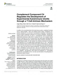
binding of C8 to C5b-7, with the a-subunit being capable of binding to C9 (Stewart & Sodetz, 1985). Despite the absolute requirement for C8 in the membrane bound C5b-8 complex to allow C9 binding, direct interaction in the membrane between C8 and C9 has not been demonstrated (Monahan et al., 1983). The C5b-8 complex is thought to catalyse the polymerization of C9 (Tschopp et al., 1985) with C8 participating directly in this process. In the present study monoclonal antibodies have been prepared to human C8 for use as tools in the investigation of C8 function. Monoclonal antibodies to C8 were prepared by standard methods (Galfre & Milstein, 1981) as previously described for C9 (Morgan et al., 1983) using purified C8 donated by Dr. A. Esser as antigen. Ten monoclonal antibodies were prepared, four of which have been reported previously (Abraha et al., 1986). By immunoblotting six were shown to react with the a-chain, two with the 1-chain and two with the y-chain. Monoclonal antibodies to y showed strong cross-reactivity with a and monoclonal antibodies to 1 weak cross-reactivity with a. No cross-reaction with 1 or y was shown by monoclonal antibodies to a. None of these antibodies interacted with C9, though in other studies antibody cross-reactions with C9 and other components of the MAC have been observed (Tschopp & Mollnes, 1986; Tschopp et al., 1986). Epitope mapping of the monoclonal antibodies was conducted using each antibody in solution phase to interfere with the binding of '251-labelled C8 to each antibody on a solid phase. These studies suggested that whereas the monoclonal antibodies to y and bound to a single epitope on each subunit, the monoclonal antibodies to a
Fig. 1. Epitope map summarizing the binding of ten mouse monoclonal antibodies to human C8 The stippled area on the a-subunit indicates cross-reactions of monoclonal antibodies to and y.
Vol. 15
BIOCHEMICAL SOCIETY TRANSACTIONS
650 were directed to three sites (Fig. 1). When cross-reacting with a the monoclonal antibodies to fi interacted with all three sites and those to y with two sites. Immunoglobulin fractions containing antibodies L6 (anti-a) B1 (anti-fi) and M1 (anti-y) were prepared from ascites fluid and coupled to cyanogen bromide-activated Sepharose. Purified C8 was obtained from any of these immunoadsorbants by first binding C8 from a 3&50% saturation ammonium sulphate fraction and subsequently eluting with 50 mM-diethylamine, subsequently neutralized with Tris/HCl. Starting with 50 ml of fresh plasma 1.5-2 mg of C8 was recovered and assessed by SDS/polyacrylamidegel electrophoresis, rocket immunoassay using polyclonal anti-C8 serum and reconstitution of C8-depleted human serum in red cell lysis assays. The ay- and fi-subunits of C8 could be separately purified simply by salt elution from the appropriate immunoaffinity columns (respectively anti-b and anti a), approx. 0.4M-NaCl giving 50% recovery. The monoclonal antibodies to C8 may be used to interfere with MAC formation (Abraha et al., 1986)and also to localize C8 on the cell surface. Evidence that MAC formation at the cell surface can result in sublytic damage playing a role in autoimmune disease has been obtained in immunohistochemical studies of muscle fibres from patients with myositis (Morgan et al., 1984). In addition to necrotic fibres showing intense staining with monoclonal antibodies to C9, it was also found that fibres which are apparently normal by other light microscopic histochemical criteria have punctate localizations of C9 and C8 on the cell surface (Luzio et al., 1987). The monoclonal antibodies to C8 should prove useful in the
further investigation of the mechanisms by which mammalian cells may recover from sub-lytic complement attack. The arthritis and Rheumatism Council, the World Health Organization and the Muscular Dystrophy Group of Great Britain and Northern Ireland provided support. Abraha, A., Bailyes, E. M., Richardson, P. J., Campbell, A. K. & Luzio, J. P. (1986) Biochem. SOC.Trans. 14, 779 Campbell, A. K. & Luzio, J. P. (1981) Experimenta 37, 11 10-1 I12 Galfre, G. & Milstein, C. (1981) Methocis Enzymol. 73, 3 4 6 Luzio, J. P., Abraha, A., Richardson, P. J., Daw, R. A,, Sewry, C. A., Morgan, B. P. & Campbell, A. K. (1987) Methodol. Surv. Biochem. 17, in the press Monohan, J. B., Stewart, J. L. & Sodetz, J. M. (1983) J . B i d . Chem. 288, 50565062 Morgan, B. P., Daw, R. A., Siddle, K., Luzio, J. P. & Campbell, A. K. (1983) J. Immunol. Methocis 64, 269-281 Morgan, B. P., Sewry, C. A., Siddle. K.. Luzio, J. P. & Campbell, A. K. (1984) Immunology 52, 181-188 Stanley, K. K., Page, M., mcampbell, A. K. & Luzio, J. P. (1986) Mol. Immunol. 23, 451-458 Steckel, E. W., York, R. G., Monahan, J. B. &Sodetz, J. M. (1980)J. B i d . Chem. 255, 1197-12005 Stewart, J. L. & Sodetz, J. M. (1985) Biochemistry 24, 45984602 Tschopp, J. & Mollnes, T.-E. (1986) Proc. Natl. Acad. Sci. U.S.A. 83, 42234227 Tschopp, J., Podack, E. R. & Muller-Eberhard, H. J. (1985) J. Immunol. 134,495499 Tschopp, J., Masson, D. & Stanley, K. K. (1986) Nature (London)322, 831-834 Received 21 November 1986
Biosynthesis of complement proteins by the U937 cell line D. GOUNDIS and K. B. M. REID M . R.C. Immunochemistry Unit, Department of Biochemistry, South Parks Road, Oxford OX1 3QU, U . K . Liver is the primary site of synthesis of most complement components (Colten, 1976). However, several other cell types have been shown to synthesize one or more of the complement components in vitro. Fibroblasts and intestinal columnar epithelial cells, for example, synthesize the first complement component C1 (Reid & Solomon, 1977; Morris et al., 1978), whereas monocyte/macrophage cells synthesize all the components of the alternative pathway as well as C2, C4 and C5 (Whaley, 1980; Cole et al., 1980; Beatty et al., 1981). The monocytic cell line (U937) has also been shown to synthesize C2, C3, C5, C7, properdin, factor H, factor B, C1 INH, and factor D (Minta & Pamburn, 1986; Malhotra & Sim, 1985; Barnum & Volanakis, 1985). We have studied the effect of y-interferon on the synthesis and secretion of Clq, C3, factor H, factor B, factor D and properdin by the U937 cells employing immunoprecipitation techniques and analysing the results by SDS/PAGE electrophoresis and fluorography. Cells were grown in 75 cm3 tissue culture flasks in RPMI 1640 medium, containing foetal calf serum (lo%, w/v), penicillin (100 i.u./ml), streptomycin (100 pg/ml), kamamycin (50pg/ml), 1 mM-sodium pyruvate and modified Eagle's medium non-essential amino acids, in an atmosphere of 5% co,/95% air at 95% humidity. Two 20ml aliquots, containing 4.5 x lo5 cells/ml, were transferred into two Abbreviations used: PAGE, polyacrylamide-gel electrophoresis; SBTI, soybean trypsin inhibitor (Sigma type I-S); IAM, iodoacetamide; DFP, di-isopropyl fluorophosphate; DTT, dithiothreitol.
75cm3 flasks. y-Interferon (a gift from Dr. T. Makoena, Sir William Dunn School of Pathology, Oxford) was then added to one of the flasks at a final concentration of 300 units/ml. After 24 h the cells were washed three times with 50ml of DAB buffer (8.2m~-NaH,P0,/1.5m~KH, PO, /139 m ~ - N a C 1 / 3m ~ - K C 1 / 5mM-glucose/l mMCaCl,/I m~-MgCl,,pH 7.4), resuspended in 6 ml of RPMI 1640 without cysteine (GIBCO select-amine kit), containing 5% (w/v) foetal calf serum and were incubated at 37°C for 30 min. 250 pCi of ~-[~'S]cysteine(478.4 mCi/mmol; Amersham International, U.K.) was added to each flask, and the cells were cultured under normal conditions for 20 h. After centrifugation the culture supernatants were stored at - 70°C and the cell pellets were washed twice in DAB buffer containing 2 mM unlabelled cysteine followed by two washes in DAB. They were then lysed by adding 2ml of lysis buffer (10m~-K,P0,/0.5m~-EDTA/l%, w/v, NP40/100 pg of SBTI/Iml 2.5 m~-1AM/2.5mM-DFP/ 1 .O mM-phenylmethanesulphonylfluoride) at 4°C for 30 min. The nuclei were pelleted and the cell extracts were stored at 70°C. The supernatants were treated with 2 mM-L-cysteine/ 2.5 m~-DFp/2.0~M-IAM/SO pg of SBTI/ml. For immunoprecipitation of proteins, the relevant antibodies were added to the supernatants of the stimulated and unstimulated cells followed by incubation at 4°C for 18h. Rabbit antibodies were used for Clq, C3, factor B, factor D, factor H and a sheep antibody for properdin. Protein A-agarose (25 pl packed volume) was then added to the antibody-antigen solution followed by incubation for 4 h at 4OC. The mixture was spun at lOOOOg for 1Omin and the pellet washed twice with 1 ml of solution A (0.5%, w/v, Triton X-l00/0.25?A0,w/v, sodium deoxycholate/0.5%, w/v, SDS/20 m~-NaN,/0.3%,w/v, ovalbumin), transferred into 1987