Segmentation of Basal Nuclei and Anatomical Brain Structures Using Support Vector Machines A. Bosnjak1, R. Villegas1, G. Montilla1, and I. Jara2 1
Centro de Procesamiento de Imágenes, Universidad de Carabobo, Valencia, Venezuela 2 Hospital Metropolitano del Norte, Valencia, Venezuela
Abstract—Segmentation of structures inside of the brain are essential for planning computer assisted surgery. Structures such as basal nuclei are difficult to detect in MR images because they have fuzzy edges, and exhibit few changes on the gray level intensities relative to the anatomical structures surroundings. The traditional techniques of image processing thus cannot be used to segment the basal nuclei. We propose a new processing pipeline conformed by five modules: 1) Image acquisition on DICOM format using MRI inside clinics or hospitals. 2) An image pre-processing for improvement on its contrast in a specific window. 3) An anisotropic filter applied to these images. 4) An automatic selection of input vectors for the Support Vector Machine applied. This selection expands radially, where the operator give only a center point of the structures to be detected. This software searches in a radial direction for the supporting vectors that belong to the basal nucleus, and for those corresponding to the background, based on the histogram. 5) Once the Support Vector Machine is trained in the previous module, the generated model serves to classify the image. Finally, the structures are properly detected and segmented, and are then validated by a neuro-anatomist by comparing the segmentation made by our software with a manual segmentation. In conclusion, we propose a new processing pipeline that includes a classifier using Support Vector Machines (SVM) for segmentation that can be used for image guided surgery.
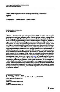
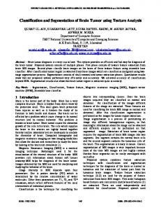
Normalization - (SyN), and one patch-label fusion technique with the manual segmentation made by an specialist. The automatic labeling of deep brain structures is compared to the manual segmentation. Haegelen [3] used the Dice – kappa metrics and center of gravity to evaluate the quality of the segmentation. These metrics are useful for comparing the quantity of common pixels among the segmented structures made by the automatic and manual methods. Basal nuclei (figure 1), also known as basal ganglia, is a set of anatomical structures located deep in the cerebral cortex, between the anterior and the medial zones of the brain. Their main components are the striatum, globus pallidus, subthalamic nucleus and substantia nigra. The striatum is composed by the caudate nucleus and putamen.
Keywords—Image Segmentation, Support Vector Machine, (SVM), Basal nuclei, Image Guided Surgery.
I.
INTRODUCTION
Image segmentation has a key role in clinical diagnosis. An ideal method of segmentation has some basic properties, such as minimal operator interaction, fast calculation, accuracy and noise-robust results through the image variability for different patients. Many studies have been done that have focused on segmentation of X-ray imaging, MR images, CT scan, and ultrasound. There has not been however, one method that is ideal according to the above properties. Some traditional methods such as regional growth, formbased and statistical methods, work well, only when the segmentation structures have a defined gray level [1] [2]. Haegelen et al. [3], compared three recently segmentation methods, such as: Automatic Nonlinear Image Matching and Anatomical Labeling (ANIMAL) Symmetric Image
Fig. 1 Localization of caudate nuclei, globus pallidus, and putamen. (source: The Brain From Top to Bottom at http://thebrain.mcgill.ca/ flash/i/i_06/i_06_cr/i_06_cr_mou/i_06_cr_mou.html)
Functionality of these structures is associated to the generation of stimulatory and inhibitory signals, which are essential to control and coordinate motor activities, as well as for regulatory processes of movements, behaviors and emotions. Basal nuclei are key elements in treatment of pathologies of the nervous system, such as Parkinson’s or Huntington’s disease, whose symptoms include the difficult performance of motor functions, producing from writhing movements, abnormal postures, to essential tremor and Tourette syndrome.
L.M. Roa Romero (ed.), XIII Mediterranean Conference on Medical and Biological Engineering and Computing 2013, IFMBE Proceedings 41, DOI: 10.1007/978-3-319-00846-2_80, © Springer International Publishing Switzerland 2014
321
322
A. Bosnjak et al.
Minimally invasive surgeries oriented to treat this kind of neurodegenerative disease, such as pallidotomy or deep brain stimulation (DBS), are based on performing therapeutic lesions or implanting electrodes for electrical stimulation in specific zones of the basal nuclei. During the planning phase of these procedures, neurosurgeons use magnetic resonance (MR) images of the patient in order to locate the nuclei and select the structures that will be the target of the surgical approach. Basal nuclei are not clearly observed on MR images because they have fuzzy borders and show fewer changes in the gray level intensities relative to neighboring anatomical structures. Therefore, traditional image processing techniques fail to detect them and generally require auxiliary assistance in order to locate and segment their edges. II.
. Equation (2) expresses all the function 1 and -1 is 2⁄ points which are projected behind the planes of distance 1 except the border vectors and the outliers. Equation (1) presents a multi-objective minimization problem that involves the magnitude of w (coefficient of smoothness or gradient) and the sum of the errors. Equations (4) - (5) provide the dual problem from the Lagrangian. Equation (4) shows the term , that represents the scalar product in the featured space. Equation (6) represents the distance function in the featured space, and it can also be drawn as the input space. This is the decision function of the classifier. The zero level surface of this function will be used in solving the 3D modeling problem.
METHODS: SINGLE BINARY CLASSIFIER (SVC)
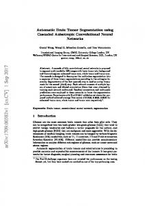
The SVM was constructed by Vapnik [4] as the learning machines which minimize the classification error, finding the hyperplane for maximum margin separating the two classes in the featured space. The binary classification is presented as follow. Given a set of points in the input space {x i } ⊂ ℜ n i = 1,, l and a function Ψ : x i → yi yi ∈ {− 1,1} which assigns to the points one of two possible values, Vapnik [4] proposed to project the problem to another space (feature space) using a transformation Φ : ℜn → ℜm . In the featured space the classes are lineally separable by a hyperplane of maximum margin. This proposal is presented in figure 2, and the optimization problem is defined by the following equations [5].
Fig. 2 Points and hyperplane in the feature space
min α
l
min w,b ,ξ
1 2 w + C ξi 2 i =1
(
)
yi w T Φ(x i ) + b ≥ 1 − ξ i
ξi ≥ 0
i = 1,..., l
(
)
l 1 l α iα j γ iγ j K xi , x j − α i 2 j =1 i =1 l
(1)
α iγ i = 0 i =1
(2)
D ( x ) = α i γ i K ( xi , x ) + b
(5)
l
(6)
i =1
(3)
Figure 2 is considered a planar function (Distance Function) in the featured space. This function is extended in the featured space and it is null over the hyperplane of maximum separation. It can assign arbitrarily a value of +1 to distance function over the nearest points to the optimum hyperplane, which we called border vectors. Vectors can also be allowed a distance function 1 , which we called outliers. The other vectors behind the two planes of distance 1 are called the interior points. The variable w (gradient of the distance function) adjusts the smoothness of the function. A minimum value of w gives the maximum smoothness, and a maximum separation between two classes, since the real distance between the two planes of distance
(4)
III.
GENERAL SCHEME OF THIS PROJECT
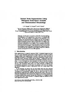
This work is divided into several modules from image acquisition to its classification as shown in the diagram of Figure 3. All studies were conducted with MRI. The general scheme of this project follows these methods: 1) We developed a DICOM image reader and DICOMDIR directory files reader because they allow organized access to images and information sets [6]. This software was developed using the DCMTK 2012 library [7], which has several years of evolution and continuous use in medical applications. It can also be attached to any other software under development that requires the handling of DICOM images and
IFMBE Proceedings Vol. 41
Segmentation of Basal Nuclei and Anatomical Brain Structures
DICOMDIR directory files. 2) We made a contrast enhancement of each image based on the histogram, because the structures of interest were blurred. This pre-processing step should be performed because these images do not use fully the gray level at 12 bits range. 3) We made a preprocessing filter corresponding to the anisotropic filter inside of the 5 x 5 pixels window. 4) It picks up the candidate vectors radially as explained below. 5) A procedure that trains the Support Vector Machine, and 6) Finally, the classification performed is based on an image test file ('test') that uses all pixels of the image and determines whether it belong to a class 1 = “belongs to basal nuclei”, or to class 2 = “corresponds to the background of the image”.
323
directions where the central pixels are these red points and those blue points shown in Figure 5.
Fig. 4 Selection of the training vectors in a radial search from the center of the structure of interest
μ= σ d2 =
Fig. 3 Developed modules for segmentation of Basal Nuclei
1 M .N
1 #∈ d
M −1 N −1
I (i, j ) i j
(7)
=0 =0
[I (i, j ) − μd ] i j d
2
, ∈
(8)
and other structures
A. Initialization of Segmentation The user selects a seed within the structure of interest, in this case inside of the center of the basal nuclei. The software calculates the mean and variance radially from the center outwardly corresponding from the selected point to the edges of the structure. The candidate vector that belongs to the edge is calculated when there is a significant variation on the profile of the histogram. A dipole is placed on the edge, which corresponds to the two training vectors. The interior vector is placed just inside the edge of the structure to be segmented. The exterior vector is placed outside the structure. Figure 4 shows the selected vectors inside and outside of the structure of interest. B. Extraction of Textured Features The model chosen in this paper for the segmentation of basal nuclei tissues is based on two indicators which measure the homogeneity and contrast of texture: the average in a window of 5 × 5 pixels, and the variance calculated at position (x, y) following the arrangements of the windows used for the Nagao filter. The average is obtained by averaging the gray scale level in a 25 pixels centered window, computed according to equation (7). The directional variance is computed at each of the selected masks in a 5 x 5 pixels window using equation (8) for each of eight possible
Fig. 5 Nine masks used to implement the Nagao filter Nagao [8] proposed those sub-regions that take into account the orientation of the contour. Nagao [8] described this filter as a rotating bar around the pixel (x, y) that detects the orientation of a mask where the variance of the gray level is minimal. For this implementation, Nagao picked a neighborhood of 5 x 5 pixels, which is subdivided into nine sub-regions as shown in figure 5. We calculate then the mean and the variance over the area represented by a nine masks of figure 5. Each of the variances calculated on the Nagao’s window are used as texture components for the Support Vector Machine as shows in figure 6. C. Training of the v-SVM Machine
According to the model previously defined, each training set is represented by a vector which contains: the position (x, y), the mean μ in a 5 x 5 pixels window, and eight textural
IFMBE Proceedings Vol. 41
324
A. Bosnjak et al.
components computed as explained above. The label of the class is also included in the vector of features. This label determines whether it belongs to basal nuclei or not. Figure 6. class
x
y
μ
σ 1 σ2 σ3 σ 4 σ5 σ6 σ7 σ8
Fig. 6 Structure of features vector for the training set The training of the v-SVM machine is done by adjusting the parameters nu (ν) and gamma (γ) of the SVM, using a Gaussian kernel. When training is complete, feature vectors associated with the pixels of the brain’s image are used to query the SVM to determine whether the new pixel belongs or not to the basal nuclei. IV. EXPERIMENTAL RESULTS To evaluate the methodology presented, several experiments were performed in order to detect the globus pallidus and putamen. The images chosen were taken from brain imaging studies of male and female patients aged between 42 and 75 years old. Sagittal, axial and coronal MR images at a resolution of 256 x 256 and 512 x 512 pixels were used. For training, a point is chosen within the structure that we wish to detect. This software handles the selection of the interior class of the structure to be segmented, and searches the outer class based on the difference of the radial gradient relative to the central point selected. We assembled the training vectors using the positions (x, y), the gray level averaged over a 5x5 pixels window placed on the position x, y, and eight (8) directional variances calculated in windows proposed by the Nagao filter. The training set was inputted to a -SVC machine with RBF kernel, provided by the LibSVM library from National Taiwan University [9]. Parameters and were adjusted in a way that the number of support vectors and the classification error is minimized.
V.
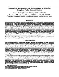
We developed a semi-automatic method of segmentation with a minimal interaction. The user selects the position inside the area corresponding to the basal nuclei and this software calculates the textured parameters, and trains the SVM machine. The preliminary results of the segmentation of the basal nuclei performed using our method is shown on Figure 7. We can see the detection of the basal nuclei and the comparison between the segmentation performed manually by a specialist. With this technique, there is a need to adjust the model’s parameters, which is done by trial and error. After adjusting the parameters for one type of acquisition, these parameters can be used in the study of other images of different patients that have been made with the same equipment. We are researching for methods of error evaluation in segmenting images with noise, applying the above processing pipeline.
REFERENCES 1.
2.
3.
4. 5. 6.
7.
8. 9.
Fig. 7 Experimental results showing the structures manually segmented (left), and segmented structures (right) obtained with C = 300 and γ= 55.0
CONCLUSIONS
Zhang J, Ma K-K, Meng Er, Chong V. (2010) "Tumor Segmentation from Magnetic Resonance Imaging by Learning via one-Class Support Vector Machine". Report of INRIA-00548532, Dec 20, 2010. LI Q-Y, Zhang S.X, Tan L.W, Qiu M.G. (2009) "Reconstruction and Application of Digital Brain Model on Chinese Visible Human (CVH)". O. Dössel and W.C. Schlegel (Eds): World Congress 2009, IFMBE Proceedings 25/IV, pp. 1119-1122. Haegelen C., Coupé P., Fonov V., Guizard N., Morando X., Collins DL. (2013) "Automated Segmentation of Basal Ganglia and deep brain structures in MRI of Parkinson's disease". International Journal of Computer Assisted Radiology and Surgery. 2013 Jan; 8 (1) pp. 99-110 DOI 10.1007/s11548-012-0675-8 Vapnik V (1998) "Statistical Learning Theory". John Wiley & Sons, Inc, New Jersey, USA. Chen P-H, Lin C-J, Schölkopf B. (2005). "A tutorial on -support vector machines". Appl. Stochastic Models Bus Ind, 21:111-136. Villegas R, Montilla G, Villegas H. "A Software Tool for Reading DICOM Directory Files". User Centered Design for Medical Visualization. DOI: 10.4018/978-1-59904-777-5.ch018 DCMTK 2012, "Digital imaging and communications in medicine tool kit". DICOM toolkit software documentation. Oldenburger Forschungs und Entwicklungsinstitut für Informatik-Werkzeuge und Systeme (OFFIS). Retrived on July 2, 2013, from http://dicom.offis.de/dcmtk.php.en Nagao M., Matsuyama T. (1979) "Edge Preserving Smoothing". Computer Graphics and Image Processing. Vol. 9, pp. 394-407. Chang C-C, Lin C-J (2010) LIBSVM: "A library for support vector machines". accessed at http://www.csie.ntu.edu.tw/~cjlin/libsvm Author: Institute: Street: City: Country: Email:
IFMBE Proceedings Vol. 41
Antonio BOSNJAK Centro de Procesamiento de Imágenes. Facultad de Ingeniería, Universidad de Carabobo. Valencia. Venezuela
[email protected]