... cerebro-spinal fluid. (CSF), white matter, and gray matter and ii) between normal .... H are described in the following equations. â. = = m c c k h c. H. 1. 1. ' (5). â. = = m ..... [2] Shan Shen, William Sandham, Malcolm Granat and Annette Sterr,.
International Journal of Soft Computing and Engineering (IJSCE) ISSN: 2231-2307, Volume-1, Issue-6, January 2012
Segmentation of Tissues in Brain MRI Images using Dynamic Neuro-Fuzzy Technique S.Javeed Hussain,T. Satya Savithri, P.V. Sree Devi 3Professor, Department of ECE, AU, Vishakapatnam, Andhra Pradesh, India Abstract- In this paper, an efficient technique is proposed for the Normally the structure of brain is complex and its accurate precise segmentation of normal and pathological tissues in the MRI segmentation is very crucial for finding the tumors, edema brain images. The proposed segmentation technique initially and necrotic tissues in order to specify proper therapy [4]. performs classification process by utilizing Fuzzy Inference The brain matters are mainly categorized as white matter, System (FIS) and FFBNN. Both classifiers are utilizing the gray matter, cerebrospinal fluid (CSF) or vasculature. extracted image features as an input for the classification process. Mostly the brain structures are clearly described by the The features that are extracted in two ways from the MRI brain boundaries of the tissue classes, so a technique to segment images. The FIS are used to make the classification process by tissues based on these categories is a major step in generating the fuzzy rules using extracted features. Five features quantitative morphology of brain [6]. Apart from other are extracted from the MRI images: they are two dynamic statistical features and three 2D wavelet decomposition features. In diagnostic methods, Magnetic resonance imaging (MRI) Segmentation, the normal tissues such as WM (White Matter), GM systems can generate many images and each image indicates (Gray Matter) and CSF (Cerebrospinal Fluid) are segmented from a different essential parameter of inner anatomical structures the normal MRI images and pathological tissues such as Edema in the same body section with multiple differences, based on and Tumor are segmented from the abnormal images. The nonthe local variations of spin–spin relaxation time (T2), spin– cortical tissues in the normal images are removed by the lattice relaxation time (T1), and proton density (PD) [5]. preprocessing stage. The implementation result shows the The presence of noise, errors in the scanners, and the efficiency of proposed tissue segmentation technique in structural variations of the imaging objects are the major segmenting the tissues accurately from the MRI images. The obstruction to the segmentation of MR images, such performance of the segmentation technique is evaluated by performance measures such as accuracy, specificity and sensitivity. obstructions are categorized into four types: The performance of segmentation process is analyzed using a thermal/electronic noise, magnetic field inhomogeneities, defined set of MRI brain image and compared against K-means biological tissue variations, and incomplete volume effects clustering and Fuzzy ANN based segmentation methods. [7]. Keyword: MRI, FFBNN, FIS
1. Introduction Segmentation of brain tissue on magnetic resonance (MR) images normally determines the type of tissue present for each pixel or voxel in a 2D or 3D data set respectively, based on the information gathered from both MR images and prior knowledge of the brain. It is one of the most vital preprocessing steps in several medical research and clinical applications, such as quantification of tissue volume, visualization and analysis of anatomical structures, multimodality fusion and registration, functional brain mapping, identification of pathology, surgical planning, surgical navigation, and brain substructure segmentation [1]. Segmentation at preliminary stage is important and necessary for the analysis of medical images for computeraided diagnosis and treatment. As the images are inherent in nature, medical image segmentation is a difficult and challenging task [2] [14] [15]. Magnetic resonance imaging (MRI) is a significant diagnostic imaging method, which is employed for the early detection of abnormal changes in tissues and organs [3] [16] as well as it is a non-invasive imaging technique, so it allows a radiologist to create an image of the inner aspects of living tissue [12]. Manuscript Details Received on December, 2011 S.Javeed Hussain, Associate Professor,Department of ECE, BCETFW, Kadapa, India T. Satya Savithri, Professor, Department of ECE, JNTUH Hyderabad, India P.V. Sree Devi, Professor, Department of ECE, AU, Vishakapatnam, Andhra Pradesh, India
Moreover, recognition and analysis of the lesions manually from MR brain images are generally time consuming, expensive and can produce unacceptably high intraobserver and interobserver variability [8]. The segmented MR images used in the medical diagnostic process depends on a combination of two, often conflicting, requirements, that is, the removal of the unnecessary information present in the original MR images and the maintenance of the significant details in the resulting segmented images [13] [17]. MRimage segmentation methods are usually evaluated based on their ability to differentiate i) between cerebro-spinal fluid (CSF), white matter, and gray matter and ii) between normal tissues and abnormalities [9]. Many techniques proposed in the recent years, which are used for the segmentation of brain tissues from MR image, are classical pattern recognition methods, rule-based systems, image analysis methods, crisp and fuzzy clustering procedures, feedforward neural networks, fuzzy reasoning, geometric models to determine lesion boundaries, connected component analysis, deterministic annealing, atlas based methods and contouring approaches [10] [11]. Lots of researches have been performed for the segmentation of normal and abnormal tissues in MRI brain images. Some of the recent related works regarding the segmentation of brain tissues are reviewed in the following section.
2. Proposed methodology for tissue segmentation in MRI brain images In this paper, we propose an efficient method to segment the normal and pathological tissues in the MRI brain images. Two major stages are involved in our proposed methodology:
416
Segmentation of Tissues in Brain MRI Images using Dynamic Neuro-Fuzzy Technique
o o
The features are extracted from each block in the block variable B s and each block is of n n dimension. Each
Classification Segmentation
Initially, the classification process is done on the given MRI brain images. In classification, the feature extraction process is performed in two ways and then these extracted features are given to the FIS and FFBNN for classification. Utilizing both classifier results, the MRI brain images (input image) are classified into normal and abnormal. Next, the segmentation process is performed over these classified images. Before the segmentation process, the non cortical tissues in the normal images are removed by performing preprocessing. Normal tissues such as WM, GM and CSF are segmented from the normal images and pathological tissues such as edema and tumor are segmented from the abnormal images.
block contains number of pixels. Particularly in each block, 5 features are extracted namely, statistical features such as mean and variance, and multilevel 2D wavelet decomposition features such as horizontal, vertical, diagonal bands of wavelet transform. The feature vector of each block is
Fk {M k ' , Ek ' , H k ' ,Vk ' , D k ' } Pixel values are represented as
mean and variance are calculated for these pixels in the blocks. Features are calculated by using the following equations
Mk'
2.1 Classification In classification stage, the MRI brain images are classified into normal and abnormal brain images. Two phases are involved in this classification that are mentioned below (i) Feature extraction (ii) Fuzzy Rules Generation (iii) Network training and testing
2. 1.1.1 Block wise Feature Extraction: Let I be the brain image that is divided into N number of
{bk } ; k 1 N . .For
feature extraction process, we have considered only a few numbers of blocks (not all blocks) by performing Euclidian distance measure. For this, we have taken one block bk and checked its neighbor blocks. If the entire neighbor blocks value is 0, then these blocks are not considered for feature extraction process or else determine the distance between the chosen block bk and neighbor blocks by exploiting Euclidian distance.
Dkl bk bl ; l 1 N , (k l ) (1) The distance value of each block Dkl is compared with the user defined threshold value t1 . During this comparison, if the distance value
Dkl of all blocks is less than this
threshold t1 , then it is adequate to store one block instead of storing all the blocks or else store the block’s values individually. As a result of the above process, we obtain the block values that are stored in a ' ' ' variable Bs {k }; k 1 N and the Feature extraction process is carried out for those stored blocks only.
1
pu
u 1
(3)
1 E k ' ( pu M k ) 2
u 1
2.1.1 Feature Extraction The features are extracted from the database MRI brain images. This process is done in two ways: (i) features that are extracted from the input images are processed blockwise; (ii) features are extracted directly from the input images. These two features extraction customs extract same features.
blocks, which is represented as B
pu
(2) and the features such as
(4) To obtain the wavelet features, here haar wavelet is applied to the blocks and performed a two level wavelet transform. The two level wavelet transform is applied to the n n size block images. After the two level wavelet transform, three features are extracted from the result image. Each feature has four pixel coefficients as m , and the computation of ' these three features H ' ,V ' , D k are described in the k k following equations 1 m (5) Hk' hc c c 1 1 m (6) Vk ' vc c c 1 1 m (7) Dk ' dc c c 1 In equations (5), (6), (7) the parameters hc , vc and d c are
the coefficients of the horizontal, vertical, and diagonal ' bands of one block k . 2.1.1.2 Direct Feature Extraction from Input Image Features are extracted directly from the input image I . Features are extracted by utilizing the equations (3), (4) (5), (6) and (7) but they are extracted for the whole image not for blocks. The extracted features are M , E, H ,V and D . In feature extraction phase, the extracted features in 3.1.1.1 FFBNN and the features from 3.1.1.2 are given to the FIS to accomplish the classification process. 2.1.2 Fuzzy Inference System (FIS) The fuzzy inference system normally contains three major operations: Fuzzification, Rules Evaluation and
417
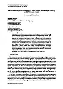
International Journal of Soft Computing and Engineering (IJSCE) ISSN: 2231-2307, Volume-1, Issue-6, January 2012 Defuzzification. Fuzzy inference is the process of creating a mapping from a given input to an output by means of a fuzzy logic. Then, the mapping provides a basis from which decisions can be made, or patterns discerned. The process of fuzzy inference involves Membership Functions, Logical Operations, and If-Then Rules. The schematic diagram of the fuzzy inference system (FIS) is shown in Fig. 2.
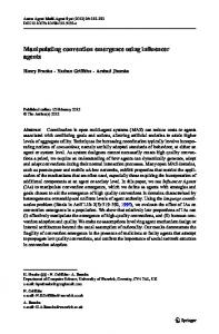
The following steps describe the function of the Neural Network: Step 1: Put the input weights to every neuron except the neurons in the input layer. Step 2: The neural network is designed with five inputs layers, H a hidden layers and one output layer. The weights are then added to the neural network and it is biased. Step 3: The planned bias function and activation function for the neural network is described below. The
Figure 1: Fuzzy Inference System Structure Fuzzification In fuzzification process, the crusty quantities are changed into fuzzy. In our proposed method, the fuzzification process is carried out by employing the features that are extracted in section 3.1.1.2. The extracted features are M , E, H ,V and D , for each feature we perform the fuzzification process. For the fuzzification process, we collect all the M , E, H ,V and D features values of training images and computed each feature minimum (min) and maximum (max) values. The fuzzification process is performed by using the following equations.
max min ML(M ) min 3 max min XL( M ) ML 3
input layer bias function is given ' ' ' ' ' ' M k ' (k n ) , Ek ' (k n ) , H k ' (k n ) , Vk ' (k 'n' ) and Dk ' (k 'n' ) are the extracted features of the ' block k . The activation function for the output layer is given in Eq. (10). Step 4: Compute the learning error for the neural network. N 1 1 a L(e) Dn ' Z n ' (10) Ha ' n 0 (e) In Eq. (11), L is the FFBNN network output, Dn ' and Z n ' are the desired and actual outputs respectively.
Dual FFBNN networks F1 and F 2 are well trained with these extracted features and different number of unknown brain MRI images is tested. After generating both networks, we compute average value between F1 and F 2 networks
(8) (9)
results f1 , f 2 .
ML( M ) and XL( M ) are the minimum and maximum limit values of the feature M . In above equations (8) & (9),
The same equations are used for the features E, H ,V and D to compute the minimum and maximum limit values.
f f2 (13) A 1 2 The result of these F1 and F 2 networks average value is represented as A and this value A is compared with threshold value t 2 .
Abnormal; A t 2 result Normal; A t 2
(14)
In this way, the brain MRI images are classified into normal and abnormal. Next, the segmentation process is performed for these classified images.
3.2 Segmentation
Figure 2: Basic Structure of the F1 FFBNN
Segmentation process is performed in both normal and abnormal images. In normal images, the normal tissues such as WM, GM and CSF are segmented and in abnormal images, the edema and tumor tissues are segmented. Following are the two steps involved in the segmentation process (i) Preprocessing (ii) Tissue Segmentation
418
Segmentation of Tissues in Brain MRI Images using Dynamic Neuro-Fuzzy Technique (a) Normal tissue segmentation (b) Pathological tissue Segmentation
value of each pixel in the image
global threshold T g and the resultant binarized image is I b .
3.2.1 Preprocessing Various preprocessing methods have been proposed to deal with the MRI brain images used for segmentation. Among all preprocessing methods, Skull stripping is used for the segmentation of brain tissues. The brain cortex can be visualized as a distinct dark ring surrounding the brain tissues in the MRI images. The distinct dark ring surrounding the brain tissues are removed by skull stripping method. In skull stripping, initially the given MRI brain image is converted into gray scale image and then a morphological operation [25] is performed in the gray scale image. Then the brain cortex in the gray scale image is stripped by using region based binary mask extraction. The preprocessing process is performed in the classified normal images, not abnormal images. Because preprocessing process helps to improve the normal tissue CSF is lightly placed in the cortex surrounding area. The normal is image obtained after skull stripping is denoted as I s .
Then the binarized image I b is subjected to morphological opening and closing operation. Opening and closing operation is utilized to remove small objects and small holes from the image I b . Finally, MRI brain image WM and GM tissues are segmented based on their intensity values. WM ; if I bi 1 I wg (18) GM ; if I bi 0 CSF segmentation To segment the cerebrospinal fluid from the brain MRI image, an Orthogonal Polynomial Transform (OPT) is applied to the skull stripped image I s . In orthogonal polynomial transformation, image 3 I s (i ) I cf Sin 100
2
0.05 * rand (| I s |)
(19)
After skull stripping, the brain MRI images are involved in the tissue segmentation process. Different methods are used to segment the WM, GM, CSF, edema and tumor tissues.
After the polynomial transform, the corresponding CSF region is segmented in the resultant image I cf .
3.2.2.1 Normal Tissue Segmentation
3.2.2.2 Pathological Tissue Segmentation
Segmentation of Normal tissues such as WM, GM and CSF are performed from the normal images. Here, segmentation process is performed in two ways namely, (i) WM and GM segmentation (ii) CSF segmentation
WM and GM segmentation The skull stripped image I s is given as input to the WM and GM segmentation process. Here, the major step is to segment the WM and GM tissues from the image I s by utilizing Gradient Method. The smoothing process is performed in the input image I s by applying Gaussian convolution filter. Smoothed image obtained from the Gaussian convolution filter is I G . After that, gradient operation is applied to the image I G . The gradient of two variables x and y is defined as follows,
(17)
Then, the binarization process is performed in the edge marked image E m . In binarization process, the gray level
Pathological tissues such as edema and tumor are segmented from the classified abnormal images and these tissues are segmented by two different methods: (i) Tumor segmentation (ii) Edema segmentation
Tumor Segmentation The tumor tissue segmentation is performed in the abnormal brain MRI images. The main objective is to segment the tumor tissue in the abnormal image I a . Here we utilize the Region Growing Method (RGM) to segment the tumor tissue. Region growing method is a region based image segmentation method; it selects the initial seed points from the input image I a . The RGM observes the neighbor pixel values with the initial seed points, that is it checks whether the neighbor pixels are included in this region or not [24]. The tumor segmentation result is represented as I T .
Edema Segmentation Edema tissue is segmented from the abnormal image I a .
(15)
Using the gradient values, the current edges in the image are marked using the Equ. (16) & (17). (16) G x(i ) 2 y( j ) 2
1 Em 1 G
I s is computed using the
following formula,
3.2.2 Tissue Segmentation
I I I G ( x, y ) G i G j x y
E m is observed by using
Before the edema segmentation process, histogram equalization process is executed over the image I a . The quality of image I a
is enhanced by the histogram
' equalization and it is denoted as I a . Then the enhanced I a' image is converted into indexed image by using multilevel thresholding function. ' converts the grayscale I a image
419
Grayslice function into
International Journal of Soft Computing and Engineering (IJSCE) ISSN: 2231-2307, Volume-1, Issue-6, January 2012 indexed image using multilevel threshold and the result '' '' image is I a . After that, the image I a is converted into HSV (Hue, Saturation and Value) color model and it is ''' represented as I a . Next, the threshold process is performed ''' in the image I a . We define separate threshold value for Hue, Saturation, and Value. Each pixel in the image is compared with these threshold values to select the pixels.
H t3 , S t 4 ,V t5 p ; p t , t & t4 X u u 3 5 (20) 0; otherwise In the above eqn [15], X is the pixel values that satisfy the
above conditions. Morphological closing operation is applied on the mask X and the resultant image is denoted (c ) (c ) as X . Now, the image X contains z number of regions and then we compute the centroid value for each (c ) region, which is represented as X h ( x, y ), h 1,2, z . Subsequently, the distance is determined between the coordinates of center pixels of the regions in X h(c) ( x, y) and the tumor centroid coordinate value t ( x, y ) .
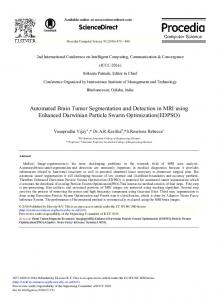
given as input to the dual FFBNN networks. These networks are trained using back propagation algorithm. The result of dual FFBNN network is evaluated by unknown testing images. The classification results of dual FFBNN networks are shown in Fig.4. Then, the segmentation process is performed on the classified images. The normal images are segmented into three normal tissues such as WM, GM and CSF and the abnormal images are segmented into two pathological tissues such as edema, tumor. Preprocessing process is performed in the classified normal images before the segmentation process. In preprocessing, skull stripping process is performed to remove the non cortical tissues from the images. Fig. 6 shows output of the preprocessed MRI normal image.
Figure 6: Preprocessed MRI normal image output Normal tissues GM and WM are segmented by the gradient method and CSF is segmented by OPT. The segmented normal tissue results are shown in Fig. 7.
Oh ( x, y) X h(c) ( x, y) t ( x, y) (21) The resultant Oh ( x, y ) is then verified with threshold value t 6 and an edema region coordinate values are obtained, O ( x, y ) t 6 Ie h 0; otherwise
Figure 7: Segmentation outputs of normal tissues (i) WM segmentation (ii) GM segmentation (iii) CSF segmentation and (iv) WM, GM and CSF in original normal image
(22)
Pathological tissues such as tumor and edema are segmented by RGM and thresholding process. Various thresholding and morphological operations are performed during the edema segmentation, which are explained in section 2.2.2.2. Figure 8 shows the intermediary result of the edema segmentation. The segmented pathological tissues are shown in Figure 9.
Then the morphological dilation and closing operations are performed in the image I e .
4. Experimental results The proposed brain tissue segmentation technique is implemented in the working platform MATLAB (version 7.10) and it is evaluated using 10 medical brain MRI images, which are collected from various medical diagnosis centers. Among 10 MRI images, 5 images are normal and the remaining is abnormal. The fig. 4 shows the given input MRI brain images used for the MRI image classification process.
Figure 8: Images obtained from (i) Histogram Equalized Image (ii) HSV color model Image result (iii) Image obtained from HSV Thresholding Process (iv) Closing Operation result image (vi) Edema region result (vi) Closing Operation and (vii) Dilation Operation Figure 4: Classification result of normal and abnormal images The input images are classified by two FFBNN networks. Input values for both FFBNN networks are five features such as mean, variance, horizontal, vertical and diagonal functions of 2D wavelet decomposition and these features is
420
Segmentation of Tissues in Brain MRI Images using Dynamic Neuro-Fuzzy Technique
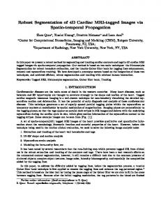
Figure 9: Segmentation result of pathological tissues (i) Tumor (ii) Edema and (iii) Tumor and Edema in abnormal image The performance measures values in Tables 1, 2 and 3 shows that the proposed segmentation method gives high percentage accuracy result than the K-means clustering and Fuzzy ANN method. The performance measures values corresponding graphs of both proposed and K-means clustering, Fuzzy ANN is listed below.
(iii) Figure 11: GM Tissues Segmentation result (i) Accuracy (ii) Sensitivity (iii) Specificity
(i)
(i)
(ii)
(ii)
(iii) Figure 12: CSF Tissues Segmentation result (i) Accuracy (ii) Sensitivity (iii) Specificity (iii) Figure 10: WM Tissues Segmentation result (i) Accuracy (ii) Sensitivity (iii) Specificity
(i) (i)
(ii) (ii)
421
International Journal of Soft Computing and Engineering (IJSCE) ISSN: 2231-2307, Volume-1, Issue-6, January 2012
(iii) Figure 13: Edema Tissues Segmentation (i)Accuracy(ii)Sensitivity(iii)Specificity
5. Conclusion In this paper, an efficient segmentation was developed to segment the normal and pathological tissues from the MRI brain images. The performance of the proposed segmentation was analyzed using defined set of MRI normal and abnormal images. Statistical measures were utilized to measure the performance of the proposed tissue segmentation method. The performance of the proposed segmentation method was analyzed and compared against the K-means clustering method and existing Fuzzy ANN based segmentation method. The comparative results showed that the proposed method outperforms in terms of accuracy and sensitivity rather than the Fuzzy ANN and Kmeans clustering. It lacked in performing in terms of specificity values for certain images. Even though there is a lack in specificity values, the greater improvement in accuracy and sensitivity values makes that tolerable. Hence the performance of the method was understood from the experimental results and analysis.
result
(i)
References
(ii)
(iii) Figure 14: Tumor Tissues Segmentation result (i) Accuracy (ii) Sensitivity (iii) Specificity The Figure 10 to 14 shows the graphical representation of WM, GM, CSF, Edema and tumor tissue segmentation performance compared to the Fuzzy ANN and K-means clustering method. It shows that the accuracy, sensitivity and specificity measure in this tissue segmentation processes are nearly same (or) higher than the Fuzzy ANN and Kmeans clustering methods. Also, the GM, CSF, Edema and Tumor tissue segmentation process has given high accuracy and sensitivity result than the Fuzzy ANN and K-means clustering methods, but these tissues performance lacks in specificity measure. However, this low performance of specificity measure will not affect the segmentation process because the specificity is only slightly lower than the Fuzzy ANN and K-means clustering as well as the accuracy level of both tissues are nearly same (or) high when compared to this low level result of Fuzzy ANN, K-means clustering methods.
[1] Chaozhe Zhu and Tianzi Jiang, "Multicontext Fuzzy Clustering for Separation of Brain Tissues in Magnetic Resonance Images", NeuroImage, Vol.18, No. 3, pp. 685-696, 2003 [2] Shan Shen, William Sandham, Malcolm Granat and Annette Sterr, "MRI Fuzzy Segmentation of Brain Tissue Using Neighborhood Attraction With Neural-Network Optimization", IEEE Transactions On Information Technology In Biomedicine, Vol. 9, No. 3, pp. 459467, September 2005 [3] Senthilkumaran and Rajesh, "Brain Image Segmentation using Granular Rough Sets", International Journal of Arts and Sciences, Vol. 3, No. 1, pp. 69 - 78, 2009 [4] Pradipta Maji, Malay K. Kundu and Bhabatosh Chanda, "Second Order Fuzzy Measure and Weighted Co-Occurrence Matrix for Segmentation of Brain MR Images", Journal of Fundamenta Informaticae, Vol. 88, No. 1-2, pp. 161-176, 2008 [5] Jzau-Sheng Lin, Kuo-Sheng Cheng, and Chi-Wu Mao, "Segmentation of Multispectral Magnetic Resonance Image Using Penalized Fuzzy Competitive Learning Network", Journal of Computers and Biomedical Research, Vol. 29, No. 4, pp. 314–326, 1996 [6] Mostafa G. Mostafa, Mohammed F. Tolba, Tarek F. Gharib and Mohammed A-Megeed, "A Gaussian Multiresolution Algorithm For Medical Image Segmentation", In Proceedings of IEEE International Conference On Intelligent Engineering Systems, Assiut-Luxor, Egypt, 2003 [7] Jagath C. Rajapakse, Jay N. Giedd and Judith L. Rapoport, "Statistical Approach to Segmentation of Single-Channel Cerebral MR Images", IEEE Transactions on Medical Imaging, Vol. 16, No. 2, pp. 176-186, April 1997 [8] W.Wells, W.Grimson, R. Kikinis, F.A. Jolesz, “Adaptive Segmentation of MRI Data”, IEEE Transaction on Medical Imaging ,Vol.15, No. 4, pp. 429-442, August 1992 [9] Nicolaos B. Karayiannis and Pin-I Pai, "Segmentation of Magnetic Resonance Images Using Fuzzy Algorithms for Learning Vector Quantization", IEEE Transactions on Medical Imaging, Vol. 18, No. 2, pp. 172-180, February 1999 [10] Fitsum Admasua, Stephan Al-Zubia, Klaus Toenniesa, Nils Bodammerb and Hermann Hinrichsb, "Segmentation of Multiple Sclerosis Lesions from MR Brain Images Using the Principles of Fuzzy-Connectedness and Artificial Neuron Networks", In Proceedings of International Conference on Image Processing, Barcelona, Spain, Vol. 3, 2003 [11] N. K. Subbanna, M. Shah, S. J. Francis, S. Narayanan, D. L. Collins, D. L. Arnold and T. Arbel, "MS Lesion Segmentation using Markov Random Fields", In Proceedings of International Conference on Medical Image Computing and Computer Assisted Intervention, London, UK, September 2009 [12] Kenneth Revett and Aurangzeb Khan, "An On-Line (Real-Time) Automated MRI Based Pathology Detection System Using Selforganised Maps", In Proceedings of Virtual Multi Conference on Computer Science and Information Systems, pp. 213-216, 2005 [13] Nahla Ibraheem Jabbar and Monica Mehrotra, "Application of Fuzzy Neural Network for Image Tumor Description", World Academy of Science, Engineering and Technology, Vol. 44, pp. 575-577, 2008
422
Segmentation of Tissues in Brain MRI Images using Dynamic Neuro-Fuzzy Technique [14] Manisha Sutar and Janwe, "A Swarm-based Approach to Medical Image Analysis", Global Journal of Computer Science and Technology, Vol. 11, No. 3, pp. 23-26, March 2011 [15] Pradipta Maji, Malay K. Kundu and Bhabotosh Chanda, "Segmentation of Brain MR Images Using Fuzzy Sets and Modified Co-Occurrence Matrix", In Proceedings of IET International Conference on Visual Information Engineering, Bangalore, India, pp. 327-332, 2006 [16] Forghani, Forouzanfar and Forouzanfar, Tehran "MRI Fuzzy Segmentation of Brain Tissue Using IFCM Algorithm with Particle Swarm Optimization", In Proceedings of International Symposium on Computer and Information Sciences, Ankara, pp. 1-4, 2007 [17] Nicolaos B. Karayiannis, "A Methodology for Constructing Fuzzy Algorithms for Learning Vector Quantization”, IEEE Transactions on Neural Networks, Vol. 8, No. 3, pp. 505-518, May 1997 [18] Zhiqiang Lao, Dinggang Shen, Dengfeng Liu, Abbas F. Jawad, Elias R. Melhem, Lenore J. Launer, R. Nick Bryan and Christos Davatzikos, "Computer-Assisted Segmentation of White Matter Lesions in 3D MR Images Using Support Vector Machine", Academic Radiology, Vol. 15, No. 3, pp. 300-313, March 2008 [19] Zhang, Wang, and Wu, "A Novel Method for Magnetic Resonance Brain Image Classification Based On Adaptive Chaotic PSO", Progress In Electromagnetics Research, Vol. 109, pp. 325-343, 2010 [20] Mina Rafi Nazari and Emad Fatemizadeh, "A CBIR System for Human Brain Magnetic Resonance Image Indexing", International Journal of Computer Applications, Vol. 7, No.14, pp. 33-37, October 2010 [21] Shafaf Ibrahim, Noor Elaiza Abdul Khalid and Mazani Manaf, "SeedBased Region Growing (SBRG) vs Adaptive Network-Based Inference System (ANFIS) vs Fuzzy c-Means (FCM): Brain Abnormalities Segmentation", International Journal of Electrical and Computer Engineering, Vol. 5, No. 2, pp. 94-104, 2010 [22] Nandita Pradhan and Sinha, "Development of a Composite Feature Vector for the Detection of Pathological and Healthy Tissues in FLAIR MR Images of Brain", Journal of ICGST-BIME , Vol. 10, No. 1, pp. 1-11, December 2010 [23] Jayashri Joshi and Phadke, "Feature Extraction and Texture Classification in MRI", In Proceedings of International Conference on Computer Technology, Vol. 2, No. 2, 3, 4, pp. 130-136, 2010 [24] Frank Y. Shih and Shouxian Cheng, "Automatic seeded region growing for color image segmentation", Journal of Image and Vision Computing, Vol. 23, pp. 877–886, 2005 [25] Soumya Maitra, "Morphological Edge Detection Using Bit-Plane Decomposition in Gray Scale Images", In Proceedings of INDIACom, New Delhi, 2011.
Dr.P.V.Sridevi obtained B.Tech in electronics &communication engineering from V.R.Siddartha Engineering college, Vijayawada in 1986 and M.E in applied electronics from P.S.G. College of technology, Coimbatore in 1988. She obtained her Ph.D degree from Andhra University in 1997.She is having teaching experience of 20 years. Her Areas of interest include Antennas, Microwaves, VLSI, Image Processing .At present she is working as Associate Professor in the department of Electronics and communication Engineering, AU College of Engineering, Visakhapatnam. She has published in 10 International Journals, 15 International Conferences.
Dr.T. Satya Savithri, Specialized in Digital Image Processing. Her Research interests include Design and Testing of VLSI and also interest in Microwave Engineering. She has 10 publications in various National and International Journals and Conferences. The total services 13 years and worked at CBIT, KSRM, JNTUCEA and JNTU CEH in Asst. Prof. and Associate Professor Levels. At present working as Associate Professor in ECE Department, JNTU CEH. She had conducted two refresher courses. She has been a Resource person for a short term course held at NRSA,. Hyderabad and workshop Mahaveer College chaired a National Conferences held at JNTUCE, Anantapur. She is Chairman for Department Academic and Planning Activities.
S.Javeed Hussain received B.Tech from JNTUniversity, & M.Tech Degree in Electronics & Communication Engg. Hyderabad, India. He is currently working towards PhD Degree in Digital Image Processing at J.N.T University,Hyderabad, India. At present he is with Bharat college of engineering for women, Kadapa, A.P., India working as an Vice Principal & HOD in Dept. of ECE. He presented many research papers in National & International Conferences. He is a member of Professional societies like IE(India), ISTE (India), BMESI (India). His research interests include Signal Processing, Time Series Analysis and Image Processing.
423