with wide pH-sensing ranges, high durability, and small drifts in potentials. ...
Miniaturized pH sensor arrays based on biocompatible materials would be of
great ...
IEEE SENSORS JOURNAL, VOL. 13, NO. 10, OCTOBER 2013
3857
Sol-Gel Iridium Oxide-Based pH Sensor Array on Flexible Polyimide Substrate Cuong M. Nguyen, Wen-Ding Huang, Smitha Rao, Hung Cao, Uday Tata, Mu Chiao, and Jung-Chih Chiao
Abstract— Iridium oxide pH sensing film is demonstrated with wide pH-sensing ranges, high durability, and small drifts in potentials. Using sol-gel process, a lower fabrication cost and less labor-intensive method, to deposit iridium oxide thin films for pH sensing is reported previously by our group with expected advantages. In this paper, we fabricate and test pH sensing characteristics of 4 × 4 anhydrous iridium oxide thinfilm electrode arrays on flexible substrates. The sensors in arrays exhibit Nernstian potential responses in the range of 57.0–63.4 mV/pH. Stability, repeatability, and hysteresis effects of the pH sensor arrays are examined. A multichannel recording system is built to demonstrate the functionality of the pH sensor arrays in monitoring spatial and temporal pH changes across a surface. Index Terms— Flexible substrate, iridium oxide, pH sensor, sensor array.
I. I NTRODUCTION
I
T has been demonstrated that hydrogen ion concentration represented as pH value can affect the activities of many biological and medical processes such as enzymatic reaction [1]–[3], acid-base homeostasis [4], exchange of nutrients and waste products in living organisms [5]. These processes may also alter pH values of milieus or solutions. Therefore, in situ assessment of pH could provide important information to further understand these processes in cells, tissues, and organs. Miniaturized pH sensor arrays based on biocompatible materials would be of great importance for uses in such biomedical applications that also require long-term monitoring and capability to map biochemical processes spatially. These applications include, but are not limited to, assessment of conditions for wound healing [6]–[8], cell culture [9]–[11] and fermentative food processes [12]. Measurement of pH levels at multiple locations using optical and electrochemical methods have been proposed [13]–[19]. Optical approaches, such as hydrogel microarray [20] and quantum dot-based ratiometric pH sensors [16], utilize Manuscript received June 7, 2012; revised November 23, 2012; accepted December 6, 2012. Date of publication December 25, 2012; date of current version August 30, 2013. The associate editor coordinating the review of this paper and approving it for publication was Prof. Yong Xu. C. M. Nguyen, W.-D. Huang, S. Rao, H. Cao, U. Tata, and J.-C. Chiao are with the Department of Electrical Engineering, University of Texas at Arlington, Arlington, TX 76019 USA (e-mail: cmnguyen@mavs. uta.edu;
[email protected];
[email protected];
[email protected];
[email protected];
[email protected]). M. Chiao is with the Department of Electrical Engineering, University of Texas at Arlington, Arlington, TX 76019 USA, and also with the Department of Mechanical Engineering, University of British Columbia, Vancouver, BC V7E5B4, Canada (e-mail:
[email protected]). Color versions of one or more of the figures in this paper are available online at http://ieeexplore.ieee.org. Digital Object Identifier 10.1109/JSEN.2012.2236551
images of pH-sensitive fluorescent indicators to determine pH values. It has been shown that the optical pH sensors have advantages of fast response, high accuracy and high precision [16], [21]. However, the photo-bleaching effect of fluorescein limits the use of optical pH sensors in long-term experiments [16], [21]–[23]. Quantum dot based pH sensors [16] overcome photo-bleaching issues but the synthesis of quantum dots involve complicated multiple-step processes. There are also potential biocompatibility issues of delivering quantum dots onto or into tissues for practical long-term clinical uses. The distribution and immobilization of quantum dots in wet surfaces or aqueous solution also face significant technical challenges. In consideration of aforementioned factors for in vivo implants or in vitro analysis in flowing liquid, electrochemical sensors are preferred owing to features of direct electrical response, mechanical stability and welldefined spatial responsivity, besides their lower material cost and simpler fabrication process. Micro-array pH sensors with needle-like features have been proposed [17]. The durable micro-scale metal tips deposited with pH-sensing materials offer the ability to measure intra- and extra-cellular pH values to study bacterial or cellular metabolism [17], [24]. However, these sensors are not suitable for in situ larger-area biomedical experiments since the rigid tips could cause irreversible tissue damage. Therefore, we propose an array of planar, miniature pH sensors on a deformable polyimide substrate. The choices for the materials of pH sensors were considered based on the requirement of biocompatibility, reliable performance and ease of fabrication. Iridium oxide (IrOx ) is preferred among other solid-state metal oxides including RuO2 , PtO2 , OsO2 , Ta2 O5 [19], [25]–[27]. Different deposition methods have been investigated such as sputtering, electrodeposition, thermal oxidation, and sol-gel processes [18], [24], [28]. Sputter deposition of iridium oxide and thermal oxidation of iridium offer conformal, high-density and highquality thin films. However, they are expensive due to the needs of high-purity iridium targets and sophisticated vacuumbased deposition systems [26], [29]. They also present thermal issues during fabrication on flexible polymer substrates. On the other hand, sol-gel processes and electrodeposition are regarded as considerably lower costs and simpler methods for coating IrOx thin films (IROFs) without creating local heating problems on polymer substrates [30]–[32]. In our previous work, we have reported the use of sol-gel processes to fabricate a flexible pH sensor with a response of 58.5 ± 2 mV/pH which matched the Nernstian response [33]. The sol-gel based IrOx thin film has also been demonstrated to have many prominent characteristics such as low temperature dependence, low
1530-437X © 2012 IEEE
3858
IEEE SENSORS JOURNAL, VOL. 13, NO. 10, OCTOBER 2013
ion-interference and low voltage drifts [33]. In this paper, we further employed the sol-gel method to fabricate a pH sensor array consisting of sixteen sensors on a single polyimide substrate with a batch fabrication process. Each sensor in the flexible array consisted of a working IrOx electrode and a silver/silver chloride (Ag/AgCl) reference electrode. We implemented individual reference sensor electrodes in order to avoid any possible interference or crosstalk issues. Electrical interfaces were also built to enable the integration of our sensor array with a telemetry circuit for wireless signal transduction in the future.
□ 3cm
□ 1 mm 2 mm Connection Pad
(a)
Ag/AgCl
II. FABRICATION
-
Hydrochloric acid HCl (35%), acetic acid CH3 COOH, sodium chloride NaCl (99%), ethanol C2 H5 OH (95%) and powder of iridium chloride IrCl4 · 2H2 O (99%) were used. Sol-gel solution was prepared by mixing 42 ml of ethanol with 10-ml acetic acid. After stirring the solution for 30 minutes, one-gram hydrated iridium chloride was added. Esterification reaction between ethanol and acetic acid formed ethyl acetate ester that bonded with iridium ions and increased the solution viscosity. The solution was stirred for one hour before use. Utilizing the fabrication processes similar to that detailed in our previous work [33], we further reduced the sensing area of each electrode to 1 × 1 mm2 in order to have a compact array. A flexible polyimide film with a thickness of 125 µm was used as the substrate. The film was cleaned and heated at 100 °C for ten minutes to remove moisture in order to improve photoresist adhesion. Layers of 7-nm and 150-nm thick Cr and Au, respectively, were deposited after patterning the working electrodes by photolithography process. Next, a layer of SU-8-100 (Microchem) acting as an insulation layer was spincoated and patterned. The sample was then dip-coated in the sol-gel solution to form IrOx thin film. The immersion and withdrawal process of the samples from the sol-gel solution were precisely controlled by a custommade dip coater. The immersion time primarily affected the film conformity and should be sufficient for sol-gel molecules to settle on the substrate. The withdraw rate primarily affected the film thickness and quality, which determined the sensor sensitivity. In our experiments, after numerous iterations of fabrication and thickness profile measurements to identify the parameters for the large arrays, we chose the immersion time and withdraw rate at 5 minutes and 10.0 cm/min, respectively, which produced satisfactory film thickness and reasonable surface uniformity consistently. The samples then underwent thermal oxidation to oxidize iridium chloride (IrCl4 ). As discussed in our previous work [33], iridium oxide film began to crack above 400 °C. Therefore, the thermal treatment process was carried out at 300 °C for one hour on a hot plate. The reaction can be described as 3 300°C 2IrCl4 + O2 −→ IrO2 + 4Cl2 2
(1)
Annealing was then carried out in a closed oven at 300 °C for five hours to complete the conversion to iridium oxide
IrOx +
Vout
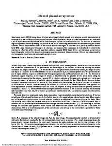
(b) Fig. 1. (a) Photo of a pH sensor array. The array includes 16 individual sensors. Each sensor consists of one working iridium oxide and one Ag/AgCl reference electrodes. (b) Entire sensing area was 3 × 3 cm2 . Each electrode has a metal line to a pad connecting to the recording circuit.
according to the following equation: 1 300°C 2IrO2 −→ Ir2 O3 + O2 (2) 2 3 300°C (3) 2IrCl4 + O2 −→ IrO2 + 4Cl2 2 The iridium oxide thin-film had thickness of 0.9 µm, measured by a profilometer after dip-coating and thermal treatment processes. The SU-8 insulation film was then removed. Due to thermal stress and cracks inside the SU-8 film yielded during annealing step, the film warped and detached at the edges of polyimide substrate. The film, therefore, was easy to remove simply by peeling the film off with a sharp tweezer. After cleaning, a 30-nm thick Ag on 3-nm thick Cr was deposited and patterned to form the Ag/AgCl reference electrodes. Next, an insulation layer of SU-8-5 was coated on the whole sample except the sensing areas and connection pads. The reference electrodes were created by chloridizing silver electrodes in 0.1-M hydrochloric acid (HCl) solution saturated with sodium chloride (NaCl). The process has been done by applying a voltage bias of 0.5 V between silver electrodes and a Pt wire electrode in a period of 5 seconds. The change of color at the tip of silver electrodes to dark grey indicated the presence of silver chloride thin film. III. E XPERIMENTAL S ETUP Fig. 1(a) shows a photo of a pH sensor array. Fig. 1(b) shows the dimensions, configuration and circuitry for measurement. The array of 4 × 4 pH sensors fits on a flexible polyimide substrate with dimensions of 6 × 6 cm2 while the sensing area for 16 devices was 3 × 3 cm2 . Electrodes were connected to contact pads through 2-cm long metal lines. The recording circuit board was connected via copper wires attached to the contact pads by conducting silver epoxy (MG Chemicals). Each pH sensor consisted of one working and one reference electrodes which had a size of 1 × 1 mm2 . The space between
NGUYEN et al.: SOL-GEL IRIDIUM OXIDE-BASED pH SENSOR ARRAY ON FLEXIBLE POLYIMIDE SUBSTRATE
two electrodes was 2 mm. It should be noted that additional length for metal lines, additional space between metal lines and electrodes, and larger contact pads were allowed in this prototype demonstration for ease to conduct experiments. The dimensions can be reduced to further miniaturize the array. Each sensor could be recorded separately or simultaneously. A differential amplifier was used between the sensor and NI-USB-6210 data acquisition card that was interfaced to a computer. This was to eliminate the loading effect which occurred when the sensor terminals were directly connected to the low impedance inputs. Precise instrumentation amplifiers AD8221 (Analog Devices) with a high common-mode rejection ratio (CMRR), low power consumption, and zero-drift output voltage were chosen to build the differential amplifier. The potentials obtained from the pH responses were displayed in real time with a graphical user interface in LabVIEW (National Instruments) and stored. This interface circuit can also be readily connected to a wireless module, such as EZ430RF2500 (Texas Instruments), for remote monitoring.
3859
pH 2 pH 4
pH 7
pH 10 pH 12
Sensor 1 Sensor 2
Sensor 3 Sensor 4
(a)
pH 2 pH 4
pH 7
IV. R ESULTS AND D ISCUSSION
pH 10
A. pH Measurement in Individual Sensors Each sensor was tested individually. Sensors from the same batch of fabrication were expected to have similar sensitivities. The measurements were performed by dripping successively five different buffer solutions of pH = 2, 4, 7, 10, and 12 onto the electrodes of each sensor using a pipette. First, pH = 2 solution was dripped sequentially on all sensors while they were continuously recorded. Before the introduction of a new solution (pH = 4), the existing solution was pipetted out. The procedure of cleaning the electrodes has been done by dripping DI water on the electrodes and then pipetting out. The electrodes were also air-dried to eliminate interference from the previous buffer solution (pH = 2). The procedure was controlled to be completed in 30 seconds for each electrode. During the cleaning, the electrodes were disconnected from the recording circuit to avoid short-circuit issues. The procedure was repeated for all buffer solutions. Fig. 2(a) and (b) show the recorded potentials of eight exampled sensors in a 30-minute window. Results showed distinct pH responses to different buffer solutions and the time points for each transition events could also be clearly identified. The response time is the period of time needed for hydrogen ions to exchange and equilibrate in the porous iridium oxide film. We previously defined the response time of a pH sensor as the transient time required for its output voltage to reach 90% of equilibrating value after dripping liquid onto the sensor [33]. According to our previous paper, the response time of the sensor was less than 2 seconds [33]. In this experiment, the response time was longer due to noises when the buffer solutions were dripped on the electrodes. The signals of sensors #1 and #5 slowly stabilized after one minute and both showed noisy outputs. The sensors #4 and #8 showed cleaner outputs with faster responses since it took less than 30 seconds to achieve stable output voltage with solutions of pH = 2, 4, 7, 10. This phenomenon was due to the manual dripping of solutions with a hand-held pipette on
pH 12 Sensor 5 Sensor 6 Sensor 7 Sensor 8
(b) Fig. 2. Individual sensor test. Experiments were conducted with five buffer solutions of pH = 2, 4, 7, 10, and 12. Each sensor was tested in 30 minutes. Sensors (a) #1–#4 and (b) #5–#8.
the electrodes. Thus, the sensors #1 and #5 had more noises as they were inside the array while sensors #4 and #8 were on the edge. The liquid dripped on the sensors experienced vibration causing micro-scale redistribution of ions on the sensing thin film due to unsteady hands. For pH = 12 solution, it was observed that the potentials slowly stabilized before overshoot to positive potentials. This phenomenon was also observed in our previous study as the surface OH− ion exchanges slowly approached to a new equilibrium point [33]. Fig. 3 showing the sensitivity responses of these eight pH sensors were plotted by using the nominal pH values of buffer solutions as calibration references. The sensitivities were in the range of 57.0–63.4 mV/pH among all eight sensors, which matched with the theoretical Nernstian response. The variations in sensitivity were due to subtle differences of nano-scale pore sizes in the iridium oxide film, quality, and uniformity of Ag/AgCl reference electrodes, as we have discussed in [33]. The standard error of the mean (SEM) of sensitivities of 0.7 mV/pH and an average linearity regression r 2 of 0.96 demonstrated acceptable uniformity of the sensor performance. In order to investigate the substrate-deforming effect on the sensor performance, the array was bent at different
3860
IEEE SENSORS JOURNAL, VOL. 13, NO. 10, OCTOBER 2013
Fig. 4. Measurement deviations in pH for four different sensors in comparison with the measured pH values, which were calculated from the calibration curves and the nominal pH values of the buffer solutions. Fig. 3. Sensitivities of eight individual sensors were obtained with five pH buffer solutions. The sensor responses were in the range of 57.0–61.2 mV/pH. TABLE I S ENSITIVITIES OF S IX S ENSORS ON THE A RRAY W HEN THE A RRAY WAS B ENDED AT D IFFERENT C URVATURE R ADII Bending Flat Curvature R = 14 cm Curvature R = 11 cm Curvature R = 8.4 cm Curvature R = 3 cm
Mean of Sensitivities (mV/pH) 59.84 58.97 58.93 59.87 55.83
Standard Error of the Mean (SEM) 0.76 1.03 1.20 1.06 2.59
curvature radii. Standard Scotch tape (3M products) was utilized to tape the edges of the sensor array on a curved surface. Six sensors across the array and on the edges were chosen to be tested. Table I illustrates that the change in the sensitivities of these six sensors was smaller than 1 mV/pH when the bending curvature radii were less than 8.4 cm. With more bending, stresses, micro-cracks and changes in electrical resistance of the metal lines, which connected the sensing area to the pads, or at the interfaces of intermetallic layers (Au and Cr) may be created. Such changes in mechanical structures thus induced more significant impacts on the performance of the sensor array. In our design, the averaged sensitivity and deviation among six sensors started to show changes with the curvature radius of 3 cm. Therefore, re-calibration is recommended when the sensor array conforms onto a surface with smaller curvature. It should also be noted that the minimal bending curvature depends on the electrode size and spacing, connection wire width and configuration, contact pad dimensions and spacing, metal and film thicknesses, and array configuration. An array with smaller bending curvature is possible with smaller sensor and spacing dimensions. Utilizing the linear sensitivity responses in Fig. 3 as the calibration standards, pH buffer solutions were used on the
sensors again to validate the performance. The solution was dripped manually on each sensor by a pipette and a similar cleaning procedure was also applied. Fig 4 shows the pH values indicated in the four exampled sensors with solutions of pH = 2, 4, 7, 10, and 12. For solutions with pH less than 7, all sensors reacted consistently with negligible errors. With alkaline buffer solutions, the sensor #4 could not distinguish the solutions of pH = 10 and 12 while the other three could distinguish them clearly. The indistinguishable responses of the sensor #4 to pH = 10 and 12 could be explained by the larger variation in potential generated in alkaline solutions during the calibration process. Our previous study shows that the residues left in the pores of iridium oxide film and ions from the alkaline buffer solutions may distort the result of calibration process [33]. The ion or radical residues in the micro- and nano-scale pores are difficult to identify from the examination of film surfaces, thus it is difficult to ensure the calibration accuracy. We particularly discussed this case (sensor #4) to show that our “calibration and re-measurement” procedure was able to be utilized to identify incorrect calibration, even for the case when the sensitivity curve showed a good linearity regression. Each sensor was tested in the same procedure with five pH levels to ensure the accuracy. In the case of incorrect calibration, the procedure was repeated again after cleaning sensors carefully. B. Temporal Responses Temporal responses were examined by repeating the same test procedure three times with controlled timing. Four sensors were chosen. Six buffer solutions of pH = 2, 4, 5, 7, 10, and 12 were applied in sequence onto the electrodes. One complete test set of six solutions was called an episode. Signals were recorded for 90 seconds with a buffer solution applied on the electrodes of the sensors. The solution was retreated by a pipette before a new solution was applied. In this experiment, sensors were not rinsed or soaked with DI water drops, which mimicked the practical scenarios in sensing pH on a surface
NGUYEN et al.: SOL-GEL IRIDIUM OXIDE-BASED pH SENSOR ARRAY ON FLEXIBLE POLYIMIDE SUBSTRATE
3861
14 pH = 12
12 pH = 10
pH
Voltage, V
10 8
pH = 7
6 1st Episode
nd
2 Episode 3rd Episode
Sensor
pH = 4
4 pH = 2
Time, s Fig. 5. Temporal responses of four exampled sensors in three episodes. The sequence of testing in the first and second episodes was with buffer solutions of pH = 2, 4, 5, 7, 10, and 12. The sequence in the third episode was with buffer solutions of pH = 7, 12, 2, 4 5, 7, and 10. In each test, liquid was pipetted out and new buffer solution was added without cleaning the electrodes with DI water.
but would introduce noises with solution residues. For the first two episodes, the sequence of testing was with buffer solution of pH = 2, 4, 5, 7, 10, and 12. Since the responses of sensors were similar, we changed the sequence of testing in the third episode to the order of pH = 7, 12, 2, 4, 5, 7, and 10. Fig. 5 shows distinct potentials corresponding to different pH values of solutions in three episodes demonstrating the temporal responses in each sensor. The response times of the sensor #1 with pH = 2, 4, 5, 7, 10, 12 in the first episode are 47, 51, 34, 23, 30, and 33 seconds, respectively. For other episodes, the response times are less than 55 seconds. In this experiment, the response time is obviously larger than the previous experiments because there is chemical reaction such as micro-scale mixing occurred between the new solution and the residues left from the previous solution. However, the response time being less than 1 minute is considered sufficiently quick to accurately monitor the pH variations on skin or tissues during wound healing processes or in food fermentation. There were slight errors between three episodes with respect to the same pH values of a solution, and longer transient periods for sensors to reach stable values. Take one example, the standard deviations in potentials of the sensor #1 are 13. 1, 9.4, 11.7, 6.9, 12.3, and 14.5 mV for pH = 2, 4, 5, 7, 10, and 12, respectively. Recorded data from all four sensors in all three episodes showed that standard deviations are less than 15 mV, which corresponds to a pH value of 0.3 for an average sensitivity of 58 mV/pH. The deviation in the response of sensors could be explained by the interference of residues from the previous buffer solution when we repeated three episodes continuously. The repeatability of sensor performance was studied in the temporal response experiment. One sensor was chosen to measure different pH buffer solutions in three episodes. In this experiment, there was no unintended interference or cross-talk from other sensors. Buffer solutions of pH = 2, 4, 7, 10, and 12 were dripped in sequence onto the sensor
2 0 0
200
400
600
800
Time, s Fig. 6. Repeatability characteristics. The pH values of a sensor was tested three times with different buffer solutions pH = 2, 4, 7, 10, and 12, repeated without cleaning of the electrodes. The values were calculated from the output voltages in the calibration curve.
and signal was recorded. Utilizing the sensitivity calibration curve, Fig. 6 shows the measured pH value of each buffer solution in three episodes. In Fig. 6, the transient periods were ignored and the data were recorded when outputs of sensors attained stability. The sensor had similar responses through three episodes, especially when tested with buffer solutions of pH = 4 and 7. In these cases, the output deviations are less than a pH value of 0.2, which demonstrated a good repeatability in performance of our sensors in the time domain. The worst case was with a deviation in the pH value of 0.4 when tested with pH = 2, which was obvious from the result of stronger interference caused by the residues of previous buffer solution (pH = 12). C. Spatial Responses To demonstrate the functionality of mapping pH values across a surface, an apparatus was designed to confine solutions with known pH values within a small area at different locations. A thick layer of PDMS polymer with 4 × 4 punched-out wells was applied on the sensor array with uniform pressure to ensure adhesion that prevents leakage, as shown in Fig. 7(a). Each well had a diameter of 5 mm which confined a single sensor. The well ensured that the test solutions stayed on top of individual sensors without mixing with each other, as if it would have happened by just applying solution on the surface of the sensor array. Six exampled sensors were chosen with sensitivities of 57.0, 57.6, 60.3, 61.0, 61.2, 63.4 mV/pH. Utilizing the sensitivity calibration curves, the voltage response of each sensor was converted into its respective pH value. Droplets of six solutions, including buffer solutions with pH = 2, 4, 7, 10, DI water and orange juice (OJ) were added
3862
IEEE SENSORS JOURNAL, VOL. 13, NO. 10, OCTOBER 2013
0.45
pH 2 @ sensor #1 0.4 OJ @ sensor #2
wells
0.35 pH 4 @ sensor #3
Voltage, V
0.3
0.25 0.2
pH 7 @ sensor #4
0.15
A sample of water @ sensor #5
0.1 0.05
pH 10 @ sensor #6 0 -0.05 200
300
400
500
600
Time, s (a) Fig. 8. Spatial response of the sensor array. Six sensors located at different places could distinguish six different pH values.
TABLE II C OMPARISON OF M EASURED P H VALUES BY A P H S ENSOR A RRAY AND R ESPONSES OF A C OMMERCIAL G LASS - P H S ENSOR Nominal Sensor No.
(b) Fig. 7. A layer of PDMS with 4 × 4 wells was placed on top of the sensor array to confine solution for each sensor. (a) The array was placed on a flat surface and (b) conformed on the outer surface of a plastic tube (3.5 cm in radius) for measurements.
respectively into six different wells. The data were recorded by a parallel driver interface when we started dripping the liquid on the sensors. Fig. 8 shows that the voltage responses beyond the 200th second. Since the solutions were added one by one while all of the sensors were recording continuously, data in the first 200 seconds contained unwanted noises and overshoots caused by dripping solution into the wells. After the solutions were all added, the potentials reached stable values quickly. The outputs were then stable with time. Six distinct voltages indicated six different pH levels in six locations, which demonstrated that the sensor array was able to distinguish solutions at different spots across the surface without interference or crosstalk. The potentials were then converted into pH values with calibration curves. The added buffer and other solutions were further tested with a commercial glassrod pH meter to find their pH values. Table II shows the comparison of pH values measured by our sensor array and the commercial meter. Comparing to the nominal pH values, the measured results of our sensor array exhibited the deviation in pH values of less than 0.2, which is as good as the
Calculated pH
pH Value
from the
Measured pH by a
of Dripping
Measured
Commercial pH Meter
Solution
Potential
1
2
1.98
2.04
2
N/A
3.39
Orange Juice (pH = 3.45)
3
4
3.88
3.98
4
7
7.17
7.04
5
7
7.04
DI water (pH = 7.1)
6
10
10.18
10.15
commercial pH meter. It should be noted that the orange juice in the experiment was a commercial product consisting of many added chemicals such as artificial flavors, acids, sugar, and starch yet our sensor electrode could still indicate a correct pH value. The experiment was repeated when the array was bent and deformed along the outer surface of a tube with a bending curvature radius of 3.5 cm, as shown in Fig. 7(b). The bulging surface mimics the surface of an organ. The measurement results remained the same without changes. When the curvature radius was further reduced, the PDMS well pieces lost adhesion and the solutions leaked across the surface. Nonetheless, it has demonstrated that bending with a curvature radius of 3.5 cm had no obvious effect on the performance of sensors since the miniature IrOx sensing and Ag/AgCl reference electrodes were both integrated closely on the flexible substrate. The demonstrations in Figs. 2, 5, and 8 indicate the sensor array functionality and performance as a pH sensitive surface that can be used to map in situ the pH variations on a surface. The feature of electrochemical sensing enables electrical signal transduction through sensor driver interfaces which can be integrated with a telemetry module for wireless transponder.
NGUYEN et al.: SOL-GEL IRIDIUM OXIDE-BASED pH SENSOR ARRAY ON FLEXIBLE POLYIMIDE SUBSTRATE
The near future work will focus on integrating the pH sensor array with a low-power 2.4-GHz transceiver, given the reasonable potential range produced by our IrOx pH sensing film, so the flexible sensor array can be implanted inside the bodies in animal experiments without limiting the object’s mobility to continuously monitor stomach tissues. The biocompatibility issues of the pH sensor arrays including possible erosion of electrodes and connection wires, delamination of films on flexible substrates in bodily fluids and/or under strains of tissues, possible degradation of sensing performance in the in vivo environment where many interfering elements exist, need to be investigated further and in detail. V. C ONCLUSION This paper developed an array of flexible pH sensor by a lower cost and less complex sol-gel process. The fabrication process of functional sensor array requires only one single step for coating IrOx thin film on the whole array. Sensor performance was tested and reasonable uniformity was demonstrated. Temporal responses of sensors with a standard error deviation of 0.7 mV/pH was demonstrated for the stability and repeatability of the sensors. Spatial performance was examined and compared with a commercial single glass-pH sensing electrode probe. The consistent responses of individual sensors on a flexible substrate offer the potentials to implement the sensor array in biomedical applications such as studying cell migration on a substrate, bacterial infection and thriving on an organic surface or tissues, and monitoring in vivo pH distribution on tissues or in organs continuously. ACKNOWLEDGMENT The authors would like to thank the technical staff and the facility support in the Nanofab Center at The University of Texas—Arlington. R EFERENCES [1] C. M. Yates, J. Butterworth, M. C. Tennant, and A. Gordon, “Enzyme activities in relation to pH and lactate in postmortem brain in alzheimertype and other dementias,” J. Neurochem., vol. 55, no. 5, pp. 1624–1630, Nov. 1990. [2] A. Cornish-Bowden, “Detecting enzyme inactivation,” in Fundamentals Enzyme Kinetics, 3rd ed. Portland: Portland Press, 1979, pp. 83–89. [3] M. Dixon, “The effect of pH on the affinities of enzymes for substrates and inhibitors,” Biochem. J., vol. 55, no. 1, p. 161, Aug. 1953. [4] V. L. Hood and R. L. Tannen, “Protection of acid-base balance by pH regulation of acid production,” N. Engl. J. Med., vol. 339, no. 12, pp. 819–826, Sep. 1998. [5] J. G. Gaertner and P. Dhurjati, “Fractional factorial study of hybridoma behavior. 2. Kinetics of nutrient uptake and waste production,” Biotechnol. Prog., vol. 9, no. 3, pp. 309–316, May 1993. [6] D. Harrison and W. Walker, “Micro-electrode measurement of skin pH in humans during ischaemia, hypoxia and local hypothermia,” J. Physiol., vol. 291, no. 1, p. 339, Jun. 1997. [7] V. Shukla, D. Shukla, S. Tiwary, S. Agrawal, and A. Rastogi, “Evaluation of pH measurement as a method of wound assessment,” J. Wound Care, vol. 16, no. 7, pp. 291–294, Jul. 2007. [8] R. G. Sawyer, M. D. Spengler, R. B. Adams, and T. L. Pruett, “The peritoneal environment during infection. The effect of monomicrobial and polymicrobial bacteria on pO2 and pH,” Ann. Surg., vol. 213, no. 3, p. 253, Mar. 1991. [9] J. B. Russell and D. Dombrowski, “Effect of pH on the efficiency of growth by pure cultures of rumen bacteria in continuous culture,” Appl. Environ. Microbiol., vol. 39, no. 3, pp. 604–610, Mar. 1980.
3863
[10] T. Chepda, M. Cadau, P. Girin, J. Frey, and A. Chamson, “Monitoring of ascorbate at a constant rate in cell culture: Effect on cell growth,” vitro Cell. Dev. An., vol. 37, no. 1, pp. 26–30, Jan. 2001. [11] S. M. Shorrock, “The exploration of tissue pH and its relationship to bacterial contamination,” M.A. thesis, Dept. Biomedical Eng., Worcester Polytechnic Inst., Worcester, MA, 2000. [12] W. D. Huang, S. Deb, Y. S. Seo, S. Rao, M. Chiao, and J. Chiao, “A passive radio-frequency pH-sensing tag for wireless food-quality monitoring,” IEEE Sensors J., vol. 12, no. 3, pp. 487–495, Mar. 2012. [13] A. Safavi, N. Maleki, A. Rostamzadeh, and S. Maesum, “CCD camera full range pH sensor array,” Talanta, vol. 71, no. 1, pp. 498–501, Jan. 2007. [14] M. Suzuki, H. Nakabayashi, Y. Jing, and M. Honda, “Micro-arrayed cellular chips with optical sensor membranes for pH and oxygen,” in Proc. IEEE 13th Int. Conf. Solid-State Sensors, Actuat. Microsyst., Jun. 2005, pp. 1716–1719. [15] A. Safavi and M. Bagheri, “Novel optical pH sensor for high and low pH values,” Sensor Actuat. B, Chem., vol. 90, nos. 1–3, pp. 143–150, 2012. [16] T. Jin, A. Sasaki, M. Kinjo, and J. Miyazaki, “A quantum dotbased ratiometric pH sensor,” Chem. Commun., vol. 46, no. 14, pp. 2408–2410, Mar. 2010. [17] W. H. Choi and I. Papautsky, “Fabrication of a needle-type pH sensor by selective electrodeposition,” J. Micro-Nanolith. MEM., vol. 10, no. 2, p. 020501, 2011. [18] S. Carroll and R. P. Baldwin, “Self-calibrating microfabricated iridium oxide pH electrode array for remote monitoring,” Anal. Chem., vol. 82, no. 3, pp. 878–885, Jan. 2010. [19] Y. H. Liao and J. C. Chou, “Preparation and characteristics of ruthenium dioxide for pH array sensors with real-time measurement system,” Sens. Actuators B, Chem., vol. 128, no. 2, pp. 603–612, Jan. 2008. [20] S. Lee, B. L. Ibey, G. L. Coté, and M. V. Pishko, “Measurement of pH and dissolved oxygen within cell culture media using a hydrogel microarray sensor,” Sens. Actuators B, Chem., vol. 128, no. 2, pp. 388–398, Jun. 2007. [21] A. Safavi and M. Bagheri, “Novel optical pH sensor for high and low pH values,” Sens. Actuators B, Chem., vol. 90, nos. 1–3, pp. 143–150, Apr. 2003. [22] Z. Jin, Y. Su, and Y. Duan, “An improved optical pH sensor based on polyaniline,” Sens. Actuators B, Chem., vol. 71, nos. 1–2, pp. 118–122, Nov. 2011. [23] J. Lin and D. Liu, “An optical pH sensor with a linear response over a broad range,” Anal. Chim. Acta, vol. 408, nos. 1–2, pp. 49–55, Mar. 2000. [24] J. Li, Y. Du, and C. Fang, “Developing an iridium oxide film modified microelectrode for microscale measurement of pH,” Electroanalysis, vol. 19, no. 5, pp. 608–611, 2007. [25] H. N. McMurray, P. Douglas, and D. Abbot, “Novel thick-film pH sensors based on ruthenium dioxide-glass composites,” Sensor Actuat. B, Chem., vol. 28, no. 1, pp. 9–15, 2004. [26] K. G. Kreider, M. J. Tarlov, and J. P. Cline, “Sputtered thin-film pH electrodes of platinum, palladium, ruthenium, and iridium oxides,” Sensor Actuat. B, Chem., vol. 28, no. 3, pp. 167–172, 1995. [27] S. Yao, M. Wang, and M. Madou, “A pH electrode based on meltoxidized iridium oxide,” J. Electrochem. Soc., vol. 148, no. 4, pp. 29–36, Dec. 2001. [28] K. Pasztor, A. Sekiguchi, N. Shimo, N. Kitamura, and H. Masuhara, “Iridium oxide-based microelectrochemical transistors for pH sensing,” Sensor Actuat. B, Chem., vol. 12, no. 3, pp. 225–230, Apr. 1993. [29] S. F. Cogan, J. Ehrlich, T. D. Plante, A. Smirnov, D. B. Shire, M. Gingerich, and J. F. Rizzo, “Sputtered iridium oxide films for neural stimulation electrodes,” J. Biomed. Mater. Res., B, vol. 89, no. 2, pp. 353–361, 2009. [30] A. Osaka, T. Takatsuna, and Y. Miura, “Iridium oxide films via sol-gel processing,” J. Non-Cryst. Solids, vol. 178, pp. 313–319, Nov. 1994. [31] K. Nishio and T. Tsuchiya, “Electrochromic thin films prepared by sol-gel process,” Sol. Energy Mater.Sol. Cells, vol. 68, nos. 3–4, pp. 279–293, 2001. [32] K. Yamanaka, “Anodically electrodeposited iridium oxide films(AEIROF) from Alkaline Solutions for Electrochromic Display Devices,” Jnp. J. Appl. Phys., vol. 28, no. 4, pp. 632–637, 1989. [33] W. D. Huang, H. Cao, S. Deb, M. Chiao, and J. C. Chiao, “A flexible pH sensor based on the iridium oxide sensing film,” Sensor Actuat. A, Phys., vol. 169, no. 1, pp. 1–11, May 2011.
3864
IEEE SENSORS JOURNAL, VOL. 13, NO. 10, OCTOBER 2013
Cuong M. Nguyen (S’11) received the B.S. degree in mechatronics with a thesis on symbolic modeling of kinetic and dynamic multibody systems from the Hanoi University of Technology, Hanoi, Vietnam, in 2008. He is currently pursuing the Ph.D. degree in electrical engineering at The University of Texas at Arlington, Arlington. His current research interests include microelectromechanical system devices fabrication, nanostructured materials, electrochemical sensors, and lowpower low-noise signal conditioning interface for
Uday Tata (S’12) received the B.S. degree in instrumentation engineering from Osmania University, Hyderabad, India, in 2006, and the M.S. and Ph.D. degrees in electrical engineering from The University of Texas at Arlington, Arlington, in 2008 and 2012 respectively. His current research interests include microfluidics, BioMEMS, micro- and nanofabrication, sensors, and wireless system for biomedical applications.
neural signal recording.
Wen-Ding Huang received the B.S. degree from the Automatic Control Engineering Department, Feng Chia University, Taichung, Taiwan, in 1999, and the M.S. and Ph.D. degrees from the Electrical Engineering Department, The University of Texas at Arlington, Arlington, in 2005 and 2010, respectively. He was a Research Engineer with the Flight Science Group, Aerospace Industrial Development Corp., Taichung, Taiwan, from 2001 to 2003. He was with the iMEMS Group, The University of Texas at Arlington, as a Post-Doctoral Research Fellow. He is currently a Process Development Scientist with TDK Co., California division. He has authored or co-authored numerous papers in peer-reviewed journals and conferences. He holds a patent in the field of medical devices and flexible sensors. His current research interests include electrochemical sensing film/devices fabrication for biomedical, chemical, magnet, semiconductor industrial applications, microelectromechanical systems, and implantable devices.
Smitha Rao received the B.E. degree in telecommunication engineering from Bangalore University, Bangalore, India, in 2000, and the M.S. and Ph.D. degrees in electrical engineering from The University of Texas at Arlington, Arlington, in 2004 and 2009, respectively. She is currently a Research Associate with the Department of Electrical Engineering, The University of Texas at Arlington, Arlington. She has authored or co-authored over 50 papers in peerreviewed journals and conferences on microelectromechanical systems, microfluidics, and cancer cell biology. Dr. Rao was a recipient of the Best Paper Award in the 2005 IEEE TEXMEMS VII Conference. She was featured several times in the UTArlington Research Magazine, UT-Arlington Magazine, and the UT-Arlington "Be a Maverick" Web site.
Hung Cao (S’06–M’12) received the B.S. degree in electronics and telecommunications from the Hanoi University of Technology, Hanoi, Vietnam, in 2003, and the M.S. and Ph.D. degrees in electrical engineering from The University of Texas at Arlington, Arlington, in 2007 and 2012, respectively. He was a Lecturer with the Department of Electrics and Electronics, Vietnam Maritime University from 2003 to 2005. He is currently a Research Associate with the Biomedical Engineering Department, Viterbi School of Engineering, University of Southern California, Los Angeles, where he has been an Instructor for various undergraduate courses. He has authored or co-authored many papers in peer-reviewed journals and conferences. His current research interests include microelectromechanical systems, micro- and nanofabrication, sensors, and wireless system for biomedical applications, particularly in neural and cardiovascular engineering.
Mu Chiao received the B.S. and M.S. degrees from National Taiwan University, Taipei, Taiwan, in 1996, and the Ph.D. degree in mechanical engineering from the University of California at Berkeley, CA, USA, in 2002. He was with Berkeley Sensor and Actuator Center, University of California at Berkeley, as a postdoctoral research fellow, from 2002 to 2003. He has been with the Department of Mechanical Engineering, The University of British Columbia, since September 2003, and is currently an Associate Professor. He has co-authored one book and contributed to seven book chapters. He has authored over 60 papers in MEMS devices for medical and industrial application. His current research interests include design and fabrication of MEMS and nanodevices for biomedical applications. Dr. Chiao is supported by the Natural Sciences and Engineering Research Council (NSERC), Canada, Canada Foundation for Innovations (CFI). He was the recipient of the Canada Research Chairs (CRC) in MEMS and Nanoengineering for biomedical applications.
Jung-Chih Chiao (M’04–SM’11) received the B.S. degree from the Electrical Engineering Department, National Taiwan University, Taipei, Taiwan, and the M.S. and Ph.D. degrees in electrical engineering from the California Institute of Technology, Pasadena, CA, USA, in 1991 and 1995, respectively. From 1995 to 2002, he was a Research Scientist with the Optical Networking Systems and Testbeds Group, Bell Communications Research, an Assistant Professor with the Department of Electrical Engineering, University of Hawaii, Manoa, and a Product Line Manager and Senior Technology Advisor with Chorum Technologies. He joined the University of Texas at Arlington, TX, USA, in 2002. Currently, he is a Greene endowed Professor and Garrett endowed Professor of Electrical Engineering, and Adjunct Associate Professor of Internal Medicine at the University of Texas Southwestern Medical Center. He has authored and edited numerous peer-reviewed technical journal and conference papers, book chapters, proceedings and books. He holds five patents in RF MEMS, MEMS optical, liquid crystal and wireless medical sensor technologies. Dr. Chiao was the recipient of 2011 Lockheed Martin Aeronautics Company Excellence in Engineering Teaching Award, 2011 Tech Titans Technology Innovator Award, 2011 Edith and Peter O’Donnell Award in Engineering by The Academy of Medicine, Engineering and Science of Texas, 2012 Research Milestone Award in Healthcare Hero, 2012 IEEE Region 5 Outstanding Engineering Educator Award, and 2012–2014 IEEE MTT-S Distinguished Microwave Lecturer.