Spatial and temporal expression of an epithelial mucin, Muc-1, during ..... does not appear to express Muc-1 mucin until week 4 after birth, when a weak signal ...
427
Development 115, 427-437 (1992) Printed in Great Britain © The Company of Biologists Limited 1992
Spatial and temporal expression of an epithelial mucin, Muc-1, during mouse development
V. M. M. BRAGA, L. F. PEMBERTON, T. DUfflG and S. J. GENDLER* Imperial Cancer Research Fund, 44 Lincoln's Inn Fields, PO Box 123, London, WC2A 3PX, UK
•Author for correspondence
Summary The Muc-1 mucin is found as a transmembrane protein in the apical surface of glandular epithelia. To provide insight into possible functions, we have assessed the timing of expression and the distribution of the Muc-1 protein during mouse embryogenesis using three different techniques: RT-PCR, northern blots and immunohistochemistry. Our results indicate that Muc-1 expression correlates with epithelial differentiation in stomach, pancreas, lung, trachea, kidney and salivary glands. Once started, Muc-1 synthesis continually increases with time, mainly due to epithelial area growth. Our data suggest that expression of the Muc-1 gene is under spatial and temporal control during organogenesis. Although Muc-1 is present in different organs, its expression is not induced systemically, but according to the particular onset of epithelial polarization and branching morphogenesis of each individual organ. It is of particular interest that Muc-1 protein can be detected lining the apical surfaces of the developing
lumens when the epithelium of these organs is still undergoing folding and branching, and glandular activity has not yet started. We speculate that Muc-1 may participate in epithelial sheet differentiation/lumen formation during early development of the organs known to express it. This speculation is based on: (1) the detection of Muc-1 expression early during organogenesis, (2) the defined apical localization in different epithelia, (3) the decrease in cell-cell interactions when Muc-1 protein is highly expressed and (4) the possible interaction of its cytoplasmic tail with the actin cytoskeleton. However, it remains to be established using in vitro systems, whether the temporal and local expression of the Muc-1 gene coincident with the morphogenetic events described here is relevant for the process.
Introduction
salivary gland, stomach, uterus and mammary gland. The presence of these same epitopes and others in malignant mammary cell lines and adenocarcinomas revealed that MUC1 is not only overexpressed in these cells, but also showed differential glycosylation (Burchell et al., 1987). Antibody studies of MUC1 expression suggest that the aberrant expression and glycosylation of this molecule can be used as differentiation markers of some malignant tissues. Similar events have also been observed in studies on the expression of Muc-1 in mouse mammary gland during pregnancy and lactation: levels of expression, composition of oligosaccharide chains and polarized distribution of the protein are all developmentally regulated (Parry et al., 1992). Although mucins have been assumed to have protective and/or lubrication roles in secretory epithelial tissues, the structure of the Muc-1 mucin and the fact that many adenocarcinomas express high levels of the protein suggest additional functions. The size of this molecule and the glycosylation may be important to its
The Muc-1 mucin is a highly glycosylated integral membrane glycoprotein which is expressed at the apical surface of a wide variety of epithelial tissues. Mucins in general share the common features of high molecular weights, high content of carbohydrate (50 to 90%) linked to the core protein mainly by O-glycosylation and the presence of tandem repeats, in which amino acid number and sequence vary according to the type of mucin molecule (Gendler et al., 1987; Gendler et al., 1990; Gum et al., 1989; Gum et al., 1990; Lan et al., 1990; Ligtenberg et al., 1990; Porchet et al., 1991; Siddiqui et al., 1988; Wreshner et al., 1990). Studies with monoclonal antibodies to carbohydrate and core protein epitopes in the extracellular domain of human MUC1 mucin (Burchell et al., 1983; Gendler et al., 1988; Siddiqui et al., 1988) revealed its expression in a number of simple secretory epithelia present in human (Zotter et al., 1988) and transgenic mice organs (Peat et al., 1992) such as pancreas, lung, oviduct, kidney,
Key words: mucin, Muc-1, PEM, epithelial polarization, mouse development, organogenesis, morphogenesis, stomach, pancreas, lung, kidney, salivary glands.
428
V. M. M. Braga and others
function, since a transmembrane mucin with a heavily glycosylated extracellular domain can be predicted to extend far above the plasma membrane and could effectively shield the surface of cells expressing it in high amounts (Jentoft, 1990). Thus, it is not surprising that mucins have been postulated to play a role in natural killer cell resistance (Bharathan et al., 1990), in the escape of tumor cells from the immune system (Codington et al., 1973), a possible involvement in inhibition of cell growth (Shimizu et al., 1990) as well as in the metastatic potential of adenocarcinoma cells (Steck and Nicolson, 1983). This latter is particularly interesting when the influence of MUC1 mucin on cell-cell interaction is analysed. Stable transfection of MUC1 cDNA into two different cell lines has shown that high levels of expression of this mucin reduces cellular interactions, possibly by preventing association between molecules in adjacent cells (Ligtenberg et al., 1992). Recent studies on apical membrane polarity, examining the distribution of MUC1 in mammary epithelial cell cultures, have shown that the cytoplasmic tail of this protein may interact either directly or indirectly with the actin cytoskeleton (Parry et al., 1990). This cytoplasmic tail of human MUC1 contains 69 amino acids and shows the highest homology (87%) with the recently cloned mouse homologue, Muc-1 (Spicer et al., 1991). The sequence conservation and the interaction with actin filaments suggest that the cytoplasmic tail of Muc-1 may be functionally important. These data, taken together with the characteristic distribution of Muc-1 protein lining the lumens of secretory epithelia, have led us to examine whether the expression of this apical glycoprotein correlates with epithelial differentiation during morphogenesis. Epithelial formation is a continual process in embryogenesis, and its polarization and subsequent branching play a fundamental role in organogenesis. Therefore, to understand more clearly the Muc-1 function in epithelia known to express it, we studied the local and temporal expression of this mucin throughout mouse embryo organogenesis. Materials and methods Animals Mouse embryos were collected at different gestational ages (vaginal plug=l day) from pregnant mice (C57B1/ICRF) killed by cervical dislocation. Embryos were dissected, and placenta and organs of interest were removed and immediately frozen in liquid nitrogen or fixed in methacarn (60% methanol, 30% chloroform and 10% acetic acid). Neonate mice (2 to 8 weeks) were also dissected and their organs fixed in methacarn. Immunostaining Paraffin blocks containing the samples were sectioned (5 ^m thick) and routinely stained with hematoxylin and eosin. Immunostaining was performed as described (Bartek et al., 1985). At least three different embryos/organs were stained separately. The polyclonal antiserum CT1, raised to a synthetic peptide corresponding to the 17 C-terminal amino acids in the cytoplasmic tail of human MUC1 (Pemberton et
al., 1992) was used at 1:50 dilution. The sections were either incubated with preimmune serum or with CT1 antiserum previously blocked with 5 mg/ml of the synthetic peptide (30 minutes at room temperature) for negative controls. Swine anti-rabbit peroxidase-conjugated immunoglobulins (Dako Immunoglobulins a/s, Denmark) (1:50 dilution) were used as second antibody and colour development was obtained by incubation with 1 mg/ml diaminobenzidine (DAB, 3,3',4,4'tetraaminobiphenyl, Sigma) and 0.03% H2O2. The slides were subsequentially counterstained with hematoxylin. RNA extraction and processing RNA from frozen embryos and embryo organs was extracted by the acid guanidinium thiocyanate-phenol-chloroform method (Chomczyski and Sacchi, 1987). RNA was resuspended in sterile water, aliquoted and kept at —70°C until used. Concentration and purity were estimated by optical density measurements (Maniatis et al., 1982). RNA samples were fractionated in 1.5% agarose (ICN Biochemicals) formaldehyde gels and blotted to Hybond N+ nylon membranes (Amersham International pic) overnight using standard techniques (Maniatis et al., 1982). After RNA fixation to the membrane following manufacturer's instructions (5 minutes in 0.1 M NaOH solution), the membrane was blocked and treated as described (Church and Gilbert, 1984). pMuc2TR, which contains the tandem repeat sequence of mouse Muc-1 gene (Spicer et al., 1991), was labelled with [
120
(0 •o
100
o
DC
E o
JS o
40
I1
20
DAYS
ill
14 17 18 19
16 18 1*20 0 16 17 20
14 17 18 20
16 17 18 19
16 18 20
16 17 18 19 20
n
Q (Q
O
as
o n>
OJ
a
o a o
•2 so
Fig. 3. Relative mRNA abundance of Muc-1 message in mouse embryo organs throughout development using RT-PCR technique. Lactating mammary gland total RNA (LMG, 1:10 dilution) was used as the positive control and Lcell total RNA as the negative control for Muc-1 expression. Muc-1 relative mRNA abundance was calculated in relation to /3-actin mRNA levels in the same samples.
&1
k o• E •a = E«E
o Q.«
OGO
(OOOO
QQ
TTf-O)
OQQ
(O00O>
QQQ
100)0
QQQ
(OCOO
2.3 Kb
•T*
2.1 Kb
Fig. 4. Northern blot of total RNA from embryo organs (10 fig) and lactating mammary gland (1 /ig) probed with labelled Muc-1 tandem repeat probe (pMuc2TR) (10 days exposure) (A) or labelled /Sactin PCR fragment (3 hours exposure) (B). Lactating mammary gland total RNA does not show any /3-actin hybridization signal due to the amount loaded (1 pig). Salivary gland RNA (20 day) appears to be either degraded or a smaller amount was loaded.
ac
•* • * ***
y \
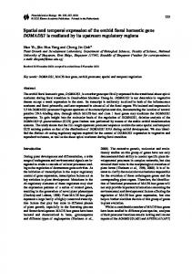
Fig. 5. Expression of Muc-1 mucin protein as revealed by immunostaining with CTl antiserum. Muc-1 is located in the apical borders of developing epithelial sheets in the different organs (arrows). The specificity of staining is shown by the photos where a 15-day embryo (liver, stomach, pancreas - 40x) section was stained by CTl (C) and with blocked CTl (B). The developmental expression of Muc-1 is shown through organogenesis of the stomach (A) 12-day (200x), (C) 15-day (40x); pancreas (D) 13-day (200x), (E) 15-day (lOOx), (F) 8-week neonate (200x); salivary glands (G) 15-day (200x), (H) 18-day (40x), (I) 4-week neonate (40x); lung (J) 14-day (200x), (K) 18-day (200x), (L) 4-week neonate (200x); and kidney (M) 14-day (200x), (N) 15-day (lOOx), (O) 8-week neonate (200x). Arrowheads in 4-week neonate lung (L) show focal expression of Muc-1 in pneumocytes. a, alveoli; ac, acini; d, duct; i, islet of Langerhans; g, glomeruli; 1, liver; m, salivary gland mucous type; p, pancreas; s, stomach; se, salivary gland serous type.
Developmental expression of Muc-1 mucin
433
that observed with embryonic pancreas. Although it is
V///W////////////A believed that the mouse stomach is differentiated at 11
Large tnL Small Int. Mmmm-gl. Sdlv*.
tzz Y/Mr///"'"'—
Long Kidney
WM0W"""""
Pancreas
\4%y//ss"""
1
Stomach Lira-
1
^
i
1
1
1
1
2
4.
6
8
10
Vaginal plug
•
mw///////////////M i
i
i
i
12
14
16
18
G e s a t l o n i d days
EpatelHilbud /cord Simple tube Prooephroj (Dty 9), mesoocphroi (D»y 10), mctancphro* (Day 12) Actnc epnbcbal duTerenuMKm
S3 11
Mimmary sprout growth m d bnnxfamg Muc-1 expression
Fig. 6. Coordinate expression of Muc-1 mucin protein during mouse organogenesis based on immunohistochemical staining data, Rugh (1990) and Sakakura (1989). Pancreas Mouse pancreas and stomach start to differentiate on day 10 (Rugh, 1990). Mucin staining is first observed in the developmental 12-day pancreas in the luminal areas of the developing tubules. The acinar structure is not clear at this stage, although it is possible to observe tubules of epithelial cells being formed (Fig. 5D - 13 day). The intensity of the stain is not as strong as in older embryos/neonates nor does the glandular structure resemble the mature organ (Fig. 5F). From gestational day 14 to 17, secretory acini with strong mucin staining in the lumen appear, but they are rather sparse and mesenchyme can be seen separating them (Fig. 5E). Although from day 18 to adulthood the pancreas gradually acquires a more compact and lobular structure, essentially no change is observed in the Muc-1 immunostaining pattern. Stomach The earliest Muc-1 expression in the stomach could be observed at day 12 (Fig. 5A). From day 13 on, an increase in the staining signal in the apical part of the stomach epithelium is observed, but it is not uniformly distributed (Fig. 5A and C). The enhancement in staining is coincident with the appearance of glandular function, as suggested by the presence of vacuoles in the stomach epithelial cells (Rugh, 1990). It is possible to detect mucin in the lumen of submucosal glands after day 16, when the formation of stained pits can also be observed (data not shown). This stomach staining pattern persists in neonate and mature mice, similar to
days in the embryo (Rugh, 1990), we could be certain of Muc-1 protein expression in the stomach only at day 12, after its expansion. The borderline between positive and negative staining is difficult to assess in 11-day or earlier embryos because of: (1) limitations in the sensitivity of the technique; (2) edge effect observed with general staining and (3) mucous secreted in the lumen or its border can arrest antibodies (brownish secretion can be seen in sections stained with blocked CT1 - Fig. 5B). Salivary glands Submandibular salivary glands have a later onset when compared to the other organs studied here: by gestational day 13, there are only epithelial cords; ducts appear at day 14 (Rugh, 1990). In our hands, staining with CT1 revealed Muc-1 mucin focally in ducts in salivary glands of embryos aged 15 days, and subsequently, the presence of Muc-1 mucin was detected in the rudimentary secretory units known as terminal tubules or primary acini (Fig. 5G,H). In developing neonate and adult salivary glands, however, we could observe the presence of Muc-1 mucin only in ducts and not in acinar cells (Fig. 51). Although there are other reports showing by immunohistochemistry the presence of mucins in both acini and duct cells (Denny and Denny, 1982; Denny et al., 1988), the antiserum used in these studies was directed to the whole biochemically purified mucin molecule. It is not clear whether this antiserum also recognizes Muc-1. However, this possibility seems remote, because their antiserum recognizes a salivary gland-specific mucin and does not cross-react with mucins from other epithelial tissues where Muc-1 is expressed (Denny and Denny, 1982). Liver and intestine Neither small nor large intestine expressed Muc-1 mucin from mouse embryogenesis to adulthood. Liver is essentially negative as well, except for the apical surfaces of the bile ducts (data not shown). Consequently, these organs were used as our negative controls (Fig. 5C). Lungs and trachea Lung buds appear in 9-day mouse embryos (Rugh, 1990). They grow and undergo branching morphogenesis so that the bronchus and bronchiolus originate at days 11 and 13, respectively. Trachea also appears at day 11. Trachea, bronchi and bronchioh have been shown to express Muc-1 gene in adult mice (Pemberton et al., 1992). In our immunostaining experiments, a weak positive signal can be observed in the lungs at day 12, and from day 13 on, this signal increases in intensity in bronchi and later in the bronchioli (Fig. 5J). Positively stained secretions can be seen inside the larger bronchi. Pulmonary alveoli do not develop until day 17, when the simple cuboidal epithelia differentiates into the alveolar type I cells and the cuboidal type II cells in the respiratory saccules (Braukner et al.,
434
V. M. M. Braga and others
1991). Positive staining of both alveoli and the secretions present in the alveolar lumens is shown on day 18 (Fig. 5K). However, the mucin expression in alveoli after birth is transient: it gradually disappears so that the 2 week neonate shows almost no alveolar staining. Adult and juvenile alveoli lumens do not stain with CT1, but some pneumocytes express Muc-1 focally (Fig. 5L - arrowheads). Detection of Muc-1 protein in the trachea was first possible at gestational day 15 (data not shown). Kidney Three distinct organs are formed sequentially during kidney organogenesis (Rugh, 1990). The first two are transient: the pronephros (gestational day 9) and mesonephros (day 10), the latter originating the ureteric bud. Finally the metanephros appears (day 12), and its development will lead to the formation of the permanent kidney. The metanephros is formed by the differentiation of fibroblast-like mesenchymal cells into epithelial cells. This process is induced by the proximity of the branching ureteric bud (reviewed by Ekblom, 1981, 1989; Klein et al., 1988). The first observation of the expression of mucin in the kidney by immunostaining with CT1 is in the day-13 embryo, in the apical surface of the ducts (Fig. 5M,N). A precise identification of which particular developing tubule expresses Muc-1 in early embryonic kidney (13-15 day) has been difficult; however, both collecting ducts and distal tubules express mucin in humans (Zotter et al., 1988) and transgenic mice (MUC-1) (Peat et al., 1992) (Fig. 5O). This is interesting because collecting ducts originate from the branching of the epithelial ureteric bud, and distal tubules are formed by epithelial cells that have differentiated from mesenchyme. So, irrespective of their origin, both tubules expresses Muc-1 mucin in adult mice. Using protein and northern blot analysis, we have observed a lower level of Muc-1 expression in the kidney when compared to the other organs studied here.
Discussion
We have described in this paper the expression of the Muc-1 transmembrane mucin protein during mouse embryogenesis and have shown that its expression correlates with the onset of epithelial sheet formation in different organs. Mouse organogenesis starts at day 8-9 (Rugh, 1990) (Fig. 6). The expression of the Muc-1 gene in mouse embryos can be first detected by gestational day 10, using RT-PCR technique with total RNA from whole embryos (data not shown). Muc-1 protein expression is coordinated both spatially and temporally with epithelial differentiation in the organs known to express it in the adult life: lungs, stomach, pancreas, salivary glands and kidney. Our RNA analysis of Muc-1 expression correlates well with the presence of mucin protein detected in the organs by immunohistochemistry. The use of the CT1 antiserum in our staining
experiments was of particular relevance. Its epitope is not subject to blockage by differential glycosylation in different organs or different physiological stages of the same organ, as has been observed with other antibodies to mucin extracellular domains (Zotter et al., 1988; Parry et al., 1992), since CT1 was raised to the human MUC1 cytoplasmic tail. The close homology observed between human and mouse cytoplasmic tail (Spicer et al., 1991) and the ability of CT1 to yield similar mucin staining patterns in different mammalian species (Pemberton et al., 1992) stress the probable importance of this intracellular domain to Muc-1 function. In general, Muc-1 was described as being expressed only in simple secretory epithelia (Peat et al., 1992; Zotter et al., 1988). Detection of Muc-1 mucin in 13-day lung was striking since, at this time, pulmonary epithelia is stratified (Braukner et al., 1991). With development, the lung epithelia changes to simple cuboidal, maintaining Muc-1 mucin at the luminal surface of the respiratory tract. Other epithelia expressing Muc-1 as determined by immunohistochemistry are nasal and tracheal (pseudostratified) and adult vagina and cervix epithelia (squamous stratified) (unpublished data). Pulmonary alveoli have a particular feature not observed in other organs: Muc-1 immunostaining can be observed both in apical borders and in secretions present inside the lumen by gestational days 18, 19 and 20. In neonates and adults, however, this alveolistaining pattern disappears completely and mucin expression is shown only focally in pneumocytes (Fig. 5K,L). Bronchi and bronchioli continue to express Muc-1 mucin in the adult. We observed that in the embryonic development of the various organs studied, Muc-1 mucin appears lining the luminal border of the epithelia that is still folding and branching and, in general, continues to be expressed throughout the life time of the mouse (Figs 5 and 6). During embryogenesis, Muc-1 expression continually increased with time, mainly due to growth in area (epithelial branching). An augmented concentration of the protein in a given lumen appears to occur in the early period of mucin expression in each organ. Pancreas and salivary gland are good examples of this increase in mucin expression, observed from embryonic development days 12 to 14 and from days 15 to 18, respectively. The development of the organs studied here varies in the time of appearance, developmental processes, and time required to become mature (Fig. 6). Lung and kidney are able to function by day 18 (Rugh, 1990), although they grow and undergo modifications postnatally. Pancreas and liver also attain functional activity before birth. On the contrary, salivary gland reaches full maturity in 4-month-old mice (Srinivasan and Chang, 1979). Therefore, the timing of Muc-1 protein appearance in the epithelia does not correlate with the completion of organ maturation, since it is present much before they attain full functional activity. In addition, the epithelial buds/cords of these organs originate at different times (Fig. 6). In general, it is possible to detect Muc-1 mucin lining the lumens of the
Developmental expression of Muc-1 mucin embryonic pancreas (12 day), salivary gland (15 day) and lung (12 day) soon after their epithelial buds start to branch and differentiate (Fig. 6). By the time of stomach expansion (12 day) and the appearance and branching of the ureteric bud from the metanephros in the embryonic kidney (13 day), mucin protein can also be observed. Our results suggest that, although the Muc-1 gene is expressed in different organs, its expression is not stimulated systemically, but it is induced concomitantly with the epithelial differentiation in each individual organ (Fig. 6). During embryogenesis, small epithelial buds undergo elongation, folding and branching through mesenchyme, resulting in distinct epithelia-containing organs (Bernfield, 1978; Bernfield, 1981). Alternatively, epithelial cells can originate by differentiation of mesenchymal cells (derived from mesoderm), which is induced by close proximity of epithelial buds (Ekblom, 1989; Hay, 1990). Similar Muc-1 expression patterns were observed in the various epithelia regardless of their origin from ectoderm or mesoderm. For instance, epithelial cells of distal tubules, which are formed by the differentiation of fibroblast-like cells in the kidney mesenchyme, express Muc-1 as well as the collecting ducts, which are derived by ramification of the primordial ureteric bud. This might indicate that polarization and branching processes are similar in both cases, and cells might respond to similar stimuli and possess similar mechanisms of turning on and off genes to produce the epithelial sheets. The timing and pattern of expression of Muc-1 mucin during epithelial differentiation and its wide distribution make the role of this large glycoprotein during organogenesis interesting to study further. It is tempting to speculate that, as it has been shown that high levels of MUC1 expression decrease cell-cell interactions, the presence of this protein in the apical part of the cells might help lumen formation in the developing epithelium, by reducing adhesive associations in the apical domain (Ligtenberg et al., 1992). The characteristic structure and biochemical properties of the Muc-1 protein may be responsible for preventing the interactions between adhesive molecules. The first feature is the rod-like structure of the extracellular domain, extending the mucin far beyond the cell glycocalyx (Jentoft, 1990). The predicted length of this mucin is ^250 nm as opposed to the expected 30 nm length of cellular adhesion molecules (Becker et al., 1989). In addition, the presence of a large number of sialic acid residues on mucin oligosaccharide chains produces a net negative charge, enhancing the steric hindrance effect caused by the large extracellular domain (Parry et al., 1992). It is not clear how many sialic acid residues are present in mucin protein during mouse embryogenesis. There have been reports showing that sialic acid levels in Muc-1 protein can be developmental^ regulated in the adult mouse (Parry et al., 1992). The temporal and local expression of the Muc-1 gene coincident with epithelial differentiation in many organs (stomach, pancreas, kidney, salivary glands and lung) might suggest a participation of this molecule in
435
the process of epithelial sheet formation. However, as Muc-1 mucin is continually expressed in adult life, additional roles must be postulated for the presence of this molecule after the completion of maturation of these organs. Although our data have shown a correlation between Muc-1 expression and the onset of epithelial differentiation in some organs during mouse embryogenesis, it is difficult to define in vivo the relevance of this correlation and hence extrapolate a possible function for this molecule. First of all, other epithelia-containing organs do not express Muc-1 (e.g. intestines). Secondly, Muc-1 immunoreactive epitopes have been found intracellularly in placenta (data not shown) and muscle (Pemberton et al., 1992), and Muc-1 mRNA expression in these tissues has been confirmed (this work and Vos et al., 1991). Thirdly, during morphogenesis, three overlapping events occur: cell polarization, epithelial sheet formation and the process of branching, making it extremely difficult to determine the precise timing of each event in vivo. It would be interesting to study the expression of Muc-1 in an in vitro system where we could deal with these events separately and where we could manipulate the experimental conditions in order to interfere in the process. The well-established in vitro systems of branching morphogenesis using embryonic lung, kidney or salivary gland rudiments could be utilized (Bernfield, 1981; Ekblom, 1981; Schuger et al., 1990). The process of cell polarization could be studied using epithelial cell lines that are able to polarize in vitro. In these ways, it should be possible to determine whether Muc-1 expression behaves similarly in vitro and to which event (if any) the expression of this gene is correlated, e.g. epithelial cell polarization, sheet formation or branching morphogenesis. Alternatively, the Muc-1 function in the mouse can be disrupted in vivo by homologous recombination. Both approaches are in progress in an attempt to elucidate the role of Muc-1. In conclusion, we have presented the first evidence that the expression of the Muc-1 mucin during mouse embryogenesis is restricted to the apical surfaces of secretory epithelial cells in stomach, pancreas, trachea, lung, kidney, and salivary gland. The induction of Muc1 gene expression is under spatial and temporal control during the epithelial differentiation in the organs where the protein is detected in the mouse. Muc-1 protein can be detected lining the apical surfaces of the developing lumens when the polarized epithelium of these organs is still folding and branching, and glandular activity has not properly started. Finally, although Muc-1 is present in different organs, its expression is not induced systemically, but according to the particular onset of epithelial polarization and branching morphogenesis of each individual organ. We would like to thank Gillian Hutchinson for the animal work, Christine Pike and colleagues for the histological preparations, and Dr Ian Goldsmith for oligonucleotide synthesis. We would also like to thank Drs E.-N. Lalani and S.Kimber for help in the analysis of immunohistochemistry
436
V. M. M. Braga and others
data and Drs J. Taylor-Papadimitriou, F. Watt, J. Williams and G. Warren for critical reading of the manuscript. V.M.M.B. has a fellowship from Conselho National de Desenvolvimento Cientffico e Tecnol6gico (CNPq), Brazil. References Bartek, J., Durban, E. M., Hallowes, B. C. and TaylorPapadlmltriou, J. (1985). A subclass of luminal epithelial cells in the human mammary gland, defined by antibodies to cytokeratins. /. Cell Sci. 75, 17-33. Becker, J. W., Erickson, H. P., Hoffman, S., Cunningham, B. A. and Edclman, G. M. (1989). Topology of cell adhesion molecules. Proc. Natl. Acad. Sci. USA 86, 1088-1092. Bernfield, M. (1978). Mechanisms of embryonic organ formation. In Abnormal Fetal Growth: Biological Bases and Consequences (ed. Naftolin, F.), pp. 101-120, Springer-Verlag, Berlin. Bernfield, M. (1981). Organization and remodeling of the extracellular matrix in morphogenesis. In Morphogenesis and Pattern Formation, pp. 139-162, Raven Press. Bharathan, S., Moriarty, J., Moody, C. E. and Sherblom, A. P. (1990). Effect of tunicamycin on sialomucin and natural killer susceptibility of rat mammary tumor ascites cells. Cancer Res. 50, 5250-5256. Braukner, J. H., Trautman, M. S. and Bernfield, M. (1991). Syndecan, a cell surface proteoglycan, exhibits a molecular polymorphism during lung development. Dev. Biol. 147, 285-292. BurcheU, J., Durbln, H. and Taylor-Papadimitriou, J. (1983). Complexity of antigenic determinants recognised by monoclonal antibodies HMFG-1 and HMFG-2, in normal and malignant mammary epithelial cells. /. Immunol. 131, 508-513. BurcheU, J., Gendler, S., Taylor-Papadimitriou, J., Girling, A., Lewis, A., Millls, R. and Lamport, D. (1987). Development and characterization of breast cancer reactive monoclonal antibodies directed to the core protein of the human milk mucin. Cancer Res. 47, 5476-5482. Cbelly, J., Concordet, J. P., Kaplan, J.-C. and Kahn, A. (1991). Illegitimate transcription: transcription of any gene in any cell type. Proc. Natl. Acad. Sci. USA 86, 2617-2621. Chomczyski, P. and Sacchi, N. (1987). Single-step method of RNA isolation by acid guanidinium thiocyanate-phenol-chloroform extraction. Analyt. Biochem. 162, 156-159. Church, G. M. and Gilbert, W. (1984). Genomic sequencing. Proc. Natl. Acad. Sci. USA 81, 1991-1995. Codington, J. F., Sanford, B. H. and Jeanloz, R. W. (1973). Cellsurface glycoproteins of two sublines of the TA3 tumor. J. Nad. Cancer Inst. 51, 585-591. Denny, P. A. and Denny, P. C. (1982). Localization of a mouse submandibular sialomucin by indirect immunofluorescence. Histochem. J. 14, 403-408. Denny, P. A., Pimprapaiporn, W., Kim, M. S. and Denny, P. C. (1988). Quantitation and localization of acinar cell-specific mucin in submandibular glands of mice during postnatal development. Cell Tissue Res. 251, 381-386. Ekblom, P. (1981). Formation of basement membrane in the embryonic kidney: an immunohistological study. /. Cell Biol. 91,110. Ekblom, P. (1989). Developmentally regulated conversion of mesenchyme to epithelium. FASEB J. 3, 2141-2150. Felnberg, A. P. and Vogelstein, B. (1983). A technique for radiolabelling DNA restriction fragments to high specific activity. Analyt. Biochem. 132, 6-13. Gendler, S., Taylor-Papadimitriou, J., Duhig, T., Rothbard, J. and Burchell, J. (1988). A highly immunogenic region of a human polymorphic epithelial mucin expressed by carcinomas is made of tandem repeats. J. Biol. Chem. 263, 12820-12823. Gendler, S. J., BurcheU, J. M., Duhig, T., Lamport, D., White, R., Parker, M. and Taylor-Papadlmltriou, J. (1987). Cloning of partial cDNA encoding differentiation and tumor-associated mucin glycoproteins expressed by human mammary epithelium. Proc. Natl. Acad. Sci. USA 84, 6060-6064. Gendler, S. J., Lancaster, C , Taylor-PapadTmltriou, J., Duhig, T., Peat, N., Peraberton, L., Lalani, E.-N. and Wilson, D. (1990).
Molecular cloning and expression of a human tumor-associated polymorphic epithelial mucin. J. Biol. Chem. 265, 15286-15293. Gum, J. R., Byrd, J. C , Hicks, J. W., Toribana, N. W., Lamport, D. T. A. and Kim, Y. S. (1989). Molecular cloning of human intestinal mucin. J. Biol. Chem. 264, 6480-6487. Gum, J. R., Hicks, J. W., SwaUow, D. M., Lagace, R. L., Byrd, J. C , Lamport, D. T. A., Slddiki, B. and Kim, Y. S. (1990). Molecular cloning of a novel human intestinal mucin gene. Biochem. Biophys. Res. Comm. 171, 407-415. Hay, E. D. (1990). Role of cell-matrix contacts in cell migration and epithelial-mescnchymal transformation. Cell Diff. Dev. 32, 367376. Jentoft, N. (1990). Why are proteins O-glycosylated? TIBS 15, 291294. Klein, G., Langegger, M., Tlmpl, R. and Ekblom, P. (1988). Role of laminin A chain in the development of epithelial cell polarity. Cell 55, 331-341. Lan, M. S., Batra, S. K., Ql, W.-N., Metzgar, R. S. and HoUlngsworth, M. A. (1990). Cloning and sequencing of a human pancreatic tumor mucin cDNA. J. Biol. Chem. 265, 15294-15299. Ligtenberg, M. J. L., Buijs, F., Vos, H. L. and Hllkens, J. (1992). Suppression of cellular aggregation by high levels of episialin. Cancer Res. In press. Ligtenberg, M. J. L., Vos, H. L., Gennlssen, A. M. C. and Hllkens, J. (1990). Episialin, a carcinoma associated mucin, is generated by a polymorphic gene encoding splice variants with alternative amino termini. J. Biol. Chem. 265, 5573-5578. Manlatis, T., Fritsch, E. F. and Sambrook, J. (1982). Molecular Cloning: a Laboratory Manual, Cold Spring Harbour Laboratories, Cold Spring Harbour. Mugrauer, G., Alt, F. W. and Ekblom, P. (1988). N-myc protooncogene expression dunng organogenesis in the developing mouse as revealed by in situ hybridization. J. Cell Biol. 107, 13251335. Parry, G., Beck, J. C , Moss, L., Bartley, J. and Ojakian, G. K. (1990). Determination of apical membrane polarity in mammary epithelial cell cultures: the role of cell-cell, cell-substratum, and membrane-cytoskeleton interactions. Exp. Cell. Res. 188, 302311. Parry, G., Li, J., Blssell, M., Schmldhauser, C , Splcer, A. P. and Gendler, S. J. (1992). Studies of Muc-1 mucin expression and polarity in the mouse mammary gland demonstrate developmental regulation of Muc-1 glycosylation and establish the hormonal basis for mRNA expression. J. Cell Sci. 101, 191-199. Peat, N., Gendler, S. J., Lalani, E.-N., Duhig, T. and TaylorPapadimitriou, J. (1992). Tissue specific expression of a human polymorphic epithelial mucin (MUC1) in transgenic mice. Cancer Res. In press. Pemberton, L., Taylor-Papadimitriou, J. and Gendler, S. G. (1992). Antibodies to the cytoplasmic tail domain of the MUC1 mucin show conservation throughout mammals. Biochem. Biophys. Res. Comm. 185, In press. Porchet, N., Van Cong, N., Dufosse, J., Audie, J. P., GuyonnetDuperat, V., Gross, M. S., Denis, C , Degand, P., Bernheim, A. and Aubert, J. P. (1991). Molecular cloning and chromosomal localization of a novel human tracheo-bronchial mucin cDNA containing tandemly repeated sequences of 48 base pairs. Biochem. Biophys. Res. Comm. 175, 414-422. Rappolee, D. A., Brenner, C. A., SchuKz, R., Mark, D. and Werb, Z. (1988). Developmental expression of PDGF, TGF-a-, and TGF-/3 genes in preimplantation mouse embryos. Science TAX, 1823-1825. Rappolee, D. A., Wang, A., Mark, D. and Werk, Z. (1989). Novel methods for studying mRNA phenotypes in single or small numbers of cells. J. Cell. Biochem. 39, 1-11. Rogh, R. (1990). The Mouse: Its Reproduction and Development, Oxford University Press. Schuger, L., O'Shea, K. S., Nelson, B. B. and Varanl, J. (1990). Organotypic arrangement of mouse embryonic lung cells on a basement membrane extract: involvement of laminin. Development 110, 1091-1099. Shimizu, M., Tanimoto, H., Azuma, N. and Yamauchl, K. (1990). Growth inhibition of BALB/c 3T3 cells by a high-molecular-weight mucin like glycoprotein of human milk fat globule membrane. Biochem. Intern. 20, 147-154.
Developmental expression of Muc-1 mucin Siddiqui, J., Abe, M., Hayes, D., Shani, E., Ynnls, E. and Kufe, D. (1988). Isolation and sequencing of a cDNA coding for the human DF3 breast carcinoma-associated antigen. Proc. Natl. Acad. Sci. USA 85, 2320-2330. Spicer, A. P., Parry, G., Patton, S. and Gendler, S. J. (1991). Molecular cloning of the mouse homologue of the tumor-associated mucin, Muc 1, reveals conservation of potential O-glycosylation sites, transmembrane and cytoplasmic domains and a loss of minisatellite-like polymorphism. J. Biol. Chem. 266, 15099-15109. Srtnivasan, R. and Chang, W. W. L. (1979). The postnatal development of the submandibular gland of the mouse. Cell Tissue Res. 198, 363-371. Steck, P. A. and Nicolson, G. L. (1983). Cell surface glycoprotein of 13762NF mammary adenocarcinoma clones of differing metastasis potentials. Exp. Cell. Res. 147, 255-267.
ATI
Vos, H. L., de Vries, Y. and Hilkens, J. (1991). The mouse epsialin (Muc-1) gene and its promoter: rapid evolution of the repetitive domain in the protein. Biochem. Biophys. Res. Comm. 181, 121130. Wreshner, D. H., Hareuvenl, M., Tsarfaty, I., Smorodlnsky, N., Horev, J., Zaretsky, J., Kotkes, P., Weiss, M., Lathe, R., Dion, A. and Keydar, I. (1990). Human epithelial tumor antigen cDNA sequences. Differential splicing may generate multiple protein forms. Eur. J. Biochem. 189, 463-473. Zotter, S., Hageman, P. C , Lossnltzer, A., Mooi, W. J. and Hilgers, J. (1988). Tissue and tumor distribution of human polymorphic epithelial mucin. Cancer Rev. 11-12, 55-101. {Accepted 3 March 1992)