TAA triplet at their 3â² end. Two long open ... polypeptide with zinc fingers homologous to the nucleic- acid-binding ... been detected in vivo with antisera raised against a fusion ..... only when the cytoplasm transmitted by the mother is reac- tive. .... decapentaplegic gene complex: suggestion of a new hybrid dysgenesis.
Development 115, 729-735 (1992) Printed in Great Britain © The Company of Biologists Limited 1992
729
Spatial and temporal expression of the I factor during oogenesis in Drosophila melanogaster PHILIPPE LACHAUME, KARIM BOUHIDEL, MICHELLE MESURE andHUBERT PINON* Laboratoire de Génétique, Université Blaise Pascal, CNRS URA 360, F-63177 Aubière Cedex, France *Author for correspondence
Summary The I factor is a functional non-viral retrotransposon, or LINE, from Drosophila melanogaster. Its mobility is associated with the I-R hybrid dysgenesis. In order to study the expression pattern of this LINE in vivo, a translational fusion between the first ORF of the I factor and the lacZ gene of Escherichia coli has been carried out and introduced in the genome of reactive (R) flies. Homozygous transgenic Drosophila lines have been established and analysed. ORF1 expression is limited to germ-line cells (nurse cells and oocyte) between stage 2 and 10 of oogenesis. No somatic expression is found. Position effects may limit the level of expression of a given transgene but do not modify its basic pattern of expression during the development of the fly. This reproducible control demonstrates both that I factor is driven by its own promoter, probably the internal one suggested by Mizrokhi et al. (Mizrokhi, L. J. , Georgevia, S. G. and
Ilying, Y. V. (1988). Cell 54, 685-691), and that tissuespecific regulatory sequences are present in the 5 untranslated part of the I factor. The nuclear localization of the fusion protein reveals the presence of nuclear localization signals (NLS) in the ORF1-encoded protein correlating with the possible structural and/or regulatory role of this protein. This expression is restricted to dysgenic and reactive females, and is similar in the two conditions. All the results obtained in this work suggest that I factor transposition occurs as a meiotic event, between stage 2 and 10 of the oogenesis and is regulated at the transcriptional level. It also appears that our transgene is an efficient marker to follow I factor expression.
Introduction
1986). Functional I factors transpose with high frequency in the germ line of SF females where cytoplasm is reactive. As a result, the progeny shows a large number of mutations and chromosomal rearrangements. Defective heterochromatic I factors present in both I and R strains are not mobilized during dysgenesis. The I factor has been cloned (Bucheton et al., 1984) and sequenced (Fawcett et al., 1986; Abad et al., 1989). The active I factors are 5.4 kb long (Fig. 1). They have no terminal repeat, as retroviruses do, but show tandem repeats of the TAA triplet at their 3′ end. Two long open reading frames (ORF) are observed. The ORF1 (1287 bp) encodes a putative polypeptide with zinc fingers homologous to the nucleicacid-binding motive found in retroviral “gag” protein. The ORF2 (3675 bp) encodes a polypeptide showing similarities to viral reverse transcriptase, RNAseH (Finnegan, 1988) and integrase (Jakubczak et al., 1990). Because of these structural and coding features, the I factor has been classified in the LINE family. LINEs, also known as non-viral retrotransposons, are found in many eukaryotic species (Fanning and Singer, 1987; Finnegan, 1988) and it has been suggested that
Hybrid dysgenesis in Drosophila melanogaster (Bregliano and Kidwell, 1983) appears as a set of unusual genetic characteristics (sterility, mutations and chromosomal rearrangements) in the progeny of crosses between particular strains (Picard and L’Héritier, 1971; Kidwell and Kidwell, 1975; Kidwell et al., 1977). Three hybrid dysgenesis systems are currently known (I-R, P-M and H-E), and are associated with the mobilization of the transposable elements I factor, P and Hobo, respectively (Fawcett et al., 1986; Rubin et al., 1982; Blackman et al., 1987). Therefore, hybrid dysgenesis in Drosophila melanogaster appears to be an efficient model to study the mechanism of transposition and its consequences. In the IR system, dysgenic symptoms appear in the female progeny (SF) obtained when inducer (I) males containing functional I factors in their genome are crossed with reactive (R) females devoid of functional I factors. In contrast, the female progeny (RSF) from the reciprocal cross is normal. SF females lay a normal number of eggs but part of them stop developing before the fourth cleavage division (Lavige,
Key words: Drosophila , LINE, oogenesis, spatial expression, I factor.
730
P. Lachaume and others
they transpose by reverse transcription of an RNA intermediate. This has been recently demonstrated for the I factor (Pelisson et al., 1991; Jansen and Heidmann, 1991) and a mouse L1 element (Evans and Palmiter, 1991). Moreover, full-length transcripts of the I factor have been detected (Chaboissier et al., 1990) and proposed as transposition intermediates. This transcription is restricted to ovaries of SF females and probably driven by the I factor itself through a promoter located downstream of the initiation of transcription (Mizrokhi et al., 1988). The control of this transcription remains unclear. Indeed its study has been difficult to carry out because of the very low level of the 5.4 kb RNA as well as the presence of transcripts from defective I factors in each strain. Moreover, proteins specific for the I factor have not been detected in vivo with antisera raised against a fusion protein containing 270 amino acids encoded by ORF1 (Evraert, 1989). The use of translational fusions with marker genes is a powerful method with which to follow the expression of any given gene. We have fused the 5′ part of a functional I factor with the lacZ gene encoding the β-galactosidase in Escherichia coli. This construct has been inserted into the genome of a reactive strain in order to study the expression and localization of the ORF1 protein. Our results demonstrate the in vivo promoter activity of the 5′ part of the I factor and suggest a precise control of the I factor expression into the germ-line cells (nurse cells and oocyte) : the fusion protein is accumulated from stage 2 to stage 10 of oogenesis. Moreover the N-terminal part of ORF1 protein probably bears a nuclear localization sequence (NLS) which may account for the efficiency of the retrotransposition process. Expression in the different context R, I, SF and RSF shows that the R and SF context are approximately equivalent. Expression of the I factor ORF1 appears correlated with the potential for I-R hybrid dysgenesis and transposition of the I factor. Material and methods Bacterial strains All plasmids were propagated in E. coli strains HB101 (Boyer and Roulland-Dussoix, 1969) or NM522 (Gough and Murray, 1983).
Drosophila melanogaster strains Except for the inducer strain of β-galn1 mutants (Knipple and MacIntyre, 1984), all the Drosophila strains used in this study are from the stock of the “Laboratoire de Génétique de Clermont-Ferrand”. They are maintained in axenic medium of David (1959) at 20°C.
P-mediated transformation P-mediated transformation (Rubin and Spradling, 1982) with pUChsneo vector (Steller and Pirotta, 1985) was conducted as described by Simon et al. (1985) using wild reactive strain Charolles. Localization of inserts were done by crosses with strain cne (II cn/cn, III e/e). Homozygous strains with insertion in the X chromosome or in the second chromosome are obtained after crosses with respectively Binscy (I yB/yB) or Valence (II CyO/Pm) strains containing a balancer chromosome.
Southern analysis Restriction endonuclease-digested genomic DNA (~5 µg) was fractionated in 1% agarose gels, transferred to nitrocellulose filters and hybridized as described by Maniatiset al. (1982).
-galactosidase staining Ovaries were dissected in PBS (50 mM Na/Na2 PO4 pH 7.4, 140 mM NaCl) and fixed for 10-15 minutes in fixing solution (4% paraformaldehyde in PBS). They were washed in PBS for 30 minutes, and then incubated for 2 days in staining solution [10 mM Na/Na2 PO4 pH 7.2; 150 mM NaCl; 1 mM MgCl2; 3.1 mM potassium ferricyanide; 3.1 mM potassium ferrocyanide; 0.5 mM Xgal (5-bromo-4-chloro-3-indolyl-β-D-galactopyranoside)] at 37°C. The Xgal was added shortly before incubation from a 10% stock solution in DMSO. After staining, the ovaries were washed in PBS and mounted in 50% glycerol solution before histological observation. For sections, coloured ovaries were washed in PBS and included in embedding medium (OCT, Miles 4583) overnight at −20°C. 14 µm thick sections were cut with a cryostat and mounted in a (50% glycerol/50% PBS) solution without other treatment. Testes were fixed for 10-15 minutes in 1% glutaraldehyde solution in PBS. Staining was performed overnight. The subsequent steps are the same as those used for staining the ovaries. Somatic tissues were fixed either in 4% paraformaldehyde or 1% glutaraldehyde before staining. The same results were observed in each case.
Construction of the 48.1 inducer stocks The inducer stocks with the 48.1 insertions were obtained during the construction of a strain with the k160 insertion and the β-galn1 allele of the β-gal-1 Drosophila gene (Knipple and MacIntyre, 1984). Inducer females galn1/galn1 were crossed with 48.1 males k160/k160. RSF females galn1/k160, thus obtained, were crossed with CyO/Pm; Dcxf/Sb males. k160,gal?/CyO; +/Dcxf progeny was selected in G418 containing medium and individually crossed with CyO/Pm; Dcxf/Sb individuals. In each progeny males and females k160 gal?/CyO; +/Dcxf were selected and crossed to obtain homozygous lines k160 gal?/k160 gal?; +/+. The β-galn1 lines were selected by rapid colouration of the flies tissues and k160 galn1/k160 galn1 stable homozygous lines were established. Three inducer strains (F4, F6 and F7) with the k160 insertions of the 48.1 line were independently obtained.
Results Obtaining of transgenic I factor-lacZ fusions Plasmid pI407 contains a full-length I factor flanked by sequences from the white gene (Bucheton et al., 1984; Fawcett et al., 1986). When introduced into the Drosophila germ line, this functional copy is able to transpose and to invade the genome (Pritchard et al., 1988). We fused the 5′ untranslated region and part of the ORF1 of this I factor in frame with the lacZ′ ORF from plasmid pMC1871 coding for Escherichia coli β-galactosidase (Casadaban et al., 1983). In this construct (Fig. 1), most of the I factor ORF1 is deleted in order to avoid both any decrease in specific activity of the resulting β-galactosidase and any possible feed-back regulation upon expression of the zinc-fingers coding domain. After insertion of our construct in the pUChsneo plasmid (Steller and Pirotta, 1985), we used P-mediated transposition
Expression of the I factor during oogenesis
731
Table 1. Transgenic lines with k160 insertion Insertion Lines 48.1 48.3 53.1 101.1 150.1 150.6 60.1 Charolles
Localization
Number
Native
in situ βgal. Test
II II X II II II II /
2 1 1 1 1 1 1 0
+ + + + + + − /
+ − + + + − − −
Charolles is a reactive line used as recipient for transgenesis and as negative control for staining experiments. The chromosomal localization of the insertions is performed by genetic analysis. The number and the integrity of the construction are tested by Southern blot analysis. As shown in the last column, expression of the transgenic fusion is observed in four transgenic lines. They derive from four unconnected events of transformation (see text).
Fig. 1. Construction of the fusion gene. The two ORFs of the I factor are shown schematically as hatched boxes and, untranslated regions as a thick line. For the k160 construction, we used the lacZ′ ORF from pMC1871 plasmid (Casadaban, 1983). The left part of the I factor comes from plasmid pI 770 (Bucheton et al., 1984) and therefore linked to a 99 bp fragment of the white gene overlaping exon and intron. The translational fusion is in the SspI(1102) site (S). An AsuII-AsuII (A) fragment containing the zinc-finger domain is deleted. The fusion gene is introduced in the pUChsneo vector (Steller and Pirrota, 1985) and this construction is inserted into the genome of the Charolles reactive line by P-mediated transposition. DNA of the transgenic lines is digested by EcoRI (E) and ClaI (C). Each line is tested by Southern blot hybridization for the integrity of the two internal fragments of 1.2 and 2.2 kb and for the number of additional fragments corresponding to the number of insertions. The totality of the plasmid is used as the probe.
to transform flies from the re-active Charolles strain with the I factor-lacZ fusion (see Material and methods). Five independent transformants, as well as two (48.1 and 48.3) derived from the progeny of a single injected embryo, have been established as homozygous lines and characterized by Southern blot analysis (Fig.1 and Table 1). This analysis has shown that only the strain 60.1 does not contain the whole internal fragments characteristic of the insertion (Fig. 1) and that five of these lines have only one native insertion of k160 fusion. The 48.1 line has two insertions; one of these presents the same external fragment of approximately 2 kb observed in 48.3. As 48.1 and 48.3 derive from the same injected embryo, the insertion of 48.3 is probably identical to one of the two detected in 48.1. All the lines are reactive; the introduction of the I factor-lacZ fusion into reactive flies does not switch them to the inducer state as full I factor does (Pritchard et al., 1988). The fusion protein is accumulated in nuclei of the female germ cells The 5.4 kb RNA is detected only in ovaries of SF females (Chaboissier et al., 1990). Consequently, we looked for the expression of the transgene first in the ovaries of the transgenic lines. Ovaries were isolated from 4- to 7-day old females and stained with Xgal for histological detection of β-
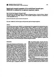
galactosidase activity (see Material and methods). No βgalactosidase activity was detected in the ovaries of a reactive line (Fig. 2I) stained in the same conditions and used as negative control. The 48.3 and 150.6 native transgenic lines as well as the rearranged one (60.1) did not express β-galactosidase in the ovaries (Table 1). This could be due to a mutation in the construct that is undetectable in Southern blot analysis or to the inhibition of its expression by position effects. The four other transgenic lines (48.1, 53.1, 101.1, 150.1) share the same pattern of expression. However, we have been able to notice quantitative differences. Among all the lines, only 48.1possesses two insertions, one of which is the same as in the 48.3 line (see above) and hence probably unexpressed. All the other lines have one active k160 insertion suggesting that the quantitative differences observed in the expression level are not dependent on the transgene copy number. Extensive observations of the 48.1 and 53.1 lines, which have the highest level of β-galactosidase expression, have been performed. Using direct observation of stained ovarioles, we can examine the expression pattern of our I-lacZ construct during oogenesis. No expression is recorded in the germarium (Fig. 2C). The β-galactosidase activity is observed in germ-line cells only (nurse cells and oocyte) and never in the surrounding somatic cells such as follicle cells (Fig. 2). Accumulation of the fusion protein is limited from stage 2 to stage 10, with a maximum in stages 5 to 9 (Fig. 2A). Most of this activity is concentrated in the nuclei of nurse cells and at lower level in their cytoplasm. At stage 7, the oocyte, which can be easily identified in the egg chamber, clearly displays a coloured nucleus (Fig. 2D). A cytoplasmic gradient builds up during oocyte development: around stage 8, when vitellogenesis begins, the cytoplasm of the oocyte alongside the nurse cells is strongly stained (Fig. 2E). Such a gradient does not result from a passive diffusion of pigments: our preparations are stable for more than six months and no diffusion of staining has been recorded either in oocytes or towards the follicle cells. Sections of total coloured ovaries confirm that the nucleus of the oocyte is coloured at stage 8 and 9 and show that the outer and granular part of the cytoplasm is less stained (Fig. 2J).
732
P. Lachaume and others
Between stage 9 and 10b, we observe a radical decrease in β-galactosidase activity in both the nucleus and the cytoplasm (Fig. 2B). After stage 10 neither nurse cells nor oocytes are coloured. This suggests a break in the expression of the transgenes, followed by the decay of the fusion protein. Variation in the percentage of positive cysts during oogenesis Some heterogeneity appears between 53.1 and 48.1; in particular we never observe 100% of the egg chambers to be stained at any given stage in 48.1. To quantify this observation more precisely, fifteen pairs of ovaries from the 53.1 and 48.1 lines have been isolated in a single experiment and stained. For each egg chamber, the stage and the coloration were recorded. The proportion of coloured cysts in the ten first stages is displayed in Fig. 3. Variations in coloration efficiency could not explain the discrepancy observed and we have to assume that the lack of ORF1 expression in the first stages of oogenesis probably persists during the whole of oogenesis in some of the egg chambers of the 48.1 line. This is confirmed by the discontinuous pattern of coloration observed in some ovarioles of the 48.1 line: one or more noncoloured cysts are bordered with two coloured ones (Fig. 2G). Approximately 10% of the analysed ovarioles of the 48.1 line show coloration discontinuity in the early stages. Absence of coloration is not particular to a specific stage but clearly shows lack of k160 expression in cysts usually coloured at these stages. No somatic expression is observed Our detection assay is so sensitive that even control testes, which are usually described as negative for endogenous βgalactosidase activity (Glaser et al., 1986), are recorded positive (see Fig. 2H). These conditions have limited the number of tissues that we have been able to test. Nevertheless, the two transgenic lines 48.1 and 53.1 have been analysed. First instar larvae, fat bodies, salivary glands, ovaries, malpighian tubules and parts of gut of third instar larvae and malpighian tubules and parts of gut in adult flies are all negative for both the control and transgenic lines. This suggests that the
expression of ORF1 is restricted to the ovaries, the only tissue where full-length I factor transcripts can be found. k160 expression is limited to reactive and dysgenic females The I factor is probably submitted to multiple regulations in the different conditions associated with I-R hybrid dysgenesis. We have tested the influence of these conditions on the k160 expression. The k160 constructs of the 48.1 line are introduced in an inducer context in the three homozygous lines F1, F4 and F6 (see Material and methods). No modifications of the inserts are observed in Southern blot analysis. However, no more β-galactosidase expression is detected in the ovaries of the females from the three inducer lines. This suggests that the inducer context inhibits expression of the ORF1. The study of the expression in dysgenic and reciprocal context is performed by crossing the 48.1 reactive line and the F6 inducer line. SF and RSF females have the same genotype. They are also homozygous for the insertions as the inducer and reactive females. The results obtained are summarized in Table 2. Expression of the construct is observed only when the cytoplasm transmitted by the mother is reactive. This expression is similar in the SF and R context, including the discontinuous pattern of coloration characteristic of the 48.1 strain. Thus expression of the I-lacZ fusion in I, SF and RSF context correlates with the ability of the I factor to give a full-length RNA (Chaboissier et al., 1990), to transpose in female germ-line and to be associated with dysgenic symptoms.
Discussion The I factor is a LINE of Drosophila melanogaster (Bucheton, 1990). Its transposition is inducible at high level in I-R dysgenic crosses. In contrast to most of the other LINEs, a functional copy of the I factor has been cloned and it has been shown to transpose by an RNA intermediate (Pelisson et al., 1991; Jensen and Heidmann, 1991). ThereTable 2. Study of the k160 expression in different genetic contexts
Fig. 3. Evolution of the percentage of egg chambers expressing the transgenic fusion during oogenesis. After coloration, percentage of coloured egg chambers at different stages have been measured in ovaries of 48.1(crosses) and 53.1 (open squares).
?
48.1 reactive line
F6 inducer line
48.1 reactive line
R females: β-galactosidase activity in the germ-line cells nuclei from the stage 2 to 10 of oogenesis
SF females: β-galactosidase activity in the germ-line cells nuclei from the stage 2 to 10 of oogenesis
F6 inducer line
RSF females: no β-galactosidase activity is observed in the germ-line cells
I females no β-galactosidase activity is observed in the germ-line cells
/
After X-gal coloration of dissected ovaries, germinal expression of k160 is recorded only when the cytoplasm of the embryos is from reactive mother (48.1 reactive line).
Expression of the I factor during oogenesis fore the I factor is a model of choice to analyse the expression and transposition of LINEs. A translational fusion of the I factor ORF1 with the lacZ reporter gene has been introduced into the genome of a reactive strain in order to study the I factor expression in flies. We have examined the transgenic β-galactosidase activity in six transformed lines displaying a native I-lacZ construct in their genome.
An internal promoter drives I factor expression Mizrokhiet al.(1988) have shown that Jockey, another LINE of Drosophila melanogaster, is transcribed from an internal promoter recognized by RNA polymerase II. Homology between I factor and Jockey 5′ ends suggests that I factor also uses the same mechanism of transcription. The homogeneity in the pattern of expression between our transgenic lines provides the most direct evidence that the 5′ part of the I factor, probably the putative RNA polymerase II promoter, is capable of promoting its in vivo expression and is not another enhancer-trap system. According to the good correlation of our results with elements already known about the I factor expression, it is unlikely that part of the pUChsneo or the short residual fragment from the fifth intron and sixth exon of the white gene can promote the I-lacZ expression observed in our transgenotes. For the initiation of translation, two ATG only (at position 187 and 586) are in frame with the ORF1 and potentially used on the full-length RNA. As ATG 586 has been removed from the k160 construct by AsuII deletion, ATG 187 should be the codon that initiates translation in vivo .
I factor expression is germ-line specific Invasion of natural populations by a transposable element accounts for the recent appearance of I and P strains around 1920 and 1950 in Drosophila melanogaster (Anxolabèhère et al., 1988; Bregliano and Kidwell, 1983). Very likely such an event is possible if transposition occurs mainly in the germ line since high levels of transposition in the soma would induce multiple damages and lethality of the host. This is typically the case for the P element (Laski and Rubin, 1989). By analysing our transgenotes we have shown that I factor expression is also limited to germ-line cells. This is consistent with the fact that no somatic mutations due to I factor insertion have been detected during dysgenic crosses (Pelisson and Bregliano, 1987) and that the potential intermediate of transposition has not been found in somatic tissues (Chaboissier et al., 1990). In addition, we did not detect any expression in the germarium or in the larval ovaries suggesting that the primordial germ cells probably do not express the I factor. This is in good agreement with the fact that mutations induced by I factor transposition are meiotic events leading to isolated mutants in the progeny of SF females (Bregliano and Kidwell, 1983). If the level of this germ-line-specific expression is sensitive to position effects, the influence of the insertion context is also illustrated by the differences between 48.1 and 53.1 and by the coloration discontinuity within a single ovary.
733
How can two cysts at the same stage, in the same ovary and with the same genotype express differently the same marker? No satisfying interpretation could explain totally this observation. Cis elements controlling I factor expression are regulated by maternal effect According to the pattern of expression in R context and if we consider the expression in I, RSF and SF females, we observe a good correlation among transcription (Chaboissier et al., 1990), our results in translation of the ORF1, strong transposition and dysgenic symptoms (Picard, 1976). This implies that probably all the cis elements responsible for the specific expression in the germ line lie within the 5′ untranslated part and the two segments of the first ORF of the I factor present in the k160 construct. This suggests also that the I factor transposition is regulated at the transcriptional level in contrast to P or Copia (Laski et al., 1986; Parkhurst and Corces, 1987). Out of the four different classes of transgenic females (R, I, SF and RSF) only those with reactive mothers, the reactive and dysgenic ones, express the I-lacZ fusion without significant differences. Influence of the maternal cytoplasm is essentially limited to the embryonic state. However, maternal reactive cytoplasm is necessary to switch on the I factor expression in the R and SF adults. Putative elements of this cytoplasm should be efficient in the pole cells of the embryos to predetermine the ability of the adult germ-line to express the I factor. The nature of these elements and their mechanism of action are still not clear. An essential result is that our transgenic construct in R context is a good indicator of the I factor expression in dysgenic conditions and can be used for further study on the control of the I factor.
The fusion protein is accumulated in germ cell nuclei Proteins assigned to nuclei contain one or two specific amino acid sequences called “nuclear localization sequences” (NLSs). These NLS are necessary and sufficient to direct protein into the nucleus (Garcia-Bustos et al., 1991; Richardson et al., 1986; Robbins et al., 1991). Since I-lacZ fusion protein is located in the nuclear compartment of nurse cells and oocytes, this protein contains NLS. The β-galactosidase does not drive the nuclear localization of chimeric protein used in many other studies (Hall et al., 1984). Therefore the NLS are located in the I factor part of the fusion protein. We have not clearly identified a NLS sequence in the ORF1 of the I factor. No region rich in basic amino acids similar to the NLS of SV40T antigen group has been found. Only a short sequence from residue 22 to 29 (PKFIIIK) corresponds partially to the group of mat α2 NLS (KIPIK; Silver and Hall, 1988). It is also possible that ORF1 proteins enter the nucleus after oligomerization with a host protein, as suggested for other proteins (Dingwall et al., 1982; Zhao and Padmanabhan, 1988). Some of the chimeric I-lacZ proteins still have a cytoplasmic localization. This cannot be explained by the size of the protein (Landford et al., 1986): in enhancer-trap experiments, the P-lacZ protein is efficiently translocated into the nucleus (Grossniklaus et al., 1989). In our construct,
734
P. Lachaume and others
partial truncation of the NLS (Welsh et al., 1986), the lack of one out of two NLS (Richardson et al., 1986), modification of the NLS, protein context (Roberts et al., 1987; Nelson and Silver, 1989), or lack of possible multimerization (Silver and Hall, 1988) can reduce the efficiency of the nuclear transport. However a role of the ORF1 protein in the cytoplasm should not be discarded. In any case, this nuclear localization is consistent with the two possible roles of the ORF1 protein suggested by the presence of zinc-finger domains: building of a retrotransposition complex and/or the regulation of I factor expression. If we assume that ORF1 protein is essential for the transposition cycle of the I factor, this transposition should be initiated during oogenesis between stage 2, when expression is first recorded, and stage 10, when accumulation of the fusion protein disappears. We thank F. Croisille for providing sections of stained ovaries, C. Vaury, Y. Croisille and C. Tatout for critical reading of the manuscript. This work was mainly supported by grants from the CNRS, ARC and FRM. Two of us, P.L. and K.B. received fellowships from the MRT, ARC and LNFC.
References Abad, P., Vaury, C., Pelisson, A., Chaboissier, M. C., Busseau, I. and Bucheton, A. (1989). A long interspersed repetitive element - the I factor of Drosophila teissieri - is able to transpose in different Drosophila species. Proc. Natl. Acad. Sci. USA 86, 8887-8891. Anxolabèhère, D., Kidwell, M. G. and Periquet, G. (1988). Molecular characterization of diverse populations are consistent with the hypothesis of a recent invasion of Drosophila melanogaster by mobile P elements. Mol. Biol. Evol. 5, 252-269. Blackman, R. V., Grimalia, R., Koeler, M. M. D. and Gelbart, W. M. (1987). Mobilization of Hobo elements residing within the decapentaplegic gene complex: suggestion of a new hybrid dysgenesis system in Drosophila melanogaster. Cell 41, 497-505. Boyer, H. W. and Roulland-Dussoix, D. (1969). A complementation analysis of the restriction and modification of DNA in Escherichia coli. J. Mol. Biol. 41, 459-472. Bregliano, J. C. and Kidwell, M. G. (1983). Hybrid dysgenesis determinants. Mobile Genetic Elements.( Ed. J. Shapiro). New York: Academic Press. pp. 363-404. Bucheton, A. (1990). I transposable elements and I-R hybrid dysgenesis in Drosophila. T.I.G. 6, 16-21. Bucheton, A., Paro, R., Sang, H. M., Pelisson, A. and Finnegan, D. J. (1984). The molecular basis of I-R hybrid dysgenesis in Drosophila melanogaster: identification, cloning and properties of the I factor. Cell 38, 153-163. Casadaban, M. S., Martinez-Arias, A., Shapira, S. K. and Chou, J. (1983). ß-galactosidase gene fusions for analysing gene expression in Escherichia coli and yeast. Method in Enzymology 100, 293-308. Chaboissier, M. C., Busseau, I., Prosser, J., Finnegan, D. and Bucheton, A. (1990). Identification of a potential RNA intermediate for transposition of the LINE-like element I factor in Drosophila melanogaster. EMBO J. 9, 3557-3563. David, J. (1959). Etude quantitative du développement de la drosophile élevée en milieu axenique. Bull. Soc. biol. France and Belgique 93, 472. Dingwall, C., Sharnick, S. V. and Laskey, R. A. (1982). A polypeptide domain that specifies migration of nucleoplasmin into the nucleus. Cell 30, 449-458. Evans, J. P. and Palmiter, R. P. (1991). Retrotransposition of a mouse L1 element. Proc. Natl. Acad. Sci. USA 88, 8792-8795. Evraert, F. (1989). Looking for translation products of the I factor, a LINE associated with hybrid dysgenesis in Drosophila melanogaster (in French). Thèse de l’Université Blaise Pascal, n° DU191, ClermontFerrand. Fanning, T. G. and Singer, M. G. (1987). Line-1: a mammalian
transposable element. Biochim. Biophys. Acta 910, 203-212. Fawcett, D. H., Lister, C. K., Kellet, E. and Finnegan, D. J. (1986). Transposable elements controlling I-R hybrid dysgenesis in Drosophila melanogaster are similar to mammalian lines. Cell 47, 1007-1015. Finnegan, D. J. (1988). I factor in Drosophila melanogaster and similar elements in other eucaryotes. In Society for general Microbiology Symposium. (Eds Kingman, A.J., Kingman, S.M. and Chatter, K.F.), pp. 271-285. Cambridge: Cambridge University press. Garcia-Bustos, J., Heitman, J. and Hall, M. N. (1991). Nuclear protein localization. Biochim. Biophys. Acta 1071, 83-101. Glaser, R. L., Wolfner, M. F. and Lis, J. T. (1986). Spatial and temporal pattern of Hsp 26 expression during normal development. EMBO J. 5, 747-754. Gough, J. A. and Murray, N. E. (1983). Sequence diversity among related genes for recognition of specific targets in DNA molecules. J. Mol. Biol. 166, 1-19. Grossniklaus, U., Bellen, H. J., Wilson, C. and Gehring, W. J. (1989). P-element-mediated enhancer detection to the study of oogenesis in Drosophila. Development 107, 189-200. Hall, M. N., Hereford, L. and Herskowitz, I. (1984). Targeting of Escherichia coli β-galactosidase to the nucleus in yeast. Cell 36, 10571065. Jakubczak, J. L., Xiong, Y. and Eickbush, T. H. (1990). Type I (R1) and Type II (R2) ribosomal DNA insertions of Drosophila melanogaster are retrotransposable elements closely related to those of Bombyx mori. J. Mol. Biol. 212, 37-52. Jensen, S. and Heidmann, T. (1991). An indicator gene for detection of germline retrotransposition in transgenic Drosophila demonstrates RNA-mediated transposition of the LINE I element. EMBO J. 10, 1927-1937. Kidwell, M. G. and Kidwell, J. F. (1975). Cytoplasm chromosome interactions in Drosophila melanogaster. Nature 253, 755-756. Kidwell, M. G., Kidwell, J. F. and Ives, P. T. (1977). Spontaneous nonreciprocal mutation on sterility in strain crosses of Drosophila melanogaster. Mut. Res. 42, 89-98. Knipple, D. C. and MacIntyre, R. J. (1984). Cytogenetic mapping and isolation of mutations of the β-gal-1 locus of Drosophila melanogaster. Mol. Gen. Genet. 198, 75-83. Landford, R. E., Kanda, P. and Kennedy, R. C. (1986). Induction of nuclear transport with a synthetic peptide homologous to the SV40 T antigen transport signal. Cell 46, 577-582. Laski, F. A., Rio, D. C. and Rubin, G. M. (1986). Tissue specificity of Drosophila P-element tranposition is regulated at the level of mRNA splicing. Cell 44, 7-19. Laski, F. A. and Rubin, G. M. (1989). Analysis of the cis-acting requirements for germ-line-specific splicing of the P-element ORF1ORF2 intron. Genes Dev. 3, 720-728. Lavige, J. M. (1986). I-R system of hybrid dysgenesis in Drosophila melanogaster: further data on the arrest of development of the embryos from SF females. Biology of the Cell 56, 207-216. Maniatis, T., Fritsh, E. F. and Sambrook, J. (1982). Molecular Cloning: A Laboratory Manual. Eds Cold Spring Harbor Laboratory. Cold Spring Harbor, New York. Mizrokhi, L. J., Georgieva, S. G. and Ilying, Y. V. (1988). Jockey, a mobile Drosophila element similar to mammalian LINEs, is transcribed from the internal promoter by the RNA polymerase II. Cell 54, 685691. Nelson, M. and Silver, P. (1989). Context affects nuclear protein localization in Saccharomyces cerevisiae. Mol. Cell. Biol. 9, 384-389. Parkhurst, S. and Corces, V. G. (1987). Developmental expression of Drosophila melanogaster retrovirus-like transposable elements. EMBO J. 6, 419-424. Pelisson, A. and Bregliano, J. C. (1987). Evidence for rapid limitation of the I element copy number in a genome submitted to several generations of I-R hybrid dysgenesis in Drosophila melanogaster. Mol. Gen. Genet. 207, 306-313. Pelisson, A., Finnegan, D.J. and Bucheton, A. (1991). Evidence for retrotransposition of the I factor, a LINE element of Drosophila melanogaster. Proc. Natl. Acad. Sci. USA 88, 4907-4910. Picard, G. and L’Héritier, P. (1971). A maternally inherited factor inducing sterility in Drosophila melanogaster. Dros. Inf. Serv. 46, 54. Picard, G. (1976). Non mendelian female sterility in Drosophila melanogaster: hereditary transmission of I factor. Genetics 83, 107123. Pritchard, M. A., Dura, J. M., Pelisson, A., Bucheton, A. and
Expression of the I factor during oogenesis Finnegan, D. J. (1988). A cloned I factor is fully functional in Drosophila melanogaster: a possible mechanisms for transposition. Mol. Gen. Genet. 214,533-540. Richardson, W. D., Roberts, B. L. and Smith, A. E. (1986). Nuclear location signals in polyoma virus large-T. Cell 44, 77-85. Robbins, J., Dilworth, S. M., Laskey, R. A. and Dingwall, C. (1991). Two interdependent basic domains in nucleoplasmin nuclear targeting sequence: identification of a class of bipartite nuclear targeting sequence. Cell 64, 615-623. Roberts, B. L., Richardson, W. D. and Smith, A. E. (1987). The effect of protein context on nuclear location signal function. Cell 50, 465-475. Rubin, G. M. and Spradling, A. C. (1982). Genetic transformation of Drosophila with transposable element vector. Science 218, 348-353. Rubin, G. M., Kidwell, M. G. and Bingham, P. M. (1982). The molecular basis of P-M hybrid dysgenesis: the nature of induced mutations. Cell 29, 987-994. Silver, P. A. and Hall, M. (1988). Transport of proteins into the nucleus.
735
In Protein Transfer and Organelles Biogenesis (Eds R.C Das and P.W. Robbins). pp 749-768. London: Academic Press. Simon, J. A., Sutton, L. A., Lobell, R. B., Glasser, R. L. and Lis, J. T. (1985). Determinants of heat shock-induced chromosome puffing. Cell 40, 805-817. Steller, H. and Pirotta, V. (1985). A transposable P vector that confers selectable G 418 resistance to Drosophila larvae. EMBO J. 4, 167171. Welsh, J. D., Swimmer, C., Cocke, T. and Shenk, T. (1986). A second domain of simian virus 40 T antigen in which mutations can alter the cellular localization of the antigen. Mol. Cell. Biol. 6, 2207-2212. Zhao, L. and Padmanabhan, R. (1988). Nuclear transport of adenovirus DNA- polymerase is facilitated by interaction with preterminal protein. Cell 55, 1005-1015 (Accepted 25 February 1992)
7089Fig. 2 tip-in Fig. 2. Expression of the β-galactosidase in the transgenic lines. The fusion protein is clearly accumulated in the nuclei of the nurse cells. It is also present in the cytoplasm of these cells. The cells with somatic origin do not express the I-lacZ gene. The same results are recorded in all transgenic lines that express the protein with only little quantitative differences. Detection of the endogenous βgalactosidase activity is a control of the coloration efficiency in total ovaries (A) general view of a X-gal-stained pair of 53.1 ovaries after spreading. (B-G, J) 48.1 line. (B) Illustration of the swift decrease of the k160 expression between stage 9 and stage 10b. (C) View of the early stages of oogenesis: no expression of the fusion gene is detected in the germarium. (D) Cyst at stage 7. The oocyte nucleus (ON) clearly accumulates the I-lacZ protein. (E) Cyst at stage 8: the cytoplasm of the oocyte is strongly coloured alongside the nurse cells. (F) Cyst at stage 9. The oocyte nucleus is still coloured. (G) Some cysts usually coloured do not express the fusion gene. They appears as a non- coloured cyst between two coloured ones. (J) Section of a cyst at stage 9. (H I) Charolles line used as control. (H) Testes stained during 12 hours: unexpected endogenous β-galactosidase prevents the efficient detection of I-lacZ expression. (I) Only the endogenous βgalactosidase is detected in this control ovaries.