[2] Michael Berger, Judith Castelino, Richard Huang, Manish Shah, Robert H. Austin, Design of a microfabricated magnetic cell separator, Electrophoresis. 2001 ...
SPINTRONIC DEVICE FOR CELL/MAGNETIC PARTICLE SORTING AND COUNTING
J.Loureiro1,2, R.Ferreira1,2, S.Cardoso1,2, J.Germano1,3, D.Snakenborg4 , J.M.S. Cabral2,5, P.P.Freitas1,2 1
INESC Microsistemas e Nanotecnologias/ IN- Institute of Nanoscience and Nanotechnology, PORTUGAL 2 Instituto Superior Técnico (IST), Physics Department ,PORTUGAL 3 INESC-Investigação e Desenvolvimento, PORTUGAL 4 DTU Nanotech, DENMARK 5 IBB- Institute for Biotechnology and Bioengineering, Centre for Biological and Chemical Engineering, IST, PORTUGAL
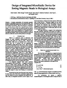
ABSTRACT This paper reports on a new platform for cell/particles sorting and counting combining a spintronic microchip with a microfluidic approach. The magnetic separation of 2 μm diameter superparamagnetic particles has been observed when applying 25 mA through current lines (due to an in-situ magnetic field gradient of 2.8x10⁵ kA/m2). By using magnetoresistive sensors with a sensitivity of 1 μV/(A/m) it was possible to monitor the movement of the particles, proving the counting capability of the system. KEYWORDS: Magnetic separation, magnetoresistive sensors, microfluidic platform INTRODUCTION Various designs have been proposed to separate cells/particles of different sizes/magnetic moments using microfluidic platforms and external magnets [1][2] or integrated current lines [3]. The detection of biological analytes targeted with magnetic particles using spin-valve (SV) sensors has been reported in recent years [4]. The novelty of this work is the capability of realizing the magnetic separation with higher magnetic field gradients created by in-situ current lines (∇H= 2.8x10⁵ kA/m2 for 25 mA) and counting the particles in a continuous flow within the same device using spin-valve sensors. ARCHITECTURE The separation process is based on a magnetophoresis effect which is dependent on the magnetic force acting on the particles and on the flow rate. A fluidic platform consisting of two microchannels implemented in an H-shaped geometry has been designed. The channels (150 μm wide, 14 μm high) are separated by a wall (40 μm wide) and connected through several gaps enabling the particles to move from one channel to the other (Fig. 1). The microfluidic component, made of PDMS, is bonded on top of a microfabricated chip which presents two main/key features: two successive sloped current lines (7μm wide, AlSi1%Cu0.5% 500 nm thick) passivated with a 500 nm thick SiO2 layer, and three spin-valve sensors (Ta2.0nm/NiFe2.5nm
Twelfth International Conference on Miniaturized Systems for Chemistry and Life Sciences October 12 - 16, 2008, San Diego, California, USA 978-0-9798064-1-4/µTAS2008/$20©2008CBMS
498
/CoFe2.5nm/Cu2.0nm/CoFe2.5nm/MnIr6.0nm/Ta2.0nm, lateral dimensions 2.5μm x 50μm and a magnetoresistive signal (MR) of 6%). The magnetic sensors are located in both channels, beyond the current lines, and are used to count the particles/cells flowing in the channels. Their capability of counting comes from the Giant Magnetoresistive effect (GMR), the sensor resistance changing due to the variation of the magnetic field (created by the magnetic particles) during the particles movement.
Figure 1. a) H-shaped fluidic channel, allowing cells/particles to be separated from one channel to the other due to the magnetic field created by the sloped current lines. Each channel contains spin valves to count the particles. EXPERIMENTAL First results were obtained with 2 μm diameter superparamagnetic particles (Micromod, Germany) diluted in a phosphate buffer solution (4x10⁴ particles/μl), injected into the upper channel while the lower channel was filled only with buffer. In the detection part of the system a small permanent magnet placed below the system magnetizes the particles with a perpendicular field of 135 kA/m. The particle presence is monitored by the magnetoresistive sensor (with a sensitivity of 1 μV/(A/m)). The predicted time span due to the particles speed is ~20 ms. To increase the amplitude resolution the sensor signal was filtered with a high-pass filter and 100x amplified (SRS-SIM910-JFET-Preamp). RESULTS AND DISCUSSION With flow rates up to 40 nL/min and applying 20 mA through the current lines, particles were observed following the line path, moving from one channel to the other, with velocities between 25 to 200 μm/s (Fig. 2, left). Although some beads were not separated, the second current line increased the efficiency of the device to a large extent, indicated by the number of beads following this line. More current lines therefore should be added to future versions. A sensor signal with amplitude of 75 μV is expected to be achieved when a sensor current of 1 mA is applied (for particles passing 2 µm above the sensor – fig. 2) [4]. As depicted in figure 2 (bottom right) particle signals of 50 μV have been amplified and measured using a voltmeter, at a sampling rate of 250 Hz (meaning that the particles were flowing at a sensor distance higher than 2 μm). Complex signals have Twelfth International Conference on Miniaturized Systems for Chemistry and Life Sciences October 12 - 16, 2008, San Diego, California, USA
499
also been obtained due to agglomerates formation implying that a signal analysis is crucial to obtain a correct number of counted particles.
Figure 2. left) Particles separation when applying 20 mA through the current line; right) Spin valve transfer curve with MR of 6% (inset: microscope image of the microfluidic channels and the expected sensor signal vs height for 2 μm beads) (top); right) Amplified sensor signal for particles moving over the sensor (bottom). CONCLUSIONS A simple prototype both for particle separation based on magnetophoresis and particle counting based on the GMR effect has been designed and demonstrated. By labelling cells with magnetic particles it will be possible to separate/count cells instead of single particles and this will extend the platform applications to biological experiments. In the future the separation and counting of human hematopoietic stem/progenitor cells will be done using umbilical cord blood treated samples. ACKNOWLEDGEMENTS J.Loureiro thanks FCT for the grant SFRH/BD/30056/2006 and for the Project PTDC/CTM/68617/2006. INESC MN acknowledges FCT funding through the Associated Lab - Instituto de Nanotecnologias0.4 and the Programa Operacional Ciência e Inovação 2010 (POCI 2010) funded by the European program FEDER. REFERENCES [1] Z.Jiang, J.Llandro, T.Mitrelias, and J.A.C. Bland, An integrated microfluidic cell for detection, manipulation, and sorting of single micron-size magnetic beads, J. Appl. Phys 99, 08S105 (2006) [2] Michael Berger, Judith Castelino, Richard Huang, Manish Shah, Robert H. Austin, Design of a microfabricated magnetic cell separator, Electrophoresis 2001, 22, 3883–3892 [3] Chengxun Liu, Liesbet Lagae and Gustaaf Borghs, Manipulation of magnetic particles on chip by magnetophoretic actuation and dielectrophoretic levitation, Appl Phys Lett 90, 184109 (2007) [4] P.P Freitas, R.Ferreira, S.Cardoso and F.Cardoso, Magnetoresistive sensors, J.Phys.: Condens. Matter 19, N16 (2007), 165221 Twelfth International Conference on Miniaturized Systems for Chemistry and Life Sciences October 12 - 16, 2008, San Diego, California, USA
500