was slowly added 2M HCl in Et2O (0.50 mL, 1.0 mmol). After the addition was complete, .... Weblogo presentation of the 11 bp consensus. (TDGâGTWACCHA) ...
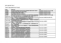
Supplementary Table S1. Oligonucleotides used in this study Oligo
Sequence 5’-3’
Usage
Reference
AE2862
GCGCGCGAATTCGCCACCATGCCACACGCCTTCAAGCCCGGG
pIRES2-eGFP-hHRP2-HA construction
This study
AE2683
CGCGCGGATCCTAGGCGTAGTCCGGGACGTCGTACGGGTAGCTGC CGCCGCTCTCCTCGTCCAGGGCCTCCGAG GCGCGCGAATTCGCCACCATGCCGCACGCCTTCAAGCCCGGG
pIRES2-eGFP-hHRP2-HA construction
This study
pIRES2-eGFP-mHRP2-HA construction
This study
AE2955
CGCGCGGATCCTAGGCGTAGTCCGGGACGTCGTACGGGTAGC TGCCGCCGCTGTCCTCATTGTCATCATTGGCA
pIRES2-eGFP-mHRP2-HA construction
This study
AE2878
AAGTTTGCCCTAAAGGTCAACAGGCCCGGACGTGAAGAGG
D489N mutagenesis
This study
AE2879
CCTCTTCACGTCCGGGCTGTTGACCTTTAGGGCAAACTTG
D489N mutagenesis
This study
AE4887
GCGTGGATCCTCCGTAGAGGAGAGACTGCAA
pGEX-4T-1-mHRP2/IBD construction
This study
AE4888
GCGCTCGAGTCACTCATCCTCTGCCAGCTC
pGEX-4T-1-mHRP2/IBD construction
This study
AE2331
GAGATATCGAGGCAGAAAGAAGACTGGGATAG
Psip1 genotyping
15
AE2802
GCATGGTGGCACAATGGCAACTGGGTC
Psip1 genotyping
15
AE2511
TTCTGTTCTCATCCATGCTCTAGTG
Hdgfrp2 genotyping
This study
AE2512
GCTGTTTATTGGCTTGCTAACCATG
Hdgfrp2 genotyping
This study
AE3747
TGTTTGACCATGTCAGTGTGTAC
Hdgfrp2 genotyping
This study
AE3748
CTTACCTTCTTCTTGCGTCCTCC
Hdgfrp2 genotyping
This study
AE2963
TGTGTGCCCGTCTGTTGTGT
LRT qPCR
15
AE4422
GAGTCCTGCGTCGAGAGATC
LRT qPCR
This study
AE2965
FAM-CAGTGGCGCCCGAACAGGGA-TAMRA
LRT qPCR
15
AE4450
GCCTGGGAGCTCTCTGGCTAA
2-LTR qPCR
33
AE4451
GCCTTGTGTGTGGTAGATCCA
2-LTR qPCR
33
AE4452
FAM-AAGTAGTGTGTGCCCGTCTGTTGTGTGACTC-TAMRA
2-LTR qPCR
33
AE2604
AGTGAGTTCCAGGACAGCCAGG
BBL-PCR
15
AE2605
GGCTGGCCTCGAACTCAGAAATC
BBL-PCR
15
AE2606
GTCTGAAGACAGCTACAGTGTAC
BBL-PCR
15
AE2954
1
AE2607
GTGAGCCACCATGTGGTTGCTGG
BBL-PCR
15
AE2608
CTCACCATCATCAGAACGCAGCACT
BBL-PCR
15
AE2609
TCGCCATCTGGTAATCTCTGAAG
BBL-PCR
15
AE3014
ATGCCACGTAAGCGAAACTCTGGCTAACTAGGGAACCCACTG
BBL-PCR; PIC assay
4
AE3013
ATGCCACGTAAGCGAAACTC
BBL-PCR; PIC assay
4
AE990
CTGACTAAAAGGGTCTGAGG
BBL-PCR; PIC assay
15
AE995
FAM-TTAAGCCTCAATAAAGCTTGCCTTGAGTGC-TAMRA
BBL-PCR; PIC assay
15
AE2413
GTTGTTCCAGTTTGGAACAAGAGTC
PIC assay
4
AE2414
ACTCAACCCTATCTCGGTCTATTC
PIC assay
4
AE3160
TCCGACTCCCGCCTCTGACTT
Hdgfrp2 qRT-PCR exon 1/2
This study
AE3161
CACGGCACCATCAGCAATGTC
Hdgfrp2 qRT-PCR exon 1/2
This study
AE2624
CCTCAAACATGACTCGCGATTTC
Psip qRT-PCR exon 2/3
15
AE2625
GCTCCATCAGGAACTTCATCTAC
Psip qRT-PCR exon 2/3
15
AE2553
TGAGTCGGAGAAGACCAGTGACC
Hdgfrp2 qRT-PCR exon 5/7
This study
AE2554
AATCCGAGGCTGATGGCACCTTC
Hdgfrp2 qRT-PCR exon 5/7
This study
AE3664
AGGTCCTGGCATCTTGTCCATGG
Ppia qRT-PCR, exon 4
This study
AE3665
GGCTTCCACAATGTTCATGCC
Ppia qRT-PCR, exon 5
This study
2
Supplementary methods. Details of BI-D synthesis.
Synthesis of (±)-BI-D
Scheme 1. Synthetic route for the preparation of BI-D (1). (±)-BI-D (1) was synthesized in 10 linear steps from commercially available 4-hydroxy2-methylquinoline S2 (Sigma-Aldrich) via slight modification of a synthetic route reported by Tsantrizos and colleagues at Boehringer Ingelheim (29). The synthesis commenced with preparation of 3-bromo-4-chloro-2-methylquinoline S3 through a selective bromination/ chlorination sequence of the quinoline ring system at the C3 and C4 positions, respectively (66,67). The aryl bromide 3 was then selectively functionalized via a Stille coupling with tributyl(vinyl)tin (68) and subsequent Upjohn dihydroxylation (69) to provide diol S5. Finkelstein-type substitution of the remaining chloride group with iodide, followed by selective protection of the primary alcohol of the diol as the pivalate provided S6. The secondary alcohol was then converted to the t-butyl ether in the presence of perchloric acid to provide intermediate S7. Introduction of the chroman group through Suzuki coupling of boronic acid S13 (Scheme 2) with iodide S7 provided the biaryl adduct S8. Finally, hydrolysis of the pivalate group of S8 and subsequent oxidation (70) of the primary alcohol to the corresponding carboxylic acid furnished 1.
Scheme 2. Synthetic route for the preparation of boronic acid S13.
3
3-Bromo-4-chloro-2-methylquinoline S3. 4-hydroxy-2-methylquinoline (S2) (1.19 g, 7.50 mmol) in acetic acid (34 mL) was treated with N-bromosuccinimide (1.33 g, 7.50 mmol) and the resulting suspension was heated to 60 °C and stirred for 2 h. The mixture was cooled to room temperature, cold water was added and the formed precipitate was filtered and collected. The solid was washed sequentially with water, saturated aqueous NaHCO3, and acetone, then dried to provide 3-bromo-4-hydroxy-2-methylquinoline (1.63 g, 91%) as an off-white solid: 1H NMR (300 MHz, DMSO-d6) δ 12.15 (s, 1H), 8.09 (d, J = 8.0 Hz, 1H), 7.68 (t, J = 7.6 Hz, 1H), 7.55 (d, J = 8.3 Hz, 1H), 7.36 (t, J = 7.5 Hz, 1H), 2.56 (s, 3H). This material was taken directly into the chlorimation reaction with no further purification. A suspension of 3-bromo-4-hydroxy-2-methylquinoline (1.55 g, 6.50 mmol) in POCl3 (5.95 mL, 65 mmol) was stirred at 80 °C for 2 h. After cooling to room temperature, the mixture was poured onto ice (~30 g) and neutralized with 50% (w/v) aqueous NaOH while maintaining the temperature at 0 °C. The resulting mixture was extracted with ethyl acetate (3 x 20 mL) and the combined organic phases were washed with brine, dried over Na2SO4, and concentrated under reduced pressure to afford chloride S3 (1.64 g, 98%) as a white solid: 1H NMR (300 MHz, CDCl3) δ 8.18 (d, J = 8.4 Hz, 1H), 8.01 (d, J = 8.4 Hz, 1H), 7.75 (t, J = 7.6 Hz, 1H), 7.60 (t, J = 7.6 Hz, 1H), 2.91 (s, 3H). 4-Chloro-2-methyl-3-vinylquinoline (S4). A mixture of 3-bromo-4-chloro-2methylquinoline (3) (880 mg, 3.43 mmol), tributyl(vinyl)tin (1.05 mL, 3.60 mmol), Pd(PPh3)4 (396 mg, 0.34 mmol) in DMF (17 mL) was stirred overnight at 100 °C. After cooling to room temperature, the reaction was quenched with 10% (w/v) aqueous KF (20 mL), filtered through Celite and washed with ethyl acetate. The filtrate was transferred to a separatory funnel and extracted with ethyl acetate (3 x 10 mL). The combined organic phases were washed with 10% aqueous KF, brine, dried over Na2SO4, and concentrated under reduced pressure. Flash chromatography (SiO2, 5% ethyl acetate in hexanes) afforded vinylquinoline S4 (459 mg, 66%) as a clear oil: 1H NMR (400 MHz, CDCl3) δ 8.22 (d, J = 8.3 Hz, 1H), 8.00 (d, J = 8.4 Hz, 1H), 7.71 (ddd, J = 8.4, 6.9, 1.4 Hz, 1H), 7.57 (ddd, J = 8.2, 7.0, 1.1 Hz, 1H), 6.87 (dd, J = 17.9, 11.6 Hz, 1H), 5.80 (dd, J = 11.6, 1.3 Hz, 1H), 5.65 (dd, J = 17.9, 1.3 Hz, 1H), 2.76 (s, 3H). Diol S5. A mixture of vinylquinoline S4 (305 mg, 1.50 mmol), N-methylmorpholine Noxide (264 mg, 2.25 mmol), osmium tetroxide polymer-bound (15 mg, 0.01 g/mmol) in acetone (15 mL), tert-butanol (3 mL), and water (1.5 mL) was stirred overnight at 85 °C. The resulting yellow solution was cooled to room temperature, filtered through Celite and washed with acetone. The filtrate was concentrated under reduced pressure and suspended in dichloromethane (5 mL). The organic phase was washed with water (10 mL). The resulting aqueous phase was extracted with dichloromethane (3 x 5 mL). The combined organic phases were washed with saturated aqueous NaHCO3, brine, dried over Na2SO4, and concentrated under reduced pressure. Flash chromatography (SiO2, 5% methanol in dichloromethane) provided diol S5 (311 mg, 77%) as a pale yellow solid: 1H NMR (300 MHz, DMSO-d6) δ 8.19 (d, J = 8.2 Hz, 1H), 7.95 (d, J = 8.3 Hz, 1H), 7.78 (t, J = 7.6 Hz, 1H), 7.67 (t, J = 7.6 Hz, 1H), 5.73 (d, J = 4.2 Hz, 1H), 5.49 (dd, J = 10.8, 6.4 Hz, 1H), 4.94 (t, J = 5.9 Hz, 1H), 2.85 (s, 3H). Pivalate S6. To a suspension of diol S5 (190 mg, 0.80 mmol) in THF (3.2 mL) at 0 °C was slowly added 2M HCl in Et2O (0.50 mL, 1.0 mmol). After the addition was complete, the ice bath was removed and the resulting pale yellow suspension was stirred at room temperature for 1
4
h before being concentrated under reduced pressure to give a pale yellow solid. The solid was suspended with NaI (600 mg, 4.0 mmol) in anhydrous acetonitrile (6.40 mL) and stirred at reflux for 18 h. After cooling to room temperature, the mixture was diluted with dichloromethane (5 mL) and water (5 mL). The phases were separated and the aqueous phase was extracted with dichloromethane (3 x 5 mL). The combined organic phases were washed with 10 % aqueous Na2S2O3, brine, dried over Na2SO4, and concentrated under reduced pressure to give a yellow solid. The crude solid was suspended in CH2Cl2 (7.1 mL), treated sequentially with pivaloyl chloride (183 µL, 1.49 mmol) and triethylamine (119 µL, 0.85 mmol), and then stirred overnight at room temperature. The clear solution was diluted with water (15 mL), partitioned, and extracted with dichloromethane (3 x 5 mL). The combined organic phases were washed with saturated aqueous NaHCO3, brine, dried over Na2SO4, and concentrated under reduced pressure. Flash chromatography (SiO2, 1% methanol in dichloromethane) afforded pivalate S6 (206 mg, 62 %) as a pale yellow solid: 1H NMR (300 MHz, CDCl3) δ 8.09 (d, J = 8.3 Hz, 1H), 7.93 (d, J = 8.3 Hz, 1H), 7.68 (t, J = 7.4 Hz, 1H), 7.55 (t, J = 7.7 Hz, 1H), 5.85 (dd, J = 8.3, 4.3 Hz, 1H), 4.70 – 4.60 (m, 1H), 4.35 (dd, J = 11.5, 3.8 Hz, 1H), 2.98 (s, 3H), 1.21 (s, 9H). tert-Butyl ether S7. Perchloric acid (235 µL, 1.65 mmol) was added to a suspension of pivalate S6 (206 mg, 0.50 mmol) in tert-butyl acetate (5 mL). The mixture was stirred at room temperature for 2 h, then quenched with water and neutralized with saturated aqueous NaHCO3. The aqueous phase was extracted with ethyl acetate (3 x 5mL). The combined organic phases were washed with saturated aqueous NaHCO3, brine, dried over Na2SO4, and concentrated under reduced pressure. Flash chromatography (SiO2, gradient of 1% from 5% to 10% ethyl acetate in hexanes) provided ether S7 (215 mg, 92%) as a pale yellow oil: 1H NMR (300 MHz, CDCl3) δ 8.09 (d, J = 8.5 Hz, 1H), 7.93 (d, J = 8.2 Hz, 1H), 7.68 (t, J = 7.6 Hz, 1H), 7.55 (t, J = 7.6 Hz, 1H), 5.64 (dd, J = 8.2, 5.3 Hz, 1H), 4.39 (dd, J = 10.8, 9.2 Hz, 1H), 4.22 (dd, J = 11.3, 4.8 Hz, 1H), 2.99 (s, 3H), 1.17 (s, 18H). Biaryl adduct S8. A mixture of ether S7 (150 mg, 0.32 mmol), chroman-6-ylboronic acid S13 (71 mg, 0.40 mmol), potassium carbonate (133 mg, 0.96 mmol), Pd(PPh3)4 (37 mg, 0.032 mmol) in DMF (4 mL) and water (0.4 mL) was heated to 110 °C and stirred overnight. After cooling to room temperature, the mixture was diluted with cold water, filtered through Celite and washed with ethyl acetate. The filtrate was partitioned and the aqueous phase was extracted with ethyl acetate (3 x 5 mL). The combined organic phases were washed with brine, dried over Na2SO4, and concentrated under reduced pressure. Flash chromatography (SiO2, 20 % ethyl acetate in hexanes) afforded biaryl adduct S8 (142 mg, 93%) as a white foam: 1H NMR (300 MHz, CDCl3) δ 8.04 (d, J = 8.3 Hz, 1H), 7.63 (dd, J = 9.8, 4.2 Hz, 1H), 7.42 – 7.30 (m, 2H), 7.04 – 6.88 (m, 2H), 6.60 (d, J = 17.9 Hz, 1H), 4.86 (ddd, J = 11.5, 8.4, 2.5 Hz, 1H), 4.35 (t, J = 9.8 Hz, 1H), 4.12 (d, J = 5.0 Hz, 1H), 3.02 (s, 3H), 2.88 – 2.79 (m, 2H), 2.72 (t, J = 6.5 Hz, 1H), 2.16 – 2.06 (m, 2H), 1.96 (dt, J = 11.7, 6.0 Hz, 1H), 1.15 (s, 9H), 0.97 (s, 9H). Alcohol S9. Biaryl adduct S8 (142 mg, 0.30 mmol) was dissolved in methanol (1.5 mL) and treated with 3N aqueous NaOH (0.5 mL, 1.49 mmol). After stirring for 2 h, the solution was concentrated under reduced pressure and diluted with water (10 mL). The aqueous phase was extracted with ethyl acetate (3 x 5 mL), the combined organic phases were washed with saturated aqueous NaHCO3, brine, dried over Na2SO4, and concentrated under reduced pressure. Flash chromatography (SiO2, 50% ethyl acetate in hexanes then flush with acetone) provided alcohol
5
S9 (99 mg, 85%) as a white solid: 1H NMR (300 MHz, CDCl3) δ 8.00 (d, J = 8.4 Hz, 1H), 7.62 (ddd, J = 8.3, 4.2, 1.5 Hz, 1H), 7.38 – 7.24 (m, 2H), 7.03 – 6.80 (m, 3H), 4.81 – 4.70 (m, 1H), 4.29 (t, J = 5.1 Hz, 2H), 3.84 (t, J = 10.5 Hz, 1H), 3.53 (t, J = 8.8 Hz, 1H), 2.98 (s, 3H), 2.94 – 2.71 (m, 2H), 2.27 (dd, J = 24.8, 10.5 Hz, 1H), 2.11 (t, J = 8.3 Hz, 2H), 1.04 (s, 9H). Chroman-6-ylboronic acid (S13). To a solution of 6-iodochroman (S12) (29) (130 mg, 0.50 mmol) in THF (3.3 mL) at -78 °C was added nBuLi (2.5M, 250 µL, 0.625 mmol), dropwise and stirred for 30 min at -78 °C. The cooled solution was treated with of B(OMe)3 (114 µL, 1.0 mmol) and stirred for 30 min at -78 °C before gradually warming the reaction to room temperature over 2 h. The reaction was quenched with water and the aqueous phase was extracted with ethyl acetate (3 x 5 mL). The combined organic phases were washed with brine, dried over Na2SO4, and concentrated under reduced pressure. The concentrate was dissolved in 3N aqueous NaOH (5 mL) and washed with ethyl acetate (3 x 5 mL). The aqueous phase was acidified to pH 1 and extracted with ethyl acetate (3 x 5 mL). The combined organic phases were washed with brine, dried over Na2SO4, and concentrated under reduced pressure to afford boronic acid S13 (75 mg, 84 %) as a white solid: 1H NMR (300 MHz, CDCl3) δ 7.95 (d, J = 8.2 Hz, 1H), 7.89 (s, 1H), 6.90 (d, J = 8.1 Hz, 1H), 4.27 (d, J = 4.8 Hz, 2H), 2.91 (t, J = 6.2 Hz, 2H), 2.13 – 2.02 (m, 2H). BI-D (1). A mixture of alcohol S9 (97 mg, 0.25 mmol) and PDC (466 mg, 1.24 mmol) in DMF (0.95 mL) was stirred overnight at room temperature. The mixture was quenched with cold water and extracted with ethyl acetate (3 x 5 mL). The combined organic phases were washed with brine, dried over Na2SO4, and concentrated under reduced pressure. The concentrate was dissolved in 3N aqueous NaOH (5 mL) and washed with ethyl acetate (3 x 5 mL). The aqueous phase was acidified to pH 5 and extracted with ethyl acetate (3 x 5 mL). The combined organic phases were washed with brine, dried over Na2SO4, and concentrated under reduced pressure to yield 1 (70 mg, 70%) as an off-white solid: mp 157.8-161.2 °C ; 1H NMR (300 MHz, CDCl3) δ 8.05 (d, J = 8.2 Hz, 1H), 7.66 (t, J = 7.4 Hz, 1H), 7.50 (t, J = 8.6 Hz, 1H), 7.44 – 7.33 (m, 2H), 7.01 (dd, J = 16.9, 7.4 Hz, 1H), 6.94 (t, J = 8.9 Hz, 1H), 5.40 (s, 1H), 4.29 (s, 2H), 2.97 – 2.69 (m, 5H), 2.09 (d, J = 5.3 Hz, 2H), 1.01 (s, 9H); IR (film): 2973, 1733, 1578, 1491, 1366, 1235, 1127, 756 cm−1; HRMS-ESI m/z (M + H)+ calcd for C25H27NO4 406.2018, found 406.2015.
6
Supplementary Figures Supplementary Figure S1. Mouse and human HRP2 interact similarly with HIV-1 IN. (A) Amino acid sequence alignment of mHRP2 (isoform b) and hHRP2 IBDs highlights amino acid identity (*) and substitutions with conservative changes (:). Yellow denotes mHRP2 residues Val493, Asp494, and Tyr534, which are analogous to LEDGF/p75 hotspot interaction residues Ile365, Asp366, and Phe406, respectively (38). (B) GST pull-down assay. Lanes 1-6 were loaded with input proteins; 5 µg bovine serum albumin (BSA; 5% of input) was included in all pull-down reactions (100 µl). Lanes 2-6 contain 50% of input proteins, with GST-hLEDGF347-471, GST-hHRP2470-593, and GST-mHRP2475-597 loaded in lanes 4-6, respectively. Entire pull-down reactions were loaded in lanes 7-10. The gel was stained with SYBR ruby; *, IN in pull down samples. (C) Level of IN binding (average ± standard error of the mean) by the noted GST-HRP2 IBD protein was compared to GSTLEDGF/IBD protein using FluorChem FC2 (Alpha Innotech) densitometry for two independent experiments.
Supplementary Figure S2. Genotypic and phenotypic characterization of double KO cells. (A) Genotype analysis of MEF cells from E13 embryos. Psip1 primers AE2331 and AE2802 detect an endogenous 803 bp product where the KO yields 324 bp. The Hdgfrp2 endogenous 535 bp product (primers AE2511, AE2512) is disrupted by gene trap insertion; primers AE3747 and AE3748, specific for the gene trap vector, detect a product in all cell types (see Figure 1A). Mass marker positions in kb are noted to the right of the gels. (B) QRT-PCR of HRP2 expression levels. Values from E13 and E9 cell sets were normalized to parallel +/+, +/g, and g/g cell samples (see Figure 1B). (C) QRT-PCR analysis of LEDGF/p75 expression levels among cell types. Values are normalized to matched WT
7
(++/+g) cells. (D) Western immunoblot of mHRP2 and mLEDGF/p75 expression; β-actin controls for gel loading. (E) The different types of E9 cells proliferate at similar rates. The dotted lines represent the average value across the four cell types at each time point.
Supplementary Figure S3. Local DNA sequence preference at sites of HIV-1 integration is unchanged by LEDGF/p75 and/or HRP2 KO. Weblogo presentation of the 11 bp consensus (TDG↓GTWACCHA) surrounding the sites of integration in WT ++/+g cells. The arrows mark the points of viral DNA joining; only the plus strand of the target DNA is shown for simplicity.
8
A
494!
534!
557!
B
input
C
pull-down 66.4 55.6
BSA
42.7
GST-IBD IN
34.6
GST
27 *
1 ! 2 ! 3 ! 4 ! 5 ! 6 ! 7 ! 8 ! 9 ! 10! !
% IN recovery by LEDGF-IBD
mHRP2 475 SVEERLQKLHSEIKFALKVDNPDVRKCLSALEELGTLQVTSQILQKNTDVVATLKKIRRYKANKDVMAKAAEVYTRLKSRVLG ! hHRP2 470 SVEEKLQKLHSEIKFALKVDSPDVKRCLNALEELGTLQVTSQILQKNTDVVATLKKIRRYKANKDVMEKAAEVYTRLKSRVLG ! ****:*************** ***::** ************************************** *************** ! 90 75 60 45 30 15 0 hHRP2
Wang et al., Figure S1
9
mHRP2
A
1.0 0.8 0.5 0.3
803 324
0.6
535
0.5
Psip1 (AE2331, AE2802) Hdgfrp2 (AE2511, AE2512)
0.3 0.5 0.3
433
B 150%
% +/+ control
120%
E13 set
Hdgfrp2 (AE3747, AE3748)
150% exon 1/2 exon 5/7
E9 set
exon 5/7
120%
90%
90%
60%
60%
30%
30%
0%
0%
exon 1/2
Wang et al., Figure S2
10
E13 set
200%
% ++/+g control
% ++/+g control
C 150% 100% 50% 0%
D
E9 set
E13 set
E9 set 150 130
mHRP2 mLEDGF/p75
70 50 30
β-actin
mHRP2 mLEDGF/p75 β-actin
E9 set 250%
Wang et al., Figure S2
200% 150% 100% 50%
g ++ /+g ++ /gg --/g g --/+ g ++ /+g ++ /gg --/g g --/+ g ++ /+g ++ /gg
--/+
g
0%
--/g
% ++/+g control at 48 h
E
180% 150% 120% 90% 60% 30% 0%
24 h
48 h
72 h
11
E9 set
150 130 70 50 30
WT ++/+g cells
HRP2 KO ++/gg
LEDGF KO -‐-‐/+g Double KO -‐-‐/gg
Double KO + hHRP2
Double KO + hLEDGF
Wang et al., Figure S3
12