0888-8809/04/$15.00/0 Printed in U.S.A.
Molecular Endocrinology 18(4):791–806 Copyright © 2004 by The Endocrine Society doi: 10.1210/me.2003-0305
Temporal and Spatial Changes in Transcription Factor Binding and Histone Modifications at the Steroidogenic Acute Regulatory Protein (StAR) Locus Associated with StAR Transcription HISAHIKO HIROI, LANE K. CHRISTENSON, LISA CHANG, MARY D. SAMMEL, SHELLEY L. BERGER, AND JEROME F. STRAUSS, III Center for Research on Reproduction and Women’s Health (H.H., L.K.C., L.C., J.F.S.) and Center for Clinical Epidemiology and Biostatistics (M.D.S.), University of Pennsylvania Medical Center, and the Wistar Institute (S.L.B.), Philadelphia, Pennsylvania 19104 We investigated the binding of transcription factors and histone modifications associated with expression of the steroidogenic acute regulatory protein (StAR) gene in cultured MA-10 Leydig cells and in granulosa cells isolated from mouse periovulatory follicles before and after in vivo human chorionic gonadotropin administration. Quantitative chromatin immunoprecipitation assays were employed to prove association of specific transcription factors (GATA-4, steroidogenic factor 1/adrenal-4 binding protein, CCAAT/enhancer-binding protein , cAMP response element binding protein/ cAMP response element modulator) and a coactivator (cAMP response element binding proteinbinding protein) with the promoter, to define patterns of binding to test hypotheses regarding interactions among these factors, and to correlate changes in histone modification at the StAR locus with transcription. Although each of the transcription factors bound to the StAR proximal promoter, we observed cell-specific binding patterns for in-
dividual factors. From these findings we infer that associations among some of the factors can be more complex than can be explained by simple models of stable protein-protein interactions. Histone modifications were also found to exhibit cellspecific, temporal and spatial differences across the StAR locus. In MA-10 cells, these modifications included increased acetylation of histone H3, increased dimethylation of lysine 4 on histone H3 in exonic/intronic sequences (a modification that marks transcriptionally permissive chromatin), and reduced dimethylation of lysine 9 on histone H3 (a modification linked with gene silencing). In mouse granulosa cells, we observed no change in histone H3 or H4 acetylation, but a rapid loss of the dimethyl K9 histone H3 mark. Our findings demonstrate that increased StAR transcription can occur in the context of different patterns of transcription factor binding and histone modification. (Molecular Endocrinology 18: 791–806, 2004)
T
focus of many studies (Refs. 1–3 and references therein). Comparison of StAR promoters across species indicates that the first 250 bases of the proximal promoter are critical for basal and hormone-stimulated StAR gene transcription, and that the transcription factor response elements are highly conserved in this region (4–13). Transient transfection studies with wildtype and mutated promoter sequences and EMSAs have implicated several transcription factors, including steroidogenic factor 1/adrenal-4 binding protein [SF1/Ad4BP (4–8)], CCAAT/enhancer-binding protein  [C/EBP (7, 9–11)], sterol regulatory element binding protein-1a (12), GATA-4 (10, 11), DAX-1 (13), Sp-1 (5), and cAMP response element (CRE)-binding protein (CREB) family members (14, 15), in the transcriptional regulation of the StAR gene. These promoter studies used plasmid DNA and may not recapitulate the processes occurring in the native chromatin environment; nor did they reveal the temporal sequence in which the transcription factors act. To date, in vivo footprinting of the StAR locus has not been carried out, which places
HE STEROIDOGENIC ACUTE regulatory protein (StAR) plays a key role in the translocation of cholesterol from the cholesterol-rich outer mitochondrial membrane to the cholesterol-poor inner mitochondrial membrane where the first enzymatic reaction in steroid hormone synthesis occurs (1). Transcriptional regulation of StAR gene expression is the primary mechanism of control of steroidogenesis and has been the
Abbreviations: Ad4BP, Adrenal-4 binding protein; 8-BrcAMP, 8-bromo-cAMP; CBP, cAMP response element binding protein-binding protein; C/EBP, CCAAT/enhancer-binding protein; ChIP, chromatin immunoprecipitation; CRE, cAMP responsive element; CREB, CRE binding protein; CREM, CRE modulator; GAPDH, glyceraldehyde-3-phosphate dehydrogenase; hCG, human chorionic gonadotropin; LRH-1, liver receptor homolog 1; SDS, sodium dodecyl sulfate; SF-1, steroidogenic factor 1; StAR, steroidogenic acute regulatory protein. Molecular Endocrinology is published monthly by The Endocrine Society (http://www.endo-society.org), the foremost professional society serving the endocrine community. 791
792
Mol Endocrinol, April 2004, 18(4):791–806
other limitations on the interpretation of the existing promoter studies. In addition, some transcription factors that are coexpressed in cells can bind to identical cis elements in the StAR promoter [e.g. SF-1/Ad4BP and liver receptor homolog-1 (LRH-1), GATA-4 and GATA-6, C/EBP␣, and C/EBP], and it is not known which of these associates with the StAR promoter in vivo. Furthermore, in vitro studies suggested that several of these transcription factors directly interact with each other including SF-1/Ad4BP with GATA-4, C/EBP and/or CREB family members, as well as GATA-4 with C/EBP in the context of the StAR promoter (7, 11, 15). However, these direct interactions have never been documented in the context of a native StAR gene. The presumed direct interactions of these factors should be reflected in a similar temporal pattern of association with the StAR promoter. Modification of histones and remodeling of chromatin structure play a critical role in the regulation of gene transcription (16, 17). The reversible posttranslational modifications of histones include ADP-ribosylation, glycosylation, methylation, phosphorylation, and acetylation. Histone acetylation, the best studied modification, is linked to transcriptional activation, and the discovery that coactivator proteins possess histone acetyl transferase activity provided a direct link for this posttranslational modification and transcriptional regulation (18). There is clear evidence that specific DNAbinding proteins recruit general transcription cofactors that acetylate nucleosomal histones associated with the gene promoter, which in turn attracts and activates the general transcription initiation complex to the specific promoter, initiating transcription. Chromatin immunoprecipitation (ChIP) allows investigators to analyze chromatin in its native environment in living cells and has increased our understanding of how epigenetic modification of nucleosomal histones impacts transcriptional regulation. The present studies were undertaken to define the temporal association of transcription factors with the StAR proximal promoter and the histone modifications that occur around and within the StAR gene in response to treatments that induce StAR gene expression (14, 19, 20). This was accomplished with a quantitative ChIP assay recently developed in our laboratory (19). Using this assay we tested hypotheses regarding the order of transcription factor binding, the loading of specific transcription factors that share identical DNA binding sites, and the postulated proteinprotein interactions among transcription factors, all of which are unapproachable using classical methods of promoter function. Our studies were carried out using the cultured mouse Leydig tumor cell line (MA-10) and granulosa cells isolated from periovulatory follicles of mice at timed intervals after in vivo human chorionic gonadotropin (hCG) treatment, allowing for comparison of findings obtained in the cultured cells with the truly in vivo model. Our observations reveal that transcription factors and coactivators are rapidly associated with the StAR proximal promoter in cell-specific
Hiroi et al. • Transcription Factors, Histone, and StAR
patterns, that the patterns of binding suggest complex associations among transcription factors that are thought to directly interact, and that cell-specific histone modifications occur in a temporal- and spatial pattern along the StAR gene either concomitantly or after initiation of transcription.
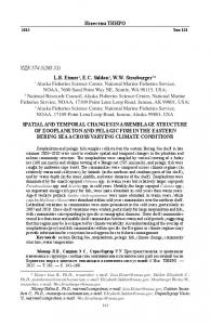
RESULTS Rapid Changes in StAR Gene Expression in MA-10 Cells after 8-Bromo-cAMP (8-Br-cAMP) Stimulation MA-10 cells stimulated with 8-Br-cAMP exhibited a pronounced increase in StAR transcription as reflected by significant increases in nascent StAR RNA levels, which were evident at 15 min, followed by the accumulation of StAR mRNA at 30 min, and StAR protein at 60 min of stimulation (Fig. 1). The changes in StAR mRNA preceded changes in mRNAs for steroidogenic enzymes including CYP11A, which increased significantly at 24 h after stimulation; 3-hydroxysteroid dehydrogenase type II, which showed no changes through 24 h of 8-Br-cAMP treatment; and CYP17, which was increased significantly after 60 min. Thus, the StAR gene is among the first steroidogenic genes to respond to tropic stimulation. Rapid Association of Transcription Factors/Coactivator with the StAR Proximal Promoter after 8-Br-cAMP Treatment of MA-10 Cells Chromatin immunoprecipitation assays were employed to assess the association of transcription factors with the StAR promoter. Association of GATA-4 with the proximal StAR promoter in MA-10 cells increased 6.5-fold within 15 min of 8-Br-cAMP treatment, remained elevated (6-fold) at 30 min, and returned to basal levels within 1 h of treatment (Fig. 2A). The distal StAR promoter (⬃3500 bp from the transcription start site represented a control because binding of transcription factors to this region was not anticipated) showed no changes in association with GATA-4 over the 4-h treatment period (Fig. 2A). Western blot analysis of GATA-4 protein revealed no differences in abundance during the 4-h 8-Br-cAMP treatment period (Fig. 2F). Treatment of MA-10 cells with 8-Br-cAMP caused a rapid (15 min) 3-fold increase in SF-1/Ad4BP association with the proximal StAR promoter, which then fell to a value intermediate to the control and 15-min levels at 30 min before progressively increasing again to its highest levels at 4 h (Fig. 2B). Distal StAR promoter association with SF-1/Ad4BP was unchanged throughout the 4-h treatment period (Fig. 2B). Levels of SF-1/ Ad4BP were not affected by 8-Br-cAMP treatment (Fig. 2F).
Hiroi et al. • Transcription Factors, Histone, and StAR
Mol Endocrinol, April 2004, 18(4):791–806
793
Fig. 1. Nascent StAR RNA, StAR mRNA, and Protein Expression in MA-10 Cells Stimulated with 8-Br-cAMP Nascent StAR RNA (A) levels and StAR mRNA (B) expression were quantified by real time RT-PCR. Results were normalized for the amount of GAPDH, and the relative amounts of both transcripts are shown as fold increase compared with the amount of transcripts in MA-10 cells before exposure to 8-Br-cAMP (0 min). C, Western blot analysis of StAR protein in MA-10 cells stimulated with 8-Br-cAMP. Whole-cell extracts from MA-10 cells at the designated time points were prepared as described in Materials and Methods. The mature 30-kDa StAR protein is identified. CYP11A (D), 3-HSD (E), and CYP17 (F) mRNAs were quantitated by RT-PCR. a,b,c,d, Means ⫾ SEM within a panel with different superscripts are different (P ⬍ 0.05).
Association of C/EBP with the proximal and distal StAR promoter in MA-10 cells is illustrated in Fig. 2C. The proximal and distal StAR promoter exhibited no significant change in association with C/EBP over the 4-h 8-Br-cAMP treatment period. C/EBP protein was slightly increased within 15 min of 8-Br-cAMP treatment and continued to rise throughout the 4-h experiment (Fig. 2F). Three putative CREs have been identified in the mouse StAR promoter between ⫺96 to ⫺64, but only one of these at ⫺80 appears to be a true CRE (14). Because both CRE binding protein (CREB) and CRE modulator (CREM) proteins have been reported to bind to the murine StAR promoter, we used an antibody that recognizes multiple members of the CREB/ CREM family for ChIP assays. Additionally, this antibody was shown to block MA-10 nuclear extracts from binding to a ⫺96 to ⫺64 StAR oligonucleotide (14). Using this antibody we found increased association of CREB/CREM proteins with the proximal promoter be-
ginning after 15 min, reaching a statistically significant increase at 30 min after 8-Br-cAMP stimulation (Fig. 2D). Western analysis with the same antibody demonstrated the presence of several immunoreactive (36- to 43-kDa) bands representing CREM and CREB proteins; no change in protein levels were observed during the time period studied (Fig. 2F). Association of CREB-binding protein (CBP) with the proximal and distal StAR promoter in MA-10 cells is illustrated in Fig. 2E. 8-Br-cAMP treatment increased CBP association with the StAR proximal promoter 2.7fold within 15 min, and this was maintained between 2.4- and 4.4-fold through the 4-h treatment period (Fig. 2E). No change in association of CBP was observed for the distal StAR promoter (Fig. 2E). Western blot analysis of CBP protein indicated that protein levels were unchanged over the 4-h 8-Br-cAMP treatment period (Fig. 2F). In addition to evaluating the patterns of transcription factor binding based on statistically significant changes
794 Mol Endocrinol, April 2004, 18(4):791–806
Hiroi et al. • Transcription Factors, Histone, and StAR
Fig. 2. Association of the Proximal and Distal StAR Promoters with GATA-4, SF-1/Ad4BP, C/EBP, CREB/CREM, and CBP in MA-10 Cells Exposed to 8-Br-cAMP Association of transcription factors/coactivator with DNA as determined by ChIP assays was quantified by real-time PCR as described in Materials and Methods, and the results are depicted as the fold increase in association of factors compared with the levels detected in the cells before exposure to 8-Br-cAMP (0 min). Panels A, B, C, D, and E show the results of ChIP analysis for GATA-4, SF-1/Ad4BP, C/EBP, CREB/CREM, and CBP, respectively. Panel F shows the results of Western Blot analysis for GATA-4 (50 kDa), SF-1 (53 kDa), C/EBP (32 kDa), CREB/CREM (36–43 kDa), and CBP (250 kDa) in MA-10 cells stimulated with 8-Br-cAMP over time as described in Materials and Methods. a,b,c, Means ⫾ SEM for the proximal StAR promoter within a panel with different superscripts are different (P ⬍ 0.05). No significant differences were observed for the distal StAR promoter. The equations define the best fit line for changes in transcription factor binding to the proximal promoter.
compared with control levels, we analyzed the overall binding patterns for each transcription factor/coactivator. This was carried out to determine whether the patterns of binding were consistent with the notion of stable, direct interactions between specific transcription factors. The CBP data had a significant quadratic profile (P ⫽
0.01, Fig. 2E), whereas a linear relationship sufficed to characterize the GATA-4, SF-1/Ad4BP, C/EBP, and CREB/CREM data sets. An additional analysis was conducted comparing the C/EBP, GATA-4, SF-1/Ad4BP, and CREB/CREM groups assuming a linear profile. In this comparison, the GATA-4 profile was significantly
Hiroi et al. • Transcription Factors, Histone, and StAR
Mol Endocrinol, April 2004, 18(4):791–806
795
different from the others (P ⫽ 0.019). The GATA-4 profile had a decreasing slope for the relationship between association and time (Fig. 2A). The association over time relationships for C/EBP and SF-1/Ad4BP were not significantly (P ⫽ 0.38) different (Fig. 2, B and C). The slope for the time relationship for CREB/CREM was decreasing, although the slope was not statistically different from zero (Fig. 2D). In addition, there was no significant increase in C/EBP binding to the proximal StAR promoter, whereas SF-1/Ad4BP binding increased. Collectively, these observations are not consistent with a model of stable direct interactions between these transcription factors, nor stable direct interactions between GATA-4 and SF-1/Ad4BP, GATA-4 and C/EBP, or CREB/CREM and SF-1/Ad4BP. StAR Gene Expression in Periovulatory Mouse Granulosa Cells after in Vivo hCG Treatment To determine whether the observations made with cultured MA-10 cells can be generalized to hormoneinduced changes in StAR gene expression in vivo, we examined StAR gene expression in granulosa cells isolated from pregnant mare’s serum gonadotropintreated mice after hCG treatment (Fig. 3). StAR nascent RNA, a measure of transcription, increased at 60 min after hCG (Fig. 3A) followed by an increase in StAR mRNA expression at 120 min (Fig. 3B). Western blot analysis revealed that mature StAR protein (30 kDa) levels were increased in cells collected at 2 h after hCG treatment and remained elevated through completion of the sampling period (Fig. 3C). The StAR preprotein levels transiently increased at 30 min concomitantly with a decline in StAR mRNA at 30 min, possibly suggesting the existence of a posttranscriptional action of gonadotropins on StAR mRNA translation. Association of Transcription Factors/Coactivator with the StAR Proximal Promoter in Mouse Granulosa Cells after in Vivo Gonadotropin Stimulation ChIP analysis revealed that in vivo administration of hCG caused a pattern of transcription factor binding that was similar in some respects to that observed in MA-10 cells, but different in others. A 2.4-fold increase in GATA-4 association with the StAR proximal promoter, but not the distal promoter, was found in granulosa cells collected 15 min after treatment. In contrast to MA-10 cells, this association was maintained (1.8to 2.7-fold) throughout the 4-h collection period after hCG administration (Fig. 4A). The amount of GATA-4 protein in mouse granulosa cells did not change after hCG treatment (Fig. 4F). Administration of hCG increased SF-1/Ad4BP association with the StAR proximal promoter in mouse granulosa cells within 15 min of hCG injection, and this was maintained through 4 h (2.0- to 2.5-fold; Fig. 4B).
Fig. 3. Nascent StAR RNA, StAR mRNA, and Protein Expression in Mouse Granulosa Cells Isolated from Mice Administered hCG Nascent StAR RNA (A) levels and StAR mRNA (B) expression were quantified by real-time RT-PCR. Results were normalized for the amount of GAPDH, and the relative amounts of nascent StAR RNA and StAR mRNA are shown as fold increase compared with the amount of transcripts measured in granulosa cells isolated from mice that did not receive hCG (0 min control). a,b,c, Means ⫾ SEM within a panel with different superscripts are different (P ⬍ 0.05). B, Western blot analysis of StAR protein in granulosa cells after in vivo administration of hCG. Whole-cell extracts (50 g) from granulosa cells at the designated time points were prepared as described in Materials and Methods. The mature 30-kDa StAR protein and 37-kDa preprotein are identified.
The distal StAR promoter showed no change in association with SF-1/Ad4BP during the entire treatment period (Fig. 4B). The levels of SF-1/Ad4BP protein in mouse granulosa cells did not change after hCG treatment (Fig. 4F). The mouse granulosa cell StAR proximal promoter exhibited a 2.8-fold increase in association with C/EBP in cells collected 1 h after hCG treatment (Fig. 4C). C/EBP remained associated (2.3- to 3.2-fold over control values) with the proximal StAR promoter in cells isolated 2 and 4 h after hCG administration, whereas the distal StAR promoter showed no change in association with C/EBP during the entire period of granulosa cell
796 Mol Endocrinol, April 2004, 18(4):791–806
Hiroi et al. • Transcription Factors, Histone, and StAR
Fig. 4. Association of the Proximal and Distal StAR Promoters with GATA-4, SF-1/Ad4BP, C/EBP, CREB/CREM, and CBP in Mouse Granulosa Cells Collected after hCG Administration Association of transcription factors/coactivator with DNA as determined by ChIP assays was quantified by real-time PCR as described in Materials and Methods, and the results are depicted as the fold increase in association of factors compared with the levels detected in the granulosa cells before in vivo exposure to hCG (0 min Control). Panels A–E show the results of in vivo ChIP analysis for GATA-4, SF-1/Ad4BP, C/EBP, CREB/CREM, and CBP, respectively. Panel F shows the results of Western blot analysis for GATA-4 (50 kDa), SF-1/Ad4BP (53 kDa), C/EBP (32 kDa), CREM/CREB (36–43 kDa), and CBP (250 kDa) in mouse granulosa cells collected from mice after hCG administration as described in Materials and Methods. a,b,c, Means ⫾ SEM for the proximal StAR promoter within a panel with different superscripts are different (P ⬍ 0.05). No significant differences were observed for the distal StAR promoter.
collection (Fig. 4C). Western blot analysis showed that C/EBP protein levels substantially increased at 30 min and remained elevated through the 4-h treatment period (Fig. 4F).
CREB/CREM binding to the StAR proximal promoter in granulosa cells was significantly increased at 2 and 4 h after hCG treatment (Fig. 4D). Western analysis using the same antibody documented the
Hiroi et al. • Transcription Factors, Histone, and StAR
presence of several immunoreactive CREB/CREM proteins in granulosa cell nuclear extracts, which did not change in abundance during the time course studied (Fig. 4F). Association of CBP with the proximal StAR promoter increased 1.5-fold (P ⬍ 0.05) within 15 min of hCG administration and then continued to increase to its maximal value (2.5-fold) at 2 h (Fig. 4E). The distal StAR promoter region did not exhibit an increase in association with CBP at any time point examined. The amount of CBP protein in mouse granulosa cells did not change after hCG treatment (Fig. 4F). To gain additional insights regarding the similarities and differences in transcription factors, we evaluated the patterns of transcription factor association with the proximal StAR promoter over time. The CBP and C/EBP data had a significant quadratic profile (P ⫽ 0.01; Fig. 4, C and E), whereas a linear relationship sufficed for the GATA-4, SF-1/Ad4BP, and CREB/ CREM data sets. There were statistically significant (P ⫽ 0.047) differences among the five profiles over time. Additional comparisons were conducted for the GATA-4, SF-1/Ad4BP, and CREB/CREM data sets assuming a linear profile. The association over time relationship for GATA-4 and SF-1/Ad4BP was not significantly different from each other (Fig. 4, A and B), which is consistent with a direct and stable interaction between these factors. The association over time relationship for CREB/CREM was increasing over time, and significantly different from the other two (P ⫽ 0.001). The differences in the binding patterns of SF1/Ad4BP and CREB/CREM proteins are not consistent with stable interactions among these proteins particularly because a significant increase in SF-1/Ad4BP binding was seen before an increase in CREB/CREM binding to the proximal promoter. Chromatin Modifications Associated with StAR Expression Induced by 8-Br-cAMP Treatment of MA-10 Cells Acetylation of histone H3 associated with the StAR proximal promoter increased 2.7-fold within 15 min of 8-Br-cAMP treatment, and this increase was maintained between 2.9- and 4.0-fold throughout the 4-h time period (Fig. 5A; see box). In contrast, the distal StAR promoter showed no changes in H3 acetylation. However, at later times (120–240 min after 8-Br-cAMP treatment) there was a broad-based increase in histone H3 acetylation in regions closer to the proximal promoter and along the StAR gene (Fig. 5A). ChIP performed with nonimmune rabbit serum showed no difference between 8-Br-cAMP-treated cells and nontreated cells (data not shown). Analysis of the MA-10 cells with an antiacetylated H4 antibody showed no differences in the amount of immunoprecipitated proximal or distal StAR promoter DNA over the 4-h 8-Br-cAMP treatment period (data not shown). Interestingly, MA-10 cells had decreased levels of phosphorylated Ser10 histone H3 associated
Mol Endocrinol, April 2004, 18(4):791–806
797
with the StAR proximal and distal promoter at 15 and 30 min of 8-Br-cAMP treatment (⬃60% of pre-8-BrcAMP values). This loss in phosphorylation of Ser10 continued to reach approximately 10% of pretreatment levels at 1 and 2 h after cAMP exposure (Fig. 6). ChIP assays on the MA-10 cells for dimethyl lysine (K) 4 histone H3, a histone modification postulated to be a mark of a transcriptionally permissive chromatin environment, revealed no significant alterations in the proximal or distal StAR promoter after 8-Br-cAMP treatment (Fig. 5B). However, dimethylation of K4 on histone H3 was increased between intron 1 through exon 7 and in the 3⬘-region of the StAR gene (Fig. 5B). The changes in this histone modification occurred well after transcription was increased (at 120–240 min), with the exception of exon 7, which showed an increase in dimethylated K4 histone H3 at 15 min (2-fold) that was maintained through 2 h (2- to 2.7-fold) before increasing again (3.6-fold) at 4 h (Fig. 5B). Trimethylation of K4 on histone H3, also thought to be a mark of active genes, increased at 240 min of 8-Br-cAMP stimulation in the StAR exonic/intronic regions; no change was observed in the 3⬘-region (Fig. 5C). Trimethylated K4 on histone H3 declined approximately 40% in the StAR proximal promoter region between 15 and 2 h, before increasing again to control levels at 4 h. MA-10 cells had reduced levels of dimethylated K9 histone H3, a modification characteristic of gene silencing, associated with the StAR proximal promoter (bp ⫺88 to ⫺153) but not putative promoter elements further upstream (i.e. bp ⫺612 to ⫺3500; Fig. 5D) 30 min after 8-Br-cAMP treatment. This histone modification was not temporally linked to the initiation of transcription (Fig. 5D). In addition, the association of dimethyl K9 histone H3 with exon 1 to intron 3 was reduced between 30 and 240 min, and with exon 4 to the 3⬘-region of the gene from 60–240 min after stimulation. Chromatin Modifications Associated with StAR Expression Induced by hCG Treatment in Mouse Granulosa Cells In contrast to MA-10 cells, we were unable to detect a significant increase in H3 acetylation in mouse granulosa cells within 4 h of hCG treatment (Fig. 7A). However, using the same antibody, we detected a statistically significant (P ⬍ 0.05, compared with 0 time) 15.4 ⫾ 8.8-fold increase in histone H3 acetylation associated with the progesterone receptor proximal promoter between 2–6 h after hCG administration. We also examined histone H4 acetylation, and as in the MA-10 cells, found no evidence of increased histone H4 acetylation in granulosa cells in either the proximal or distal StAR promoter in the 4 h after hCG treatment (data not shown). In contrast to our observations in MA-10 cells, we found a rapid decrease in dimethyl K9 histone H3 (a mark of gene silencing) associated with the StAR proximal promoter within 15 min of in vivo
798 Mol Endocrinol, April 2004, 18(4):791–806
Hiroi et al. • Transcription Factors, Histone, and StAR
Fig. 5. Histone Modifications Associated with the Promoter, Exonic/Intronic Regions, and the 3⬘-Region of the StAR Gene in MA-10 Cells Exposed to 8-Br-cAMP ChIP assays were quantified by real-time PCR with a probe specific to the mouse proximal StAR promoter or with SyBr green for all others. All data were standardized with the amount of starting (input) chromatin material as described in Materials and Methods and is expressed as the fold increase compared with the levels detected in cells collected before 8-Br-cAMP treatment (0 min). A, Acetylated histone H3; B, lysine (K) 4 dimethylated histone H3; C, trimethylated K4 histone H3; and D, dimethylated K9 histone H3 results for each primer pair as denoted by the line figure at the bottom of the figure are shown. The fine line denotes the promoter and intronic regions whereas the wide bars with white numbers denote the specific exons. The box marks the results for the proximal StAR promoter. *, Means ⫾ SEM for an individual group (i.e. A, B, C, or D) at a specific region of the StAR gene locus are different (P ⬍ 0.05).
hCG treatment (Fig. 7B). A trend for reduced association of dimethyl K9 histone H3 with the distal promoter was also observed, although it was not statistically significant. These observations suggest that removal of a gene-silencing mark broadly covering the StAR 5⬘-flanking region is an important part of the chromatin modification program involved in initiation of StAR gene transcription in luteinizing granulosa cells. This may explain why the lag time between the first detectable increase in binding of transcription factors (15-min time point) and the increase in transcription (60-min time point) is greater than that observed in MA-10 cells.
DISCUSSION Steroidogenic potential is dictated by StAR protein levels, which in turn are primarily regulated by StAR
gene transcription. To analyze the events occurring at the StAR gene promoter after cAMP/hormonal stimulation, we determined, using a quantitative ChIP assay, which transcription factors are associated with the StAR promoter and are temporally correlated with transcription and histone modifications thought to be important for transcription. This methodology allowed us to apply statistical analysis to the amount of immunoprecipitated target sequence and thus construct a map of the temporal and spatial changes in the binding of transcription factors and histone modifications at the StAR locus for both MA-10 cells and mouse granulosa cells. From these data we could 1) establish that specific factors among families of proteins capable of recognizing a common DNA-binding site are indeed associated with the StAR promoter, 2) evaluate models of protein-protein interactions based on similarities or differences in the patterns of binding, and 3)
Hiroi et al. • Transcription Factors, Histone, and StAR
Mol Endocrinol, April 2004, 18(4):791–806
799
Fig. 6. Phosphorylation of Ser10 of Histone H3 Associated with the Proximal and Distal StAR Promoter in MA-10 Cells a,b,c , Means ⫾ SEM for the proximal and distal StAR promoters with different superscripts are significantly different (P ⬍ 0.05).
assess the temporal correlation between transcription factor association, histone modifications, and transcription. Our results indicate that these two mouse steroidogenic cells utilize similar transcription factors, but in different binding patterns, to control StAR gene expression. Additionally, our analysis of some of the many possible combinations of histone modifications at the StAR locus indicated that these modifications are also specific to the cell type. In MA-10 and mouse granulosa cells, the association of GATA-4 and SF-1/ Ad4BP with the StAR proximal promoter increased concomitantly (MA-10) or before (granulosa cells) there was a significant increase in the abundance of nascent StAR RNA, a measure of transcription. Previously, it was reported that GATA-4 was a potent activator of the mouse StAR promoter (21), and mutation of a GATA element (⫺61 bp) in the StAR proximal promoter led to a decrease in basal and cAMPinduced StAR promoter activity in both MA-10 cells and granulosa cells (8, 10). Likewise, two SF-1/Ad4BP binding sites located in the proximal promoter at positions ⫺97 and ⫺42 are highly conserved across a variety of species and appear to be critical for cAMP/ hormonal responsiveness of this gene (22, 23). Like GATA-4, we found a significant increase in association of SF-1/Ad4BP with the proximal StAR promoter within 15 min of 8-Br-cAMP/hCG stimulation of MA-10 and granulosa cells, respectively. Our results show, for the first time, that GATA-4 and SF-1/Ad4BP are associated with the StAR promoter in vivo in a temporal pattern that would reflect their possible involvement in the onset of StAR gene expression. Increased binding of CREB/CREM family members to the StAR proximal promoter occurred only after increases in GATA-4 and SF-1/Ad4BP binding were detected. The lack of a change in GATA-4, CREB/CREM, and SF-1/Ad4BP protein levels in MA-10 and granulosa cell nuclear extracts throughout the 4-h 8-Br-cAMP/ hCG treatment suggests that hormonal/cAMP treatment must either cause 1) a posttranslational modification to the transcription factor, 2) recruitment by another transcription factor, and/or 3) the loss of a repressor that prevents binding of these transcription
factors to their target promoters. All of these factors undergo posttranslational modifications that appear to increase their ability to transactivate genes (11, 14, 24, 25). Indeed, Tremblay and colleagues (11) recently reported that GATA-4 phosphorylation in response to cAMP treatment is correlated with StAR gene expression. Thus, it is possible that the phosphorylation of GATA-4 facilitated the increased association of GATA-4 with the StAR proximal promoter, and that loss of phosphorylation may be linked to the decline in binding. In addition to posttranscriptional modifications, other alternatives such as ligand binding or cytoplasmic to nuclear relocation of SF-1/Ad4BP protein are possible but not as likely (23, 26). The LRH-1/NR5A2 transcription factor also binds to SF-1/Ad4BP response elements, raising questions as to whether SF-1/Ad4BP is involved in StAR gene expression, particularly because recent studies identified LRH-1 in granulosa cells (26, 27). However, our Western blot analysis of nuclear extracts revealed that LRH-1 was not detected in MA-10 cells and was barely detectable in mouse granulosa cells (Hiroi, H., L. K. Christenson, and J. F. Strauss, unpublished observations). In contrast, SF-1/Ad4BP was clearly detectable (Figs. 2 and 4). These findings are consistent with the conclusion of Falender et al. (26), who examined SF-1/ Ad4BP and LRH-1 in mouse granulosa cells. Combined with our ChIP data, which demonstrate the selective pull down of the proximal StAR promoter with a SF-1/ Ad4BP-specific antibody, we can argue that SF-1/ Ad4BP is involved in StAR gene expression, and it is likely that SF-1/Ad4BP is the predominant transcription factor that binds to SF-1/Ad4BP response elements found in the StAR gene. Although both MA-10 cells and mouse granulosa cells exhibited a rapid (15 min) increase in GATA-4 association with the StAR proximal promoter, thereafter, GATA-4-StAR promoter association diverged in these two cell types. The disparity in transcription factor-binding patterns between these two steroidogenic cells was greatest for C/EBP as evidenced by the lack of significantly increased association of C/EBP with the StAR proximal promoter of MA-10 cells vs. the significant increase observed in granulosa
800 Mol Endocrinol, April 2004, 18(4):791–806
Hiroi et al. • Transcription Factors, Histone, and StAR
Fig. 7. Histone Modifications Associated with the Proximal and Distal Promoters of the StAR gene in Mouse Granulosa Cells before and after hCG Administration ChIP assays were quantified by real-time PCR with a probe specific to the mouse proximal StAR promoter or with SyBr green for the distal promoter. All data were standardized with the amount of starting (input) chromatin material as described in Materials and Methods and is expressed as the fold increase compared with the levels detected in cells collected before hCG treatment (0 min). Panels A and B show the results of ChIP analysis for acetylated histone H3 and dimethylated K9 histone H3, respectively. a,b , Means ⫾ SEM for the proximal StAR promoter with different superscripts are significantly different (P ⬍ 0.05).
cells before the increase in nascent StAR RNA. C/EBP␣ and C/EBP are expressed in steroidogenic cells, including Leydig cells and ovarian granulosa cells (28, 29). Our results confirm that 8-Br-cAMP/hCG treatment of MA-10 cells or granulosa cells causes an increase in C/EBP protein levels that was most pronounced in the granulosa cells. Two putative C/EBPresponse elements in the StAR proximal promoter have been identified immediately adjacent to the two SF-1/Ad4BP sites, and these sites were shown to be important in promoter activation. Additionally, the distal site located at ⫺110 bp upstream of the transcription initiation site has been confirmed to bind C/EBP in ovarian extracts by electrophoretic mobility (super) shift assays (7, 9, 10). It was also reported that SF-1/ Ad4BP transactivation of the StAR promoter is dependent on the presence of functional C/EBP binding sites, which suggested that SF-1/Ad4BP and C/EBP might form a complex on this promoter (7, 10). Cellfree in vitro studies have shown the ability of C/EBP to bind to SF-1/Ad4BP (7). GATA-4 has also been
proposed to be a binding partner for C/EBP as well as SF-1/Ad4BP (30, 31). Our studies, although not ruling out this possibility (because ChIP assays measure population changes, not events at a single genetic template), suggest that if stable interactions between these factors exist in vivo, they most likely occur after SF-1/Ad4BP or GATA-4 first bind to the promoter because the association of C/EBP with the StAR proximal promoter was slower than that of SF-1/ Ad4BP and GATA-4. Statistical analysis of the patterns of transcription factor binding confirmed that GATA-4 binding displays a significantly different pattern than SF-1/Ad4BP and C/EBP in MA-10 cells, but is similar to that of SF-1/Ad4BP in mouse granulosa cells. A straightforward interpretation of these findings (with the caveat that the data reflect population changes) is that protein-protein interactions on the StAR promoter are more complex than predicted by a model of a stable complex forming before or at the time of binding to the StAR promoter. Otherwise, a greater similarity among the patterns of GATA-4, SF-
Hiroi et al. • Transcription Factors, Histone, and StAR
1/Ad4BP, and C/EBP binding would have been expected. The differences in CREB/CREM protein binding and SF-1/Ad4BP association with the mouse StAR proximal promoter also raise questions regarding stable direct interactions among these transcription factors, which has been previously suggested (15). The differences in transcription binding patterns observed in MA-10 and mouse granulosa cells also suggest that the transcription factor interactions on the StAR promoter cannot be generalized across different steroidogenic cell types. Initiation of transcription by RNA polymerase II requires cooperation of transcription factors with the basal transcriptional machinery. The coactivators p300 and CBP are thought to be key molecules involved in the communication between transcription factors and the transcriptional machinery (32). They participate in the activities of many transcription factors, including SF-1/Ad4BP (33), C/EBP (34), and GATA-4 (35). CBP may also play a role in the activation of the StAR gene by members of the CREB/CREM family (14, 20). Current models suggest that the binding of these coactivators to transcription factor activation domains positions histone acetyltransferases near specific nucleosomes in target gene promoter regions (36). We demonstrated a significant increase in the association of CBP with the StAR proximal promoter within MA-10 cells and granulosa cells that peaked simultaneously with the peak in transcription (nascent StAR RNA). The fold increase in association was greater for the MA-10 cells (4.5-fold) compared with the granulosa cells (2.5-fold), which might explain, in part, the differences in the histone H3 acetylation associated with the StAR proximal promoter in these two cell types. Core histones can be reversibly modified by acetylation, methylation, phosphorylation, ubiquitination, or ADP ribosylation, and these modifications have consequences for gene activation, gene repression, and chromosome replication (37, 38). Histone acetylation and deacetylation are thought to influence gene expression by altering accessibility of nucleosomal DNA to DNA-binding transcription activators, other chromatin modifying enzymes, or multisubunit chromatin remodeling complexes capable of displacing nucleosomes (39, 40). 8-Br-cAMP treatment increased the acetylation of histone H3 associated with the StAR proximal promoter within 15 min in MA-10 cells, and later along the structural gene. Thus, histone H3 acetylation is not restricted to the proximal promoter region, but appears to be a more global modification. It occurs coincident in time with recruitment of the histone acetyltransferase, CBP. Surprisingly, we failed to detect an increase in acetylation of histone H3 associated with the StAR proximal promoter in mouse granulosa cells up to 4 h after hCG treatment using the same antibodies and protocols that documented changes in histone H3 acetylation in MA-10 cells. These findings suggest that increased histone H3 acetylation is not an obligatory event for initiation of
Mol Endocrinol, April 2004, 18(4):791–806
801
StAR gene transcription in granulosa cells. This histone modification could occur at later time points, which would be consistent with our previous observation of increased histone H3 acetylation associated with the monkey and human StAR proximal promoters 24–36 h after hCG treatment in vivo (19). Unlike histone acetylation and deacetylation, which are generally associated with gene activity and gene repression/silencing, respectively, histone methylation occurs in association with transcription and silencing in a histone site-specific manner (i.e. lysine (K) 4 vs. K9 of histone H3), a regional manner within the gene (i.e. promoter vs. coding sequence), and with different levels of methylation (i.e. mono-, di-, and trimethylation) (41). This is particularly true for dimethylated K4 histone H3, which has been shown to be present in both active and inactive euchromatic genes (42, 43). The StAR promoter exhibited no increase in levels of dimethylated-K4 on histone H3 in the promoter region in MA-10 cells, but we did observe a significant increase in dimethyl-K4 histone H3, with the greatest increment observed farthest from the promoter. These site-specific changes in the pattern of methylation of K4 histone H3 across different nucleosomes within the same gene suggest that this chromatin mark is more closely associated with a permissive state or the elongation phase of transcription rather than the initiation phase, consistent with recent observations in yeast (42, 44). Trimethylation of histone H3 K4, also postulated to be a mark of active chromatin, follows a similar pattern with respect to the exonic/intronic regions of the StAR gene, and likewise, this modification was observed only at 4 h, a time point after initiation of transcription (42). The proximal StAR promoter exhibited an approximately 40% decline in detectable trimethylated K4 on histone H3. This is the first description of changes in this histone mark in a promoter region, and its significance remains to be explored. Dimethylation of K9 of histone H3 declined in the promoter and later in the coding regions of StAR gene in MA-10 and granulosa cells. However, the rapidity and degree of decline were much greater for the granulosa cells than the MA-10 cells. Because this mark is thought to cause the recruitment and binding of repressor proteins, such as HP1 (heterochromatin protein 1) that establish highly compacted and transcriptionally inactive regions of chromatin known as heterochromatin, it was not unexpected that it would decline in the StAR proximal promoter as gene activity increased (37). Methylation of K9 on H3 can inhibit acetylation of the H3 tail by p300/CBP and methylation of histone H3 K4 by histone methyltransferases (45). Likewise, histone H3 K4 methylation inhibits histone H3 K9 methylation and promotes acetylation of H3 by p300. Thus, loss of histone H3 K9 methylation might be permissive to StAR gene activation by influencing histone acetylation of the StAR gene, at least in the case of MA-10 cells. This would not appear to be the case for granulosa cells, as we failed to detect a change in histone H3 acetylation associated with the
802 Mol Endocrinol, April 2004, 18(4):791–806
StAR promoter. Although our findings on histone H3 K4 and K9 lysine methylation reveal reciprocal changes in chromatin marks associated with transcription and silencing, they take place after there is a detectable rise in nascent StAR RNA in MA-10 cells and, therefore, are likely to facilitate rather than initiate transcription. In contrast, in granulosa cells the early decline in methylation of K9 on histone H3 may be the initiator of StAR gene expression. This change may extend to more distal regions of the StAR promoter. A question raised by the differences in histone modifications observed in MA-10 cells and granulosa cells is whether they result from the differences in patterns of transcription factor binding, cell-specific factors, the nature of the stimulus (8-Br-cAMP vs. hCG), in vitro vs. in vivo conditions, transformed vs. normal cells, or a combination of these factors. Nonetheless, our findings demonstrate that increased StAR gene transcription can occur in the context of different patterns of transcription factor binding and histone modification. Histone phosphorylation, particularly that of histones H1 and H3, has been implicated in chromosome condensation during mitosis (37). The H3 phosphorylation at Ser10 we examined has also been implicated in transcriptional activation and in mitotic condensation (46, 47). We demonstrated that 8-Br-cAMP treatment of MA-10 cells reduced the phosphorylation of Ser10 in histone H3 associated with the StAR proximal and distal promoter at 15 and 30 min. The global decline in Ser10 phosphorylation continued through 2 h of 8-Br-cAMP treatment. Because 8-Br-cAMP treatment of MA-10 cells induces cellular differentiation and reduced proliferation, our results are considered to be consistent with the notions discussed above regarding this particular histone mark and its cellular function. Recent studies in mammalian cells indicate that H3-Ser10 phosphorylation can inhibit methylation of K9 on histone H3 but also precedes and promotes acetylation of K14 on histone H3 (47, 48). Our results are contrary to these observations and indicate that H3-Ser10 in the context of the StAR promoter may be more closely associated with the proliferative responses of these cells. Simultaneously, the acetylation of histone H3 was increased and phosphorylation of Ser10 on histone H3 was reduced in MA-10 cells; both of these chromatin modification events have been linked to gene activation. Thus, although acetylation of histone H3 increases during gene induction, it does not appear to be linked to histone H3 phosphorylation. Thus, it appears that the histone code is distinct for StAR relative to other induced genes, such as interferon- (49, 50). Although ChIP technology is a valuable tool for analyzing the interaction of endogenous proteins with native chromatin in vivo, which cannot be achieved with more classical methods of promoter analysis, it too has limitations. ChIP data represent the average status of chromatin in multiple cells. Thus, transcription factor binding or histone modifications may be occurring in some, but not all, cells. The study of a
Hiroi et al. • Transcription Factors, Histone, and StAR
homogeneous cell line (MA-10 cells), cultured to a uniform density, may limit heterogeneity in cellular response. Recent reports suggest that transcription factors rapidly cycle on and off chromatin. Very rapid changes, particularly if they did not occur in a synchronous pattern (51), could not easily be detected by ChIP technology. ChIP assays are also limited by the ability of antibodies to recognize their cognate epitopes and cause immunoprecipitation. Finally, ChIP assays cannot prove that individual transcription factors or histone modifications occur on the exact same chromatin template. This is an inference that must be drawn from the current data. In summary, our findings, using a quantitative ChIP assay and recognizing its strengths and limitations, indicate that after tropic stimulation transcription factors and coactivators rapidly and sequentially associate with the StAR proximal promoter, that the patterns of association depend on cell context, and support some, but not all, proposed models of stable direct interactions among transcription factors on the StAR promoter. Finally, there are diverse histone modifications, which also occur in cell-specific patterns along the StAR gene. Some of these modifications are temporally linked with initiation of transcription; others are demonstrable later, suggesting that they play a facilitating role.
MATERIALS AND METHODS Cell Collection and Culture The mouse Leydig cell tumor cell line MA-10 (a generous gift of Dr. Mario Ascoli) was studied because of its consistent steroidogenic response to 8-Br-cAMP stimulation and wellcharacterized changes in StAR gene expression. MA-10 cells were cultured in Waymouth medium containing 15% horse serum and 50 g/ml gentamicin as previously described (52). Two days before experiments were initiated 1 ⫻ 106 MA-10 cells were plated onto 10-cm dishes. On the day of an experiment, the cell culture medium was replaced with serumfree Waymouth medium containing 50 g/ml gentamicin. Randomly chosen plates were treated with 1 mM 8-Br-cAMP (Sigma Chemical Co., St. Louis, MO) for 15, 30, 60, 120, or 240 min. Mouse granulosa cells were collected from periovulatory follicles from 3-wk-old (21–24 d) female mice (Mus musculus; CF1 strain, Harlan Sprague Dawley, Inc., Indianapolis, IN) given a follicular stimulation protocol. The mice used in these experiments were maintained in a University Laboratory Animal Resource facility, and all animal protocols were approved by the University of Pennsylvania’s Animal Care and Use Committee in accordance with the NIH Guide for the Care and Use of Laboratory Animals. All mice were injected ip with 5 IU of pregnant mare serum gonadotropin (Calbiochem, San Diego, CA) to stimulate follicular growth and promote the development of multiple preovulatory follicles. After 44–48 h, all mice except the control group (0 h, no hCG) were injected with 5 IU of hCG (Sigma), to trigger ovulation and luteinization (standard ovulatory stimulus). Timed injections of three animals each at 15 min, 30 min, 1 h, 2 h, and 4 h with hCG were performed in each experiment; several mice were also killed at 6 and 8 h post-hCG. The mice were killed by cervical dislocation, and the ovaries were carefully cleaned
Hiroi et al. • Transcription Factors, Histone, and StAR
and placed into 4 C Dulbecco’s PBS (Life Technologies, Inc., Gaithersburg, MD) containing 0.1% BSA (Sigma). To release the granulosa cells from the follicles, the ovaries were repeatedly punctured with a 30-gauge needle. The expressed granulosa cells and minor contaminants (i.e. oocytes, red blood cells, and stromal cells) were collected and centrifuged at 770 ⫻ g to pellet the cells in a 1.5-ml Eppendorf tube. The isolation process took approximately 20–25 min/treatment group, which means that the nominal time of collection described in Results represents the minimum time at which the events were initiated by hCG. The pooled granulosa cells from the three mice at each time point were processed for RNA, protein, or ChIP assays as described below. The experimental protocol was replicated a minimum of three times each for each type of analysis (i.e. quantitative RT-PCR, ChIP, Western blots). ChIP We used a modification of the technique described by Kuo and Allis (53) as previously described by our laboratory (19). Briefly, formaldehyde (Fisher Scientific, Pittsburgh, PA) was added directly to the MA-10 cell culture medium or to mouse granulosa cells suspended in PBS containing 0.1% BSA (final concentration: 0.5% for anti-C/EBP antibody and anti-CBP antibody and 1% for all other antibodies) for 10 min at 37 C to cross-link DNA and its associated proteins. MA-10 cells were washed twice in PBS before scraping cells in PBS containing protease inhibitors (1 g/ml aprotinin, 1 g/ml leupeptin, 1 mM phenylmethylsulfonyl fluoride), and 20 mM sodium fluoride and 1 mM sodium orthovanadate. Mouse granulosa cells were pelleted (770 ⫻ g) and resuspended twice in PBS containing 0.1% BSA and inhibitors. Both MA-10 and granulosa cells were then resuspended in lysis buffer [1% sodium dodecyl sulfate (SDS); 10 mM EDTA; and 50 mM Tris-HCl, pH 8.1] containing inhibitors. Sonication of cell lysates was performed three times for 10 sec with a Ultrasonic Dismembrator 500 (Fisher Scientific) equipped with a 2-in. diameter cup horn containing four samples in 1.5-ml Eppendorf tubes. The cup horn was cooled with recirculating ice-cold water, and 10 sec elapsed between each pulse. After sonication, the samples were centrifuged (12,000 ⫻ g) for 15 min at 4 C before supernatant was transferred to a new tube. After thorough mixing with a pipette, 5 l of the supernatant were transferred to a tube and saved as input DNA for normalization of chromatin input, and the remainder was diluted 1:10 in ChIP dilution buffer (0.01% SDS, 1.1% Triton X-100, 1.2 mM EDTA, and 16.7 mM TrisHCl) containing the additives as described above. Samples were then immediately used for ChIP or stored at ⫺80 C for later analysis. The chromatin solution was cleared with a salmon sperm DNA/protein A-agarose 50% slurry (Upstate Biotechnology, Inc., Lake Placid, NY) for 30 min before overnight incubation (4 C) with 5 g of specific antibodies to acetylated histone H3 (Upstate), acetylated histone H4 (Upstate), dimethyl-Lys4 histone H3 (Upstate), dimethyl-Lys9 histone H3 (Upstate), phospho-specific-Ser10 histone H3, C/EBP (sc-150x, Santa Cruz Biotechnology, Inc., Santa Cruz, CA), GATA4 (sc-1237x, Santa Cruz Biotechnology), CREB/CREM (CREM-1 sc-440x, Santa Cruz Biotechnology), CBP (Upstate), and SF-1/Ad4BP (generous gift from Dr. Ken-ichirou Morohashi). The amount of antibody added was determined from preliminary doseresponse studies which established the quantity at which immunoprecipitation of proximal promoter target from 8-BrcAMP-stimulated MA-10 cells was maximal. Salmon sperm DNA/protein A-agarose was added and incubated for 1 h. The chromatin-antibody-protein A-agarose complexes were washed sequentially once each (2 min on a rocker plate), in low-salt, high-salt, lithium chloride, and Tris/EDTA buffer, followed by two incubations with freshly made elution buffer (1% SDS and 50 mM NaHCO3). The eluates were pooled and NaCl was added to a final concentration of 10 mM before heating the
Mol Endocrinol, April 2004, 18(4):791–806
803
mixture at 65 C for 4 h to reverse the formaldehyde cross-links. The samples were digested with proteinase K for 1 h at 45 C, and the DNA from the samples was obtained by phenol/chloroform extraction and ethanol precipitation. DNA pellets were then resuspended in 20 l of sterile water, and 1-l aliquots were used in the PCR reactions. Quantitative Real-Time PCR Primers and probes for the ChIP analysis of the mouse StAR gene promoter are shown in Table 1. All primers including the proximal (bases ⫺88 to ⫺153) promoter and a distal region of the promoter (bases ⫺3492 to ⫺3568), which we designate here as the “distal promoter,” were designed with the Primer Express software package that accompanies the Applied Biosystems model 7900 sequence detector (PerkinElmer Life Science, Norwalk, CT) as previously described (19). Briefly, the proximal primers (900 nM) and the FAM-labeled probe (200 nM) and the TaqMan Universal Master Mix (Applied Biosystems, Foster City, CA) were used to quantitate levels of the proximal promoter-immunoprecipitated chromatin. To detect other promoter regions and the exonic/intronic and the 3⬘-untranslated regions of the StAR gene, the SyBr green reagent PCR Master Mix (Applied Biosystems) was used. The average threshold cycle (Ct) for the triplicate was used in all subsequent calculations. Quantitative Real-Time RT-PCR Total RNA (5 g) isolated with Trizol reagent (Life Technologies, Inc.) was treated with DNase I (Promega Corp., Madison, WI) followed by cDNA synthesis using the Maloney murine leukemia virus reverse transcriptase (Promega) and oligo dT primer (Promega). The resulting cDNAs were diluted 1:100 in sterile water, and 1-l aliquots were used in the quantitative real-time PCRs. The primers used to quantitate nascent StAR RNA, StAR mRNA, CYP11A mRNA, 3hydroxysteroid dehydrogenase mRNA, and CYP17 mRNA are shown in Table 1. To account for differences in starting material, the rodent glyceraldehyde-3-phosphate dehydrogenase (GAPDH) primers and probe reagents from Applied Biosystems were used as described by the manufacturer. To quantify differences, the samples were compared with standard curves for each target amplicon, and the average value for the triplicate was used in all subsequent calculations. Western Blot Analysis Cells were washed twice in PBS before scraping cells in PBS containing protease inhibitors. To prepare the whole-cell extracts, cells were resuspended in lysis buffer (PBS, 1% Nonidet P-40, 1 mM EDTA) containing protease inhibitors. Nuclear extracts were prepared as described below. Cells were resuspended in the buffer (10 mM KCl; 0.1 mM EDTA; 0.1 mM EGTA; 1 mM dithiothreitol; 10 mM HEPES, pH 7.9) containing protease inhibitors for 15 min on ice after added Nonidet P-40 (final concentration at 0.625%). After centrifugation the supernatants were removed and the pellets (nuclear fraction) were resuspended with the buffer (400 mM NaCl; 1 mM EDTA; 1 mM EGTA; 1 mM dithiothreitol; 20 mM HEPES, pH 7.9) containing protease inhibitors and incubated for 15 min on ice. The samples were then centrifuged, the nuclear protein extracts were collected, and the protein concentrations were determined with Bio-Rad Protein Assay reagents (Bio-Rad Laboratories, Inc., Hercules, CA). Whole-cell extracts and nuclear extracts (50 g of each) were used for Western blot analysis. Proteins were loaded on to 10% SDS-PAGE gels and then transferred to Immobilon polyvinylidene difluoride membranes (Millipore Corp., Bedford, MA). Western blot analysis was carried out using the same antibodies described for the ChIP analysis at the fol-
5⬘-CAAGATTTTGTTCCTGCTTTTGTG-3⬘
5⬘-AGTGAATCGCTCCAAGTTCCA-3⬘
5⬘-TGTTAGCCTTGTGTGGGATGAG-3⬘
5⬘-CCCGATGCTGTTAGCTGAAGA-3⬘
5⬘-TTAAGGACTGTCCACCACATTGAC-3⬘
5⬘-TGGAGGCCACTATCCGAGAA-3⬘
5⬘-TTCTGTCCGTTGCCTATCCAA-3⬘
5⬘-ACCCACTAAGCTGCCTTGTCA-3⬘
5⬘-CCAACCCAGACTCACCTTTCA-3⬘ 5⬘-GCCAGTGGATGAAGCACCAT-3⬘ 5⬘-GCATGGTCCTTCCAGGTCTTAG-3⬘ 5⬘-TGGCACACTGGCTTGGATAC-3⬘
5⬘-TCTGTCCATGGGCTGGTCTAC-3⬘
5⬘-GGGACGAAGTGCTAAGTAAGATGGT-3⬘
5⬘-CATGTTCCTCGCTACGTTCAAG-3⬘ 5⬘-CCGGAGCAGAGTGGTGTCA-3⬘ 5⬘-CCAGTGTCCCCATGCTCAAC-3⬘ 5⬘-CCAGGCAGACCATCCTAGATG-3⬘
5⬘-GGAGGAAAGGTTATCTGAGAACACA-3⬘
5⬘-GCCAGGCCCGCATAGTAAT-3⬘
Nascent StAR RNA StAR mRNA CYP11A mRNA 3-Hydroxysteroid dehydrogenase mRNA CYP17 mRNA
5⬘-TCTCTTTTCCATCATGGAAAATAGG-3⬘
5⬘-GCGGCCCAGGGAAACTT-3⬘
Reverse Primer Sequence
5⬘-CCAACCCAGACTCACCTTTCA-3⬘
5⬘-CATGTTCCTCGCTACGTTCAAG-3⬘
Forward Primer Sequence
5⬘-TGGTTCAGGAAACTCCCTCACT-3⬘ 5⬘-GGGCTAGGGCAATATAGGACAA-3⬘ 5⬘-CAGTCTGCTCCCTCCCACC-3⬘
5⬘-GGAGGAGCAGGCTTCTGTCTT-3⬘ 5⬘-GCTCAGGAGGCCCCACATA-3⬘ 5⬘-AGAGGGTCAAGGATGGAATGATT-3⬘
Reverse Primer Sequence
5⬘-CCCCGTGTGTTTCTGAGATGT-3⬘
Forward Primer Sequence
5⬘-TAGCTGCAGGCCACAGGTT-3⬘
oligo dT (15 mer)
5⬘-TGGGACCCTTGGTACAGACTTAA-3⬘ oligo dT (15 mer) oligo dT (15 mer) oligo dT (15 mer)
Reverse Transcription Primer
6-FAM-CCTCATCCTGCAGTGCTGGCCA-TAMRA
Probe (for Taqman PCR)
Mol Endocrinol, April 2004, 18(4):791–806
RNA Transcript
Distal promoter (⫺3568 to ⫺3492, Ref. 17) ⫺1985 to ⫺1912 ⫺686 to ⫺612 Proximal promoter (⫺88 to ⫺153, Ref. 17) Exon 1/intron 1 (⫹67 to ⫹146) Intron 1 (⫹689 to ⫹766) Intron 3 (⫹1696 to ⫹1759) Exon 4 (⫹2461 to ⫹2533) Intron 6 (⫹3642 to ⫹3718) Exon 7 (⫹4382 to ⫹4472) 3⬘ region (⫹5477 to ⫹5560)
Location within StAR Gene Locus (bp)
Table 1. Primers for Quantitative StAR Promoter ChlP Analysis and for Quantitative RT-PCR
804 Hiroi et al. • Transcription Factors, Histone, and StAR
Hiroi et al. • Transcription Factors, Histone, and StAR
lowing dilutions: C/EBP (1:2000), GATA4 (1:2000), SF-1/ Ad4BP (1:2000), CREB/CREM (1:1000), and CBP (1 g/ml). For detection of antibody protein complexes the SuperSignal West Femto Kit (Pierce Chemical Co., Rockford, IL) was used according to the manufacturer’s instruction.
Mol Endocrinol, April 2004, 18(4):791–806
805
Reproduction and Women’s Health, 1354 Biomedical Research Building, 421 Curie Boulevard, Philadelphia, Pennsylvania 19104. E-mail:
[email protected]. This work was supported by NIH Grant HD06274.
Data Analysis and Statistical Analysis
REFERENCES To determine relative differences among the treatment groups for the ChIP assays we used the ⌬⌬Ct method as outlined in the Applied Biosystems protocol for reverse transcriptase-PCR. The input DNA Ct value (e.g. StAR promoter) was used to normalize the ChIP data in place of typical GAPDH values used to normalize RT-PCRs. Because both the normalizer (i.e. input) and target DNA are being tested with the same PCR primers/probe (i.e. proximal or distal StAR promoter), the concerns over PCR amplification efficiency differences between two different amplicons is not relevant. The relative amounts of experimental cDNA (i.e. StAR cDNA) and GAPDH cDNA were determined by comparison to a standard curve generated by serial dilution of a sample containing high levels of the target amplicons that were also run in triplicate. An arbitrary value of template was assigned to the highest standard and corresponding values to the subsequent dilutions (1:5, 1:10, 1:100, 1:1000, 1:5000), and these relative values were plotted against the threshold value for each dilution to generate a standard curve. The relative amount for each experimental triplicate (i.e. StAR) and GAPDH triplicate was assigned an arbitrary value based on the slope and y-intercept of the standard curve. The average of the experimental triplicate was divided by the average of the GAPDH triplicate, and the resulting normalized values were used for statistical analysis. ANOVA was used to compare the normalized levels of the StAR promoter values or cDNA as well as natural log-transformed values within MA-10 and granulosa cells over time. Statistical tests were performed with the JMP 3.1.5 computer program and SAS version 8.1 (SAS Institute, Inc., Cary, NC). Results were considered significant if P values were less than 0.05. Given statistical significance for differences among the mean values across time, comparisons among times were evaluated using Duncan’s multiple range tests. To describe the shape of the transciption factor/StAR promoter association values over time, three stages of statistical modeling were employed. First, estimated profiles for the transcription factor/StAR promoter association values over time in minutes were estimated for each of the four treatment groups separately using linear regression methods. Statistical tests for the shape of each profile were conducted by evaluating the coefficient estimates for linear and quadratic terms for time (time and time squared). Groups with a significant quadratic profile were described and compared together, whereas a linear model in time was appropriate for other groups. Therefore, a quadratic term for time (time squared) was included in stage 2. To evaluate the similarities/differences in profile among the groups, a joint linear model was estimated that included statistical interaction terms of time and time squared by group. A joint test to evaluate the statistical significance of interactions terms was conducted using appropriate contrast statements. In the final stage of modeling only the groups with a linear trend were compared for similarities and differences among the linear trend as in stage 2.
Acknowledgments We thank Dr. Ken-ichiro Morohashi for the gift of anti SF-1/Ad4BP antibody.
Received August 4, 2003. Accepted January 6, 2004. Address all correspondence and requests for reprints to: Jerome F. Strauss, III, M.D., Ph.D., Center for Research on
1. Christenson LK, Strauss III JF 2000 Steroidogenic acute regulatory protein (StAR) and the intramitochondrial translocation of cholesterol. Biochim Biophys Acta 1529:175–187 2. Stocco DM 2001 Tracking the role of a star in the sky of the new millennium. Mol Endocrinol 15:1245–1254 3. Manna PR, Huhtaniemi IT, Wang XJ, Eubank DW, Stocco DM 2002 Mechanisms of epidermal growth factor signaling: regulation of steroid biosynthesis and the steroidogenic acute regulatory protein in mouse Leydig tumor cells. Biol Reprod 67: 1393–1404 4. Sugawara T, Kiriakidou M, McAllister JM, Kallen CB, Strauss III JF 1997 Multiple steroidogenic factor 1 binding elements in the human steroidogenic acute regulatory protein gene 5⬘-flanking region are required for maximal promoter activity and cyclic AMP responsiveness. Biochemistry 36:7249–7255 5. Sugawara T, Saito M, Fujimoto S 2000 Sp1 and SF-1 interact and cooperate in the regulation of human steroidogenic acute regulatory protein gene expression. Endocrinology 141: 2895–2903 6. Rust W, Stedronsky K, Tillmann G, Morley S, Walther N, Ivell R 1998 The role of SF-1/Ad4BP in the control of the bovine gene for the steroidogenic acute regulatory (StAR) protein. J Mol Endocrinol 21:189–200 7. Reinhart AJ, Williams SC, Clark BJ, Stocco, DM 1999 SF-1 (steroidogenic factor-1) and C/EBP  (CCAAT/enhancer binding protein-) cooperate to regulate the murine StAR (steroidogenic acute regulatory) promoter. Mol Endocrinol 13: 729–741 8. Wooton-Kee CR, Clark BJ 2000 Steroidogenic factor-1 influences protein-deoxyribonucleic acid interactions within the cyclic adenosine 3,5-monophosphate-responsive regions of the murine steroidogenic acute regulatory protein gene. Endocrinology 141:1345–1355 9. Christenson LK, Johnson PF, McAllister JM, Strauss III JF 1999 CCAAT/enhancer-binding proteins regulate expression of the human steroidogenic acute regulatory protein (StAR) gene. J Biol Chem 274:26591–26598 10. Silverman E, Eimerl S, Orly J 1999 CCAAT enhancer-binding protein  and GATA-4 binding regions within the promoter of the steroidogenic acute regulatory protein (StAR) gene are required for transcription in rat ovarian cells. J Biol Chem 274:17987–17996 11. Tremblay JJ, Hamel F, Viger RS 2002 Protein kinase A-dependent cooperation between GATA and CCAAT/enhancer-binding protein transcription factors regulates steroidogenic acute regulatory protein promoter activity. Endocrinology 143: 3935–3945 12. Christenson LK, Osborne TF, McAllister JM, Strauss III JF 2001 Conditional response of the human steroidogenic acute regulatory protein gene promoter to sterol regulatory element binding protein-1a. Endocrinology 142:28–36 13. Sugawara T, Abe S, Sakuragi N, Fujimoto Y, Nomura E, Fujieda K, Saito M, Fujimoto S 2001 RIP 140 modulates transcription of the steroidogenic acute regulatory protein gene through interactions with both SF-1 and DAX-1. Endocrinology 142:3570–3577 14. Manna PR, Dyson MT, Eubnank DW, Clark BJ, Lalli E, SasoneCorsi P, Zeleznik AJ, Stocco DM 2002 Regulation of steroidogenesis and steroidogenic acute regulatory protein by a member of the cAMP response-element binding protein family. Mol Endocrinol 16:184–199 15. Manna PR, Eubank DW, Lalli E, Sassone-Corsi P, Stocco DM 2003 Transcriptional regulation of the mouse steroidogenic
806 Mol Endocrinol, April 2004, 18(4):791–806
16. 17.
18.
19.
20.
21.
22.
23.
24.
25.
26.
27.
28.
29.
30.
31.
acute regulatory protein gene by the cAMP response-element binding protein and steroidogenic factor 1. J Mol Endocrinol 30:381–397 Grunstein M 1997 Histone acetylation in chromatin structure and transcription. Nature 389:349–352 Kadonaga JT 1998 Eukaryotic transcription: an interlaced network of transcription factors and chromatin-modifying machines. Cell 92:307–313 Brownell JE, Zhou J, Ranalli T, Kobayashi R, Edmondson DG, Roth SY, Allis CD 1996 Tetrahymena histone acetyltransferase A: a homolog to yeast Gcn5p linking histone acetylation to gene activation. Cell 84:843–851 Christenson LK, Stouffer RL, Strauss III JF 2001 Quantitative analysis of the hormone-induced hyperacetylation of histone H3 associated with the steroidogenic acute regulatory protein gene promoter. J Biol Chem 276:27392–27399 Stocco DM, Clark BJ, Reinhart AJ, Williams SC, Dyson M, Dassi B, Walsh LP, Manna PR, Wang XJ, Zeleznik AJ, Orly J 2001 Elements involved in regulation of the StAR gene. Mol Cell Endocrinol 177:55–59 Tremblay JJ, Viger RS 2001 GATA factors differentially activate multiple gonadal promoters through conserved GATA regulatory elements. Endocrinology 142:977–986 Sugawara T, Holt JA, Kiriakidou M, Strauss III JF 1996 Steroidogenic factor 1-dependent promoter activity of the human steroidogenic acute regulatory protein (StAR) gene. Biochemistry 35:9052–9059 Christenson LK, McAllister JM, Martin KO, Javitt NB, Osborne TF, Strauss III JF 1998 Oxysterol regulation of steroidogenic acute regulatory protein gene expression. Structural specificity and transcriptional and posttranscriptional actions. J Biol Chem 273:30729–30735 Hammer GD, Krylova I, Zhang Y, Darimont BD, Simpson K, Weigel NL, Ingraham HA 1999 Phosphorylation of the nuclear receptor SF-1 modulates cofactor recruitment: integration of hormone signaling in reproduction and stress. Mol Cell 3:521–526 Desclozeaux M, Krylova IN, Horn F, Fletterick RJ, Ingraham HA 2002 Phosphorylation and intramolecular stabilization of the ligand binding domain in the nuclear receptor steroidogenic factor 1. Mol Cell Biol 22:7193–7203 Falender AE, Lanz R, Malenfant D, Belanger L, Richards JS 2003 Differential expression of steroidogenic factor-1 and FTF/ LRH-1 in the rodent ovary. Endocrinology 144:3598–610 Boerboom D, Pilon N, Behdjani R, Silversides DW, Sirois J 2000 Expression and regulation of transcripts encoding two members of the NR5A nuclear receptor subfamily of orphan nuclear receptors, steroidogenic factor-1 and NR5A2, in equine ovarian cells during the ovulatory process. Endocrinology 141:4647–4656 Nalbant D, Williams SC, Stocco DM, Khan SA 1998 Luteinizing hormone-dependent gene regulation in Leydig cells may be mediated by CCAAT/enhancer-binding protein-. Endocrinology 139:272–279 Sirois J, Richards JS 1993 Transcriptional regulation of the rat prostaglandin endoperoxide synthase 2 gene in granulosa cells. Evidence for the role of a cis-acting C/EBP  promoter element. J Biol Chem 268:21931–21938 Tremblay JJ, Viger RS 1999 Transcription factor GATA-4 enhances Mullerian inhibiting substance gene transcription through a direct interaction with the nuclear receptor SF-1. Mol Endocrinol 13:1388–401 Carlone DL, Richards JS 1997 Functional interactions, phosphorylation, and levels of 3⬘,5⬘-cyclic adenosine monophosphate-regulatory element binding protein and steroidogenic
Hiroi et al. • Transcription Factors, Histone, and StAR
32. 33. 34. 35. 36. 37. 38. 39.
40. 41. 42.
43.
44.
45.
46. 47.
48. 49.
50. 51. 52. 53.
factor-1 mediate hormone-regulated and constitutive expression of aromatase in gonadal cells. Mol Endocrinol 11:292–304 Vo N, Goodman RH 2001 CREB-binding protein and p300 in transcriptional regulation. J Biol Chem 276:13505–13508 Monte D, DeWitte F, Hum DW 1998 Regulation of the human P450scc gene by steroidogenic factor 1 is mediated by CBP/ p300. J Biol Chem 273:4585–4591 Mink S, Haenig B, Klempnauer KH 1997 Interaction and functional collaboration of p300 and C/EBP. Mol Cell Biol 17: 6609–6617 Dai YS, Markham BE 2001 p300 Functions as a coactivator of transcription factor GATA-4. J Biol Chem 276:37178–37185 Sterner DE, Berger SL 2000 Acetylation of histones and transcription-related factors. Microbiol Mol Biol Rev 64:435–459 Strahl BD, Allis CD 2000 The language of covalent histone modifications. Nature 403:41–45 Ng HH, Bird A 2000 Histone deacetylases: silencers for hire. Trends Biochem Sci 25:121–126 Guschin D, Wade PA, Kikyo N, Wolffe AP 2000 ATP-Dependent histone octamer mobilization and histone deacetylation mediated by the Mi-2 chromatin remodeling complex. Biochemistry 39:5238–5245 Fuks F, Burgers WA, Brehm A, Hughes-Davies L, Kouzarides T 2000 DNA methyltransferase Dnmt1 associates with histone deacetylase activity. Nat Genet 24:88–91 Turner BM 2003 Memorable transcription. Nat Cell Biol 5:390–393 Santos-Rosa H, Schneider R, Bannister AJ, Sherriff J, Bernstein BE, Emre NC, Schreiber SL, Mellor J, Kouzarides T 2002 Active genes are tri-methylated at K4 of histone H3. Nature 419:407–411 Bernstein BE, Humphrey EL, Erlich RL, Schneider R, Bouman P, Liu JS, Kouzarides T, Schreiber SL 2002 Methylation of histone H3 Lys 4 in coding regions of active genes. Proc Natl Acad Sci USA 99:8695–8700 Ng HH, Robert F, Young RA, Struhl K 2003 Targeted recruitment of Set1 histone methylase by elongating Pol II provides a localized mark and memory of recent transcriptional activity. Mol Cell 11:709–719 Wang H, Cao R, Xia L, Erdjument-Bromage H, Borchers C, Tempst P, Zhang Y 2001 Purification and functional characterization of a histone H3-lysine 4-specific methyltransferase. Mol Cell 8:1207–1217 Cheung P, Allis CD, Sassone-Corsi P 2000 Signaling to chromatin through histone modifications. Cell 103:263–271 Lo WS, Trievel RC, Rojas JR, Duggan L, Hsu JY, Allis CD, Marmorstein R, Berger SL 2000 Phosphorylation of serine 10 in histone H3 is functionally linked in vitro and in vivo to Gcn5mediated acetylation at lysine 14. Mol Cell 5:917–926 Edmondson DG, Davie JK, Zhou J, Mirnikjoo B, Tatchell K, Dent SY 2002 Site-specific loss of acetylation upon phosphorylation of histone H3. J Biol Chem 277:29496–29502 Lo WS, Druggan L, Tolga NC Emre, Belotserkovskya R, Lane WS, Shiekhattar R, Berger SL 2001 Snf1—a histone kinase that works in concert with the histone acetyltransferase Gcn5 to regulate transcription. Science 293:1142–1146 Turner BM 2002 Cellular memory and the histone code. Cell 111:285–291 Cosma MP 2002 Ordered recruitment: gene-specific mechanism of transcription activation. Mol Cell 10:227–236 Ascoli M 1981 Characterization of several clonal lines of cultured Leydig tumor cells: gonadotropin receptors and steroidogenic responses. Endocrinoloogy 108:88–95 Kuo MH, Allis CD 1999 In vivo cross-linking and immunoprecipitation for studying dynamic protein:DNA associations in a chromatin environment. Methods 19:425–433
Molecular Endocrinology is published monthly by The Endocrine Society (http://www.endo-society.org), the foremost professional society serving the endocrine community.