script, and members of the Rockefeller University Protein Sequenc- ing Facility, especially Joseph Fernandez, for protein sequencing. D.K. was supported by a ...
Proc. Natl. Acad. Sci. USA Vol. 91, pp. 1519-1523, February 1994
Cell Biology
The human CAN protein, a putative oncogene product associated with myeloid leukemogenesis, is a nuclear pore complex protein that faces the cytoplasm (wheat germ agglutinin-binding protein/monospecific antibodies/immunofluorescence/immunoelectron microscopy)
DoRis KRAEMER, RICHARD W. WOZNIAK, GUNTER BLOBEL, AND AURELIAN RADU Laboratory of Cell Biology, The Rockefeller University, Howard Hughes Medical Institute, 1230 York Avenue, New York, NY 10021
Contributed by Gunter Blobel, November 12, 1993
nupl55, does not react with WGA (5). The WGA-reactive nups share repetitive sequence motifs with each other that are conserved across various species, including yeast (2-4, 16-21). These sequence motifs appear to be part of the epitope that is recognized by monoclonal antibodies (mAbs) (4, 7, 14, 16-22), which may explain the observed polyspecificity of these mAbs. To molecularly characterize additional nucleoporins, we focused on one of the previously described WGA- and mAb 414-reactive proteins of the rat liver nuclear envelope that has an apparent molecular mass (based on mobility in SDS/PAGE) of =-225 kDa [previously estimated to have a molecular mass of -210 kDa (4)]. In subcellular fractionation, p225 cofractionated with nuclear envelopes (4) and, like p62, nupl53, and nup155, was extractable by urea/EDTA (5). Based on these criteria, p225 therefore appeared to be a strong candidate for an NPC protein. In preparation for cDNA cloning, we obtained partial amino acid sequence of internal peptides. Surprisingly, these amino acid sequences matched the cDNA-deduced amino acid sequence of the human CAN protein, a putative oncogene product of 213,790 Da that is associated with leukemogenesis (23). Although not previously noted, the CAN protein sequence showed degenerate XFXFG pentapeptide motifs. This motif can be considered a nucleoporin signature since it is also found in p62 (2, 3, 16), nuplS3 (4), and several yeast nucleoporins (17-20), clearly supporting the candidacy of CAN as a nucleoporin. However, although no data have been published on the cellular localization of the CAN protein, it was suggested to be a cytoplasmic protein (23). To definitively sublocalize the CAN protein, we obtained antibodies that react monospecifically with the WGA- and mAb 414reactive rat p225. In immunofluorescence of HeLa cells these antibodies gave the punctate nuclear surface staining pattern characteristic for nucleoporins (1). Immunogold electron microscopy showed exclusive decoration of NPCs, establishing that the CAN protein is a nucleoporin. In keeping with previous suggestions for nomenclature, we propose the alternative term nup214 (for nucleoporin of 214 kDa). Interestingly, we found that nup214 is as3ymmetrically localized on only the cytoplasmic side of the NPC, suggesting that it might be a component of the NPC-attached short fibers projecting an estimated 35-50 nm into the cytoplasm (24, 25).
ABSTRACT We have carried out partial amino acid sequence analysis of a putative nuclear pore complex protein (nucleoporin) of rat that reacts with wheat germ agglutinin and with the polyspecific monoclonal antibody 414. Surprisingly, these partial amino acid sequence data revealed a high degree of similarity with the human CAN protein, the complete cDNA-derived primary structure ofwhich was reported by Von Lindern et al. [Von Lindern, M., Fornerod, M., van Baal, S., Jaegle, M., de Wit, T., Bulis, A. & Grosveld, G. (1992) Mol. CeU. Biol. 12, 1687-1697]. The CAN protein has been proposed to be a putative oncogene product associated with myeloid leukemogenesis. Its subcellular localization was not established. To confirm that the putative rat nucleoporin is indeed a homolog of the human CAN protein and to determine its subcellular localization, we expressed a 39-kDa internal segment of the 213,790-Da human CAN protein in Escherichia coli and raised monospecific antibodies, which reacted with the putative rat nucleoporin. Immunofluorescence microscopy of HeLa cells gave a punctate nuclear surface staining pattern characteristic of nucleoporins, and immunoelectron microscopy yielded specific decoration of the cytoplasmic side of the nuclear pore complex. This suggests that the protein is part of the short fibers that emanate from the cytoplasmic aspect of the nuclear pore complex. In agreement with previously proposed nomenclature for nucleoporins, we propose the alternative term nup214 (mcleoporin of 214 kDa) for the CAN protein.
Only 3 of the estimated 100 or more nuclear pore complex (NPC) proteins [collectively referred to as nucleoporins (1)] have so far been molecularly characterized in vertebrate cells. These are p62 (2, 3), nuplS3 (4), and nuplS5 (5). By using monospecific antibodies, nuplS5 (5) and p62 (2) were found by immunoelectron microscopy to be located symmetrically on the nucleoplasmic and cytoplasmic aspects of the NPC. In contrast, nuplS3 was demonstrated to be located asymmetrically on only the nucleoplasmic side of the NPC (4), specifically on the terminal ring of the nuclear basket (6). NuplS3 has been shown to contain four Cys-Cys-type zinc finger motifs and to bind to DNA in a zinc-dependent fashion (4). It has been proposed to function in the three-dimensional organization of the chromatin (4). The functions of p62 and nupl55 are unknown. Two groups of vertebrate nucleoporins have so far been distinguished. One of them comprises about a dozen proteins, including p62 and nuplS3, that contain single N-acetylglucosamine (GlcNAc) residues attached to serine or threonine residues (refs. 7-14; for review, see ref. 15). As a consequence, these proteins react with wheat germ agglutinin (WGA) (1, 4, 7, 9-13). The other group, comprising the bulk of the vertebrate nucleoporins and so far represented by
MATERIAL AND METHODS Protein Sequencing and Analysis. WGA-binding proteins from rat liver nuclear envelopes were prepared as described (5). The proteins were concentrated in a Centricon microconcentrator (Amicon), loaded on a 5% SDS/polyacrylamide gel (26), electrophoretically transferred (27) for 20 hr at 60 V to poly(vinylidene difluoride) membrane (Millipore), and
The publication costs of this article were defrayed in part by page charge payment. This article must therefore be hereby marked "advertisement" in accordance with 18 U.S.C. §1734 solely to indicate this fact.
Abbreviations: WGA, wheat germ agglutinin; NPC, nuclear pore complex; mAb, monoclonal antibody.
1519
1520
Cell Biology: Kraemer et al.
detected with amido black. A strip of the membrane with the 225-kDa protein was cut out and digested with endoproteinase Lys C (Sigma), and the resulting fragments were separated by reversed-phase HPLC and subjected to automated Edman degradation (28). Five peaks of the HPLC profile were analyzed; one yielded a single sequence, one produced a double sequence, and three produced triple sequences. After protein homology searches with the single sequence in the GenBank data bases using the BLAST program (29), a high similarity to the human protein CAN (23) was found. After this the mixed double and triple sequences could also be separated and all ofthem could be aligned to the sequence of CAN. Some of the sequences appeared in different HPLC peaks. Similarity was measured by using the similarity matrix of Henikoff and Henikoff (30). Characterization of the cDNA-deduced protein sequences was performed on a Macintosh personal computer using software obtained from DNAstar (Madison, WI) and Mac Pattern (31). Expression and Purification of a 39-kDa Fragment of CAN. For the purpose of expressing a 38,906-Da fragment of the human CAN protein we have synthesized a 1093-bp PCR product encoding amino acids 818-1175. Nondegenerate sense (5'-CATCAGTATGTGAAATTTGCT-3') (nt 25462566) and antisense (5'-GGAGATGGCTTAAGGGAATTTAT-3') (nt 3599-3619) oligonucleotide primers containing additional restriction sites for EcoRI (5') and HindIII (3') were synthesized according to the published sequence of can (23). Together with these primers, a human Agtll cDNA library (32) was used as template to synthesize the corresponding region of the can gene using the PCR. Amplification was conducted as described (5) except that the annealing temperature was increased to 480C. The reaction product of the expected size was gel purified, cut with EcoRI and HindIII (New England Biolabs), and subcloned into the pET21b plasmid (Novagen) in frame with six C-terminal histidine residues. The resulting plasmid was designated pET21b/p39. Sequencing of the insert was performed according to the dideoxy chain-termination method (33) using Sequenase 2.0 (United States Biochemical). The p39/6xHis fusion protein was expressed in the Escherichia coli cell line BL21(DE3) (Novagen). Cells were cultured in 50 ml of LB medium containing 50 ,ug of ampicillin (Sigma) per ml at 37TC until the OD60 reached 0.6-1.0. For induction, 1 mM isopropyl P-D-thiogalactoside (Boehringer Mannheim) and, 15 min later, 250 p.g of rifampicin (Boehringer Mannheim) per ml were added and the incubation was continued at 370C for 2.5 hr. The cells were harvested by centrifugation at 5000 x g for 5 min at 40C and the pellet was suspended in 12 ml of buffer A (5 mM imidazole/0.5 M NaCl/20 mM Tris-HCl, pH 7.9). Lysozyme (100 pg/ml) (United States Biochemical) and 0.05% Triton X-100 (Boehringer Mannheim) were also added. After incubation at 300C for 15 min the cells were disrupted by sonication. All of the following steps were carried out at 40C. After centrifugation for 20 min at 12,000 x g the pellet was suspended in 10 ml of 6 M urea in buffer A and the suspension was incubated for 1 hr. Insoluble cell debris was removed by centrifugation at 40,000 x g for 20 min. The supernatant was loaded on 2.5 ml of Ni2+-nitrilotriacetic acid-agarose (Qiagen, Chatsworth, CA), which was previously equilibrated with 7.5 ml of 6 M urea in buffer A. The column was washed with 25 ml of 6 M urea in buffer A and 15 ml of 20 mM imidazole/0.5 M NaCl/20 mM Tris-HCl, pH 7.9/6 M urea, and the protein was eluted with 5 ml of 0.5 M imidazole/0.5 M NaCl/20 mM Tris HCl, pH 7.9/6 M urea (Novagen pET system manual). The protein concentration was determined according to Bradford (34). Anti-p39 Antibodies. Two hundred micrograms (first injection) and 300 pg (in three more injections) of purified p39/ 6xHis were injected into rabbits to raise polyclonal antibodies
Proc. Nad. Acad. Sci. USA 91
(1994)
200-
,4
nup 153
116 9766
___
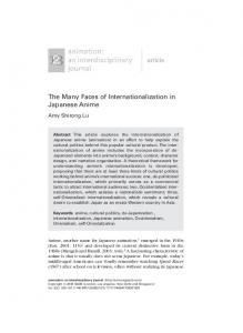
FIG. 1. Isolation of p225, a putative nucleoporin. Proteins of a 2 M urea/i mM EDTA extract of purified rat liver nuclear envelopes were fractionated on WGA Sepharose. The WGA-reactive proteins were separated by SDS/PAGE on a 5% acrylamide gel and stained with Coomassie blue. Lane 1, size marker proteins; lane 2, WGAreactive proteins. The masses of marker proteins in lane 1 are indicated to the left in kDa. The arrow indicates p225 and arrowheads point to the nucleoporins p62 and nuplS3.
(Rockland, Gilbertsville, PA). The serum was affinity purified using p39/6xHis bound to nitrocellulose (35). For immunolocalization the affinity-purified antibodies were concentrated 5-fold in a Centricon microconcentrator (Amicon). Immunoblot Analysis. After electrophoretical transfer of SDS/PAGE-separated proteins to nitrocellulose membrane (27), blots were incubated with primary p39 antibodies [diluted 1:10 in 5% nonfat dry milk/0.1% Tween 20 in phosphate-buffered saline (PBS)] and visualized with alkaline phosphatase-conjugated anti-rabbit IgG (Promega) under the same conditions as described previously (4), except that solutions were buffered with PBS. Imnunofluorescence Microscopy. HeLa cells were grown on coverslips in modified Eagle's medium (16) until they were subconfluent. After washing for 2 min with ice-cold PBS they were fixed for 10 min with 2% paraformaldehyde in PBS, permeabilized for 2 min with 0.5% Triton X-100 in PBS, and EAYNQK II I I
131
EAKOOK
rat
136
rat
KXDDSLPVGVTIDYTNFV
356
11111:11 :1111
KSDDSLPMGVVVDYTNOV
373
554
11 KPTLEST PVPSVS KXXEVRSTAP
955
111111 KTPPVRSTAP KXFFSFGNQQT
1111:111
1436 KTSFSFGSQQT KXPFSSASXGFGSTPTLN
human rat
KXXLEXTSPVXXXS
11
human
562
human rat
9641 human rat
1447 human rat
111111 11111 1900 KNPFSSASGGFGSTATSN
1917 human
KXFGFGAAPVFAS 11111111 1966 KTGGFGAAPVFGS
1978 human
KXFGEGTAXGXXGAF 111111
2010 KVFGEGTAAASAGGF
rat
rat
2024 human
FIG. 2. Comparison of partial amino acid sequences of rat p225 with similar sequences of the human CAN protein. Numbers to the left and right of human sequences indicate their positions in the complete amino acid sequence of the human CAN protein; 1, identical amino acids;:, similar amino acids. Five peaks ofthe HPLC profile of p225 digestion with endoproteinase Lys C were analyzed.
Cell Biology: Kraemer et al.
Proc. Natl. Acad. Sci. USA 91 (1994)
XFXFG: 481 519 545 1213
VFSFG TFSFV SFSFG PFSFS 1222 GFNFG
SVFG $2LKUATVTGEPPSYUGSDI1KAAPGPGPS PPSKASLAPTPAASPVAPSAA
PSGT
ITPTP5sNFTAAQGATPSTKEMOPDAFjGGGSKPS
YEA I PE.UPPSG I TSASNTTPGEPAASSMRPVAPSGTA
LSTT.SKLETPPSKLGELLFPS3LAGETLGSFSGLRVG OADDSTKPTNKA,5TSLTSTOPTKTSGVPSGFNFTAPP
VLGKHTEPPVTS&ATTTSVAPPAATSTSST SLPVTSAG3IGV SFGGTSLSAGKT 1438 SFSFG
1409 AVFG
SOOTNSTVPPSAPPPTTAATPLPTSFPTLSFGSLLj$A
TTPSLPMSAGRSTEEATI5ALPEKPGDSEVSASAASLL EEQOSAOLPOAPPOTSDSVKKEPVLAOPAVSNSGTAAS TSLTVALSAEATPATTGVPDARTEAVPPAS.F OTAVTAAA I£AGPVAVET.STP I AgTTS I VAPGPSA
1589 SVPG 1643 SVFA 1672 PVFG
EAAAFGTVTSGS
OPPAA=SAFNOLTNNTATAPSAT
OVAASTAPSLFG0OTGSTASTAAATPOV.UGFWPAF GTTAP
TTFGQA OSASAA
1783 SFSFG
1833 GFSFC
FSOPGFSSVPAFG0PASTPTSTSG AASTSM OQ$PNTGGGLFGOSNAPAFGQSPGFGOGG
GTSAATTTAATS
OA ,NTG OAASTGG OOs5 G
SGNTGRGGGFFSGLGGKPSODAANKNPF,5ASGGFGS
1719 1730 1741 1770
GVFG SVFG SVFS
SVFG
1817 SVFG
1840 1849 1860 1872
SGFG SVFG I VFG SVFG
TATSNTSNLFGNSGAKTFGGFASFGEQKPTGTF£G
1959 GFGFS
GGSVASO SPNKTGGFGAA
SPPTFGGSPGFGGVPAFGSAPAFTSPLGSTGG EGTAAASAG
2023 GFGFG
2072 GFSFG
$&UNTTSFGTLASONAPTFGSLSOOT TOUGF SGTG SNNS
FGGWRS
1974 PVFG 2010 KVFG 2054 SGFG 2064 SGFG 2081 SVOG
1521
bovine serum albumin/0.05% Tween 20 in PBS. Antibody binding was detected with Texas red-labeled goat anti-rabbit IgG (Organon). Coverslips were washed and mounted in a solution of 1 mg of p-phenylenediamine per ml in 90%o glycerol (pH 8.0) and examined as described (5). Immunoelectron Microscopy. HeLa cells were processed to prepare frozen ultrathin sections (4, 36). The sections were incubated first with the concentrated affinity-purified antip39 antibodies and then with 10-nm gold-conjugated secondary antibodies as described (4). RESULTS As previously described (5), purified rat liver nuclear envelopes contain dozens ofperipheral membrane proteins. Many of them are potential nucleoporins. These proteins can be extracted by urea/EDTA and further subfractionated on WGA Sepharose. About a dozen of these potential nucleoporins bind to WGA (5). The proteins also react polyspecifically with various mAbs (4). These mAbs decorate the NPC in immunoelectron microscopy. Fig. 1 shows the WGAreactive proteins separated by SDS/PAGE and stained with Coomassie blue (lane 2). Among them are the already molecularly characterized nucleoporins p62 (2, 3) and nupl53 (4) (lane 2, arrowhead). In preparation for the molecular cloning of a protein of -225 kDa (p225) (indicated by arrow in lane 2), p225 was subjected to proteolytic cleavage with endopro-
FIG. 3. Repetitive peptide motifs in the human CAN protein. The pentapeptide XFXFG is a nucleoporin "signature" motif. The tetrapeptide motif SVFG (on the right) and the tripeptide motif FGG (indicated in bold letters in the middle column) have so far not been described for other nucleoporins. FGQ is part of the hexapeptide motif GGLFGQ in NUP100 and NUP116 (20, 21). Polyserines are underlined. Numbers indicate the first amino acid position of XFXFG and SVFG motifs, respectively, in the human CAN protein sequence.
blocked for 1 hr with 2% bovine serum albumin/0.05% Tween 20 in PBS. The cells were probed further with the concentrated affinity-purified p39 antibodies for 1 hr at room temperature and washed five times for 5 min with 0.1%
66
A
B
1 2 3
1
- 200
-
45 -
_
- 116
FIG. 4. Expression of a 39-kDa internal segment of the human CAN protein in E. coli and characterization of antibodies raised against the expressed protein. (A) Lane 1, Coomassie blue-stained size markers; lane 2, Coomassie blue-stained 39-kDa internal segment of the CAN protein expressed in E. coli and subsequently purified; lane 3, nitrocellulose blot of protein in lane 2 probed with affinity-purified rabbit anti-p39 antibodies and visualized with alkaline phosphatase-coupled anti-rabbit IgG. (B) Lane 1, nitrocellulose blot of SDS/PAGE-separated polypeptides of rat liver nuclear envelopes probed with affmiity-purified rabbit anti-p39 antibodies. The arrowhead indicates p225. The positions of markers are labeled (in kDa).
FIG. 5. Immunofluorescence microscopy of HeLa cells. Cells grown on coverslips were fixed in paraformaldehyde, permeabilized with Triton X-100, and probed with affinity-purified anti-p39 antibodies. Focusing on the nuclear periphery (Upper) or the nuclear surface (Lower) yielded nuclear rim staining and a punctate nuclear surface staining pattern, respectively, characteristic for nucleoporins. (Bar = 5 Am.)
1522
~
Proc. Nadl. Acad. Sci. USA 91 (1994)
Cell Biology: Kraemer et al.
teinase Lys C. The resulting peptides were separated by HPLC and some of them were subjected to amino acid sequencing. Surprisingly, a comparison of the obtained amino acid sequences with those in the data banks showed a high degree of similarity with the cDNA-deduced amino acid sequence of the human CAN protein throughout the entire length of the molecule (Fig. 2). This suggested that the rat p225 is the homolog of the human CAN protein (23). Although the subcellular localization of the human CAN protein has not been established, it has been suggested to be a cytoplasmic protein (23), clearly at odds with it being a nucleoporin. An analysis of the human CAN protein sequence (Fig. 3) revealed the presence of 11 redundant XFXFG motifs that are present in vertebrate p62 (2, 3, 16) and nuplS3 (4) as well as in various yeast nucleoporins (17-19). This motif is likely to be part of the epitope reactive with mAb 414 (20). In addition, there is one other tetrapeptide motif SVFG (see Fig. 3) and the repetitive motif FGG that have so far not been reported for other molecularly characterized nucleoporins. The tripeptide motif FGQ is part of the GGLFGQ motif in NUP100 and NUP116 (20, 21). Especially the C-terminal
portion (amino acids 1213-2090) is unusually rich in serines (21%), threonines (12%), and prolines (9,%) (23). Serines and threonines, which are located less than three residues from a proline, have been proposed to be potential sites for GlcNAc addition (8). The amino acid sequence of CAN contains 32 potential phosphorylation sites (7 for protein kinase C, 24 for casein kinase II, and 1 for cAMP-dependent protein kinase). Although the presence in the human CAN protein of the XFXFG motif was consistent with it being a nucleoporin, the low abundance of this motif (relative to that in other nucleoporins) as well as its considerable degeneracy made it far from certain that the human CAN protein is a nucleoporin. To definitively localize the human CAN protein it was therefore necessary to prepare monospecific antibodies. To obtain these we expressed a 39-kDa segment of the protein (residues 818-1175) in E. coli (Fig. 4A, lane 2). This segment lacks the XFXFG motif that is part of the mAb 414-reactive epitope and the other repetitive motifs. Antibodies to this segment should therefore be monospecific for the human CAN protein. Indeed, the obtained antibodies reacted with the E. coli-expressed CAN protein segment (Fig. 4A, lane 3) and recognized a single 225-kDa protein in rat liver nuclear
a
i..
sI
FiG. 6. Immunoelectron microscopy of HeLa cells. Frozen ultrathin sections of HeLa cells were probed with affinity-purified anti-p39 and binding was detected with 10-nm gold-coupled secondary antibodies. Nucleoplasm (N) and cytoplasm (C) are indicated. (a) Section through a part of a HeLa cell. Arrows point to gold particles decorating the cytoplasmic side of the NPCs. (b-d) Transverse sections with their cytoplasmic and nucleoplasmic sides aligned up and down, respectively. Gold exclusively decorated the cytoplasmic side of the NPCs. (Bars = 200 nm in a and 100 nm in b-d.)
Proc. Natl. Acad. Sci. USA 91 (1994)
Cell Biology: Kraemer et al. envelopes (Fig. 4B, lane 1). In addition, this antibody specifically recognized the mAb 414-reactive p225 polypeptide isolated from rat liver nuclear envelopes (data not shown). These monosoedific antibodies were next used for indirect immunofluorescence microscopy of paraformaldehyde-fixed and Triton X-100-permeabilized HeLa cells grown on coverslips (Fig. 5). Focusing on the nuclear rim (Fig. 5 Upper) or the nuclear surface (Fig. 5 Lower) revealed a punctate nuclear rim and surface staining pattern that is characteristic for nucleoporins (1). Cytoplasmic staining was not detected. To definitively localize p225 to the NPC we carried out immunoelectron microscopy. Frozen ultrathin sections of HeLa cells were incubated with the monospecific antibodies and then with 10-nm gold-conjugated secondary antibodies. The gold particles decorated NPCs (Fig. 6). Most strikingly, they decorated only the cytoplasmic side of the NPC (Fig. 6). Thus we conclude that the human CAN protein is a nucleoporin that is asymmetrically localized on the cytoplasmic side of the NPC.
DISCUSSION
nup
b
NH3
1l
DEK
COOH 2090
nup
214
349
SET!
C NH, 1
270
i COOH
2090
nup
We thank Helen Shio for preparation of the immunoelectron microscopy specimens, Mike Rout for critical reading of the manuscript, and members of the Rockefeller University Protein Sequencing Facility, especially Joseph Fernandez, for protein sequencing. D.K. was supported by a postdoctoral fellowship (Kr 1412/1-1) from the Deutsche Forschungsgtmeinschaft.
31-47.
214
812 A 813
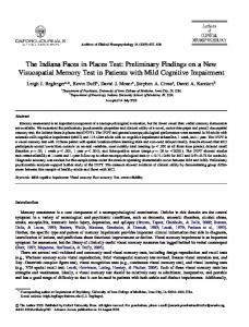
mogenesis. In Fig. 7 are summarized previously published data (23, 37). This work showed that two distinct chromosome aberrations, a translocation and a fusion (both characteristic for myeloid leukemogenesis), yielded two chimeric proteins. Because each of these chimeric proteins shares a common C-terminal part derived from the C terminus of nup214 (Fig. 7), nup214 (rather than the N-terminal DEK and SET proteins) was suggested to be the putative oncogene product (23, 37). If nup214 were to play an important role in the control of nucleocytoplasmic traffic, it is conceivable that an imbalance in traffic control could yield a proliferative advantage to the cell (39).
1. Davis, L. I. & Blobel, G. (1986) Cell 45, 699-709. 2. Cordes, V., Waizenegger, I. & Krohne, G. (1991) Eur. J. Cell Biol. 55,
The human CAN protein, a putative oncogene associated with myeloid leukemogenesis (23, 37), has been shown here by immunoelectron microscopy to be a nucleoporin. Consistent with previous nomenclature, we therefore suggest the alternative term nup214 for this protein. nup214 is only the fourth vertebrate nucleoporin that has been localized to the NPC (see Introduction). Its exclusive localization to the cytoplasmic side of the NPC suggests that. it may be part of the short "fibers" that extend 35-50 nm from the cytoplasmic aspect of the NPC (assuming that the protein composition of the cytoplasmic and the nucleoplasmic rings is identical). These fibers are the only macromolecular structures so far known to be asymmetrically associated with the NPC on its cytoplasmic side. nup214 and nuplS3 cofractionate quantitatively with nuclear envelopes. Thus, although nupl53 is located at the "extreme" nucleoplasmic end of the NPC and nup214 at its cytoplasmic perimeter, neither of them appears to be lost from the NPC during isolation of nuclear envelopes. This knowledge will be useful for further structural and functional analysis of components of the NPC in the context of isolated nuclear envelopes. The function of nup214 remains to be determined. One possibility is that this protein serves as a docking site in the receptor-mediated import of substrates across the NPC (38, 39). In fact, binding of gold-decorated nuclear import substrate to what could be such fibers has been reported previously (40). An understanding of the function of nup214 may also help in elucidating its presumed assc~iation with myeloid leukea NH3
1523
214
^,COOH
FIG. 7. Schematic representation of nup214 (CAN) and the two chimeric proteins that result from chromosome translocation or fusion associated with myeloid leukemogenesis. As a result of chromosome translocation or fusion, the C-terminal part of nup214 (CAN) (a) on chromosome 9 is linked to the N-terminal part of the DEK protein (chromosome 6) (b) or fused to the SET protein (chromosome 9) (c). Numbers indicate the positions of residues at the N and C termini and at the breakpoints.
3. Starr, C. M., D'Onofrio, M., Park, M. K. & Hanover, J. A. (1990) J. Cell Biol. 110, 1861-1871. 4. Sukegawa, J. & Blobel, G. (1993) Cell 72, 29-38. 5. Radu, A., Blobel, G. & Wozniak, R. W. (1993) J. Cell Biol. 121, 1-9. 6. Pant6, N. & Aebi, U. (1993) J. Cell Biol. 122, 977-984. 7. Davis, L. I. & Blobel, G. (1987) Proc. Natl. Acad. Sci. USA 84, 7552-7556. 8. Haltiwanger, R. S., Kelly, W. G., Roquemore, E. P., Blomberg, M. A., Dong, L. D., Kreppel, L., Chou, T. & Hart, G. W. (1992) Biochem. Soc. Trans. 20, 264-269. 9. Hanover, J. A., Cohen, C. K., Willingham, M. C. & Park, M. K. (1987) J. Biol. Chem. 262, 9887-9894. 10. Holt, G. D. & Hart, G. W. (1986) J. Biol. Chem. 104, 1157-1164. 11. Holt, G. D., Snow, C. M., Senior, A., Haltiwanger, R. S., Gerace, L. & Hart, G. W. (1987) J. Cell Biol. 104, 1157-1164. 12. Park, M. K., D'Onofrio, M., Willingham, M. C. & Hanover, J. A. (1987) Proc. Natl. Acad. Sci. USA 84, 6462-6466. 13. Schindler, M., Hogan, M., Miller, R. & DeGaetano, D. (1987) J. Biol. Chem. 262, 1254-1260. 14. Snow, C. M., Senior, A. & Gerace, L. (1987) J. Cell Biol. 104,1143-1156. 15. Hart, G. W., Haltiwanger, R. S., Holt, G. D. & Kelly, G. (1989) Annu. Rev. Biochem. 58, 841-874. 16. Carmo-Fonseca, M., Kern, H. & Hurt, E. C. (1991) Eur. J. Cell Biol. 55, 17-30. 17. Davis, L. I. & Fink, G. R. (1990) Cell 61, 965-978. 18. Loeb, J. D. J., Davis, L. I. & Fink, G. R. (1993) Mol. Biol. Cell 4, 209-222. 19. Nehrbass, U., Kern, H., Mutvei, A., Horstmann, H., Marshallsay, B. & Hurt, E. C. (1990) Cell 61, 979-989. 20. Wente, S. R., Rout, M. P. & Blobel, G. (1992)J. Cell. Biol. 119,705-723. 21. Wimmer, C., Doye, V., Grandi, P., Nehrbass, U. & Hurt, E. C. (1992) EMBO J. 11, 505,1-5061. 22. Aris, J. P. & Blobel, G. (1989) J. Cell Biol. 108, 2059-2067. 23. Von Lindern, M., Fornerod, M., van Baal, S., Jaegle, M., de Wit, T., Buijs, A. & Grosveld, G. (1992) Mol. Cell. Biol. 12, 1687-1697. 24. Jarnik, M. & Aebi, U. (1991) J. Struct. Biol. 107, 291-308. 25. Ris, H. (1991) EMSA Bull. 21, 54-56. 26. Laemmli, U. K. (1970) Nature (London) 227, 680-685. 27. Towbin, H., Staehelin, T. & Gordon, J. (1979) Proc. Natl. Acad. Sci. USA 76, 4350-4354. 28. Fernandez, J., DeMott, M., Atherton, D. & Mische, S. M. (1992) Anal. Biochem. 201, 255-264. 29. Altschul, S. F., Gish, W., Miller, W., Myers, E. W. & Lipman, D. J.
(1990) J. Mol. Biol. 215, 403-410.
30. Henikoff, S. & Henikoff, J. G. (1992) Proc. Natl. Acad. Sci. USA 89, 10915-10919. 31. Fuchs, R. (1991) Appl. Biosci. 7, 105-106. 32. Fisher, D. Z., Chaudhary, N. & Blobel, G. (1986) Proc. Natl. Acad. Sci. USA 83, 6450-6454. 33. Sanger, F., Nicklen, S. & Coulson, A. R. (1977) Proc. Natl. Acad. Sci. USA 74, 5463-5467. 34. Bradford, M. M. (1976) Anal. Biochem. 72, 248-254. 35. Drenckhahn, D. & Frahz, H. (1986) J. Cell Biol. 102, 1843-1852. 36. Tokuyasu, K. T. (1973) J. Cell Biol. 57, 551-565. 37. Von Lindern, M., van Baal, S., Wiegant, J., Raap, A., Hagemeijer, A. & Grosveld, G. (1992) Mol. Cell. Biol. 12, 3346-3355. 38. Moore, M. S. & Blobel, G. (1992) Cell 69, 939-950. 39. Moore, M. S. & Blobel, G. (1993) Nature (London) 365, 661-663. 40. Richardson, W. D., Mills, A. D., Dilworth, S. M., Laskey, R. A. & Dingwall, C. (1988) Cell 52, 655-664.