Pharmaceutics 2011, 3, 538-571; doi:10.3390/pharmaceutics3030538 OPEN ACCESS
pharmaceutics ISSN 1999-4923 www.mdpi.com/journal/pharmaceutics Review
The Application of Microencapsulation Techniques in the Treatment of Endodontic and Periodontal Diseases Asteria Luzardo Álvarez 1,*, Francisco Otero Espinar 2 and José Blanco Méndez 1,2 1
2
Departamento de Farmacia y Tecnología Farmacéutica, Facultad de Ciencias, Universidad de Santiago de Compostela, 27002 Lugo, Spain; E-Mail:
[email protected] Departamento de Farmacia y Tecnología Farmacéutica, Facultad de Farmacia, Universidad de Santiago de Compostela, 15782 Santiago de Compostela, Spain; E-Mail:
[email protected]
* Author to whom correspondence should be addressed; E-Mail:
[email protected]; Tel: 0034-9828-24151; Fax: 0034-9822-85872. Received: 5 July 2011; in revised form: 9 August 2011 / Accepted: 24 August 2011 / Published: 26 August 2011
Abstract: In the treatment of intracanal and periodontal infections, the local application of antibiotics and other therapeutic agents in the root canal or in periodontal pockets may be a promising approach to achieve sustained drug release, high antimicrobial activity and low systemic side effects. Microparticles made from biodegradable polymers have been reported to be an effective means of delivering antibacterial drugs in endodontic and periodontal therapy. The aim of this review article is to assess recent therapeutic strategies in which biocompatible microparticles are used for effective management of periodontal and endodontic diseases. In vitro and in vivo studies that have investigated the biocompatibility or efficacy of certain microparticle formulations and devices are presented. Future directions in the application of microencapsulation techniques in endodontic and periodontal therapies are discussed. Keywords: microencapsulation; periodontal diseases; endodontic failure; periodontal pocket; root canal; controlled release Abbreviations: PD: periodontal disease; EF: Endodontic failure; SRP: scaling and root planning; GCF: gingival crevicular fluid; PLA: poly(lactide); PLGA: poly(lactide-coglycolide); PCL: poly(ε-caprolactone); CMC: carboxymethyl cellulose; PHBV: polhydroxybutirate-co-hydroxyvalerate; PEO-PPO-PEO: polyethylene
Pharmaceutics 2011, 3
539
oxide/polyprolylene; ICTP: pyridinoline cross-linked carboxy-terminal telopeptide of type I collagen; MβCD: methyl β-cyclodextrin; HPβCD: hydroxypropyl β-cyclodextrin; rhBMP2: recombinant human bone morphogenetic protein-2; PRP: platelet-rich plasma; TGF-β: transforming growth factor β family; IGF-1: insulin-like growth factor-1; bFGF: basic fibroblastic growth factor; DMP1: dentin matrix protein 1
1. Introduction Endodontic and periodontal pathologies are closely related to each other and as primary diseases each may have secondary effects on the other. The appropriate treatment and route of administration are selected according to the differential diagnosis and etiopathogenesis of each disease. Periodontal and endodontic therapies can be grouped into three broad categories: mechanical debridement and cleaning to eliminate bacteria; treatment aimed at killing or affecting the metabolism of the infectious organism, such as antiseptics and antibiotics, and treatment that affects the environment of the infectious organism [1,2]. Antiseptic or antibiotic drug-loaded devices can be used to treat infection, or as a pharmacological complement to mechanical treatment by introducing particulate formulations into the targeted sites. New approaches involve the use of local drug delivery systems based on microparticles made from biocompatible polymers. Such devices enable the introduction of antimicrobial agents or other drugs directly in the periodontal pocket, or inside the root canal, and the prolonged release of constant concentrations of these agents for better control of infections. Complementary use of drug-loaded microparticle formulations can reduce periodontal and endodontic failures, and even partly replace surgical or mechanical treatment, especially in refractory patients. Biodegradable microparticles have been widely studied as drug delivery systems for many bioactive compounds such as low molecular weight drugs, macromolecular substance, DNA and antigens [3-5]. Synthetic or natural polymers have been considered for preparation of microspheres for administration by the nasal, pulmonary, oral or parental route [6-8]. Because of their excellent biocompatibility characteristics, natural polymers such as chitosan and other polysaccharides, collagen, hyaluronic acid and cellulose are often used for microsphere preparation. Poly(D,L-lactide-co-glycolide) (PLGA) biodegradable polymers also have a long history as drug delivery systems in many biomedical fields. Their biodegradability, excellent biocompatibility and ability to provide controlled-drug release have led to them being used in microsphere preparation [9-11]. Other examples of biodegradable polymers used in microparticle preparations include polyanhydrides, poly-ε-caprolactone and polyphosphazenes [12-14]. Biodegradable polymers, of natural or synthetic origin, can be degraded into acceptable biocompatible products by chemical or enzymatic hydrolysis. Microencapsulation of therapeutic agents has traditionally been performed by three main techniques and their modifications: spraydrying, solvent extraction-evaporation and coacervation or phase separation. These and other methods are described by [9,11,15-20]. Other reviews dealing with diverse aspects of microspheres for drug delivery are available [21-23]. Localized administration of medication by use of polymeric microspheres offers the advantage of providing rate-controlled release of drug at the site of action, maintaining the concentration in the
Pharmaceutics 2011, 3
540
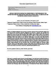
therapeutic window (thus avoiding systemic side-effects), and protection of the substance of interest from degradation and physiological clearance. The main factors responsible for controlling the rate of drug release from microparticles include physicochemical characteristics such as size, chemical composition, morphology and the rate of degradation of polymers and drug. The full therapeutic potential of microparticles in pharmaceutical applications lies in their particular properties: large surface area, ability for site-controlled drug delivery, the wide variety of biocompatible polymers that can be used, simplicity of implantation at the site of delivery, and charge density. Moreover, all these parameters can be modified to design customized microparticulate systems adapted to the requirements of each clinical case. This review focuses on localized controlled-release polymer microspheres used in periodontic and dental applications. We begin with a general description of periodontal diseases (PD) and endodontic failure (EF) and the conventional treatments used. We present the most recent therapeutic strategies based on microencapsulated drugs, with the main emphasis on periodontal and dental routes of administration. The properties of drug-loaded polymeric microparticles enable prolonged delivery of large amounts of drugs, mainly antibiotics, through the periodontal and dental route of administration, thus reducing the side effects of systemically applied antimicrobials in the treatment of PD and EF. The current status of studies and recently reported approaches evaluating microparticulate formulations for endodontic and periodontal applications, advantages over other systems for infection inhibition, practical difficulties, localized delivery for periodontal and dental tissue regeneration and safety issues are further aspects that will be discussed. 1.1. Periodontal Diseases (PD) and Periodontal Treatment Periodontal disease is a wide term that covers a combination of multiple inflammatory diseases affecting the tissues surrounding teeth: gum tissue, alveolar bone and the periodontal ligament, which is composed of collagen fibers (Figure 1). The prevalence of peridontitis worldwide has been determined on the basis of differences in clinical diagnosis between different forms of periodontitis (gingivitis, chronic or aggressive periodontitis), although according to the epidemiological data, the occurrence of this PD is related to oral hygiene status and socio-economic class. In the USA, the prevalence of chronic periodontitis in adults is around 35% [24], whereas in Western Europe 13–54% of the population is affected by periodontal diseases [25]. In low income countries, the percentage of the population affected is around 45%. Several factors affect clinical signs of PD, such as tobacco use, general health, pregnancy, diabetes and social conditions [26-28]. In periodontal pathologies, the supporting structures become infected and necrotic, leading to loss of attachment of teeth. Periodontal pathology is an infectious process with different degrees of severity: gingivitis, mild periodontitis, moderate periodontitis and advanced periodontitis, with extensive periodontal breakdown. Clinical signs of periodontitis include gum swelling, bleeding, pus discharge, tooth mobility by changes in gingival tissues and formation of a periodontal pocket. Periodontitis is essentially irreversible and if left untreated, moderate periodontitis can lead to the loss of tooth by the degradation of collagen by bacterial enzymes. The periodontal pocket where the tooth connective tissue attachment has been gradually destroyed provides an excellent environment for pathogenic bacteria. The periodontal microbiota adheres to the tooth inducing inflammation of the
Pharmaceutics 2011, 3
541
surrounding soft tissues. The inflammatory and immune mediators produced by the host in response to bacterial activity also enhance destruction of the periodontal tissue [29-31]. Periodontal diseases involve the formation of a biofilm on the tooth surface. Dental biofilm is also known as “plaque”. In this context, a biofilm is broadly defined as sessile and mixed-bacterial cells that adhere to each other and to a surface forming enclosed matrices composed of polymers of their production [32-35]. Growth of microbial biofilms is always associated with resistance to antimicrobial agents [36]. Thus, the plaque biofilm is considered the main factor that causes several localized diseases in the oral cavity, including periodontal disease, dental caries and endodontic failure. The aim of periodontal procedures is mainly to eliminate bacterial deposits or dental plaque from the tooth surface by mechanical treatment in combination with good oral hygiene to prevent re-infection of the subgingival area by pathogens and consequently, to preserve the tooth. Elimination of periodontopathic microorganisms by systematically administered antimicrobial agents has been widely used as adjunctive therapy to mechanical procedures used to treat periodontitis such as scaling and root planning (SRP). Figure 1. Representation of the structure of a healthy tooth and how it is typically affected by periodontal disease (picture source: American Academy of Periodontology; http://www.perio.org).
However, antibiotics applied locally in the periodontal pocket may further eliminate pathogens and thus enhance the effect of conventional surgical therapy without the side effects of systemically administered antibiotics. Local administration of antibiotics necessarily involves insertion of the drug-loaded device in the periodontal pocket, to achieve effective concentrations at the target site. The enhancement of antibiotic penetration into biofilms is of particular importance to prevent microbial colonization in periodontal pockets and dental root canals, since the drug should reach sites of difficult access. The use of sustained drug delivery systems may also provide drug delivery over a prolonged period in periodontal pockets. In this context, polymeric microparticles may be of particular interest by virtue of their ability to achieve sustained delivery of effective biocidal levels of drug, to inhibit microbial adhesion and to be retained or adhered on the dentin or biofilm surface, as well as their biocompatibility and large surface area to volume ratio. In addition, the small size of microspheres enables them to penetrate into deeper lesions in severe periodontitis. It is also possible to modify the physicochemical characteristics of microparticles, such as surface charge, size, porosity,
Pharmaceutics 2011, 3
542
hydrophobicity and even introduce ligands with functional groups on the surface [37,38]. Drug-microparticles in periodontal pockets may well provide useful antimicrobial activity to control or inhibit infection in both hard and soft tissues. 1.2. Dental Disease and Endodontic Re-Treatment Partial or total tooth loss through apical periodontitis caused by acidogenic bacteria is a major health problem. Endodontic disease is a common chronic disease that affects all age groups, has a significant impact on quality of life and is costly to treat [39] accounting for between 5–10% of health care expenditure [25,39]. Epidemiological surveys of dental health conditions in different countries have shown that the level of severity of the prevalence of caries is about 50% in well-developed countries, whereas in low income countries, the prevalence is around 90% [40]. Periodontal infections are the most frequent type of periodontal diseases, and are responsible for 75% of adult tooth loss. Endodontics or root canal therapy is the part of dental science that is indicated for patients who have already undergone endodontic therapy, but who suffer from persistent infections. It is of particular concern that resistant microbial species such as Enterococcus faecalis (the most common) [41,42], Staphylococcus aureus, Pseudomonas aeruginosa, Bacillus subtilis, Streptococcus spp., Actinomyces spp. and Candida spp., among others may remain in root canals [43-45]. Bacteria can colonize the enamel of the tooth surface, as well as the supporting structures of teeth. Several microbial species can attach to the tooth surface and form a dense polymeric structure involved in surface adhesion and host invasion [46-50]. Endodontic biofilms represent a common cause of persistent infections in the root canal because of their resistance to antimicrobial compounds [47]. If not detected early, caries leads to erosion of the enamel and the tooth pulp. The tooth pulp forms the dentin and contains the nervous and vascular system that supplies nutrients to the tooth. The pulp also includes cells such as fibroblasts, odontoblasts, mast cells, macrophages and plasma cells. Over time, the irreversible infection affects the pulp tissue and the tooth become non-vital as a result of necrosis. Management of the injured tooth to preserve it as a healthy and functional unit (so-called root canal treatment) includes removal of the damaged tissue, and cleaning and drying the root canal cavity. This step does not involve the use of intracanal medication. The root canal is then dressed with irrigant antimicrobial solutions (sodium hypochlorite, chlorhexidine, iodine-based irrigants, EDTA) and a provisional cap is inserted to separate the root canal from the oral cavity. This procedure is repeated every 7–10 days until the treatment is completed. The final step consists of sealing the root canal with a permanent cap to prevent further contamination by microorganisms (Figure 2). The success of endodontic treatment basically depends on the effective elimination of inflamed and/or infected pulp tissue from the root canal system. Most of the infecting bacteria may be reduced by the endodontic procedure.
543
Pharmaceutics 2011, 3 Figure 2. Inflammation and infection of pulp tissue (a) and example of completed root canal treatment. The temporary filling is placed into the empty pulp chamber; (b) Adapted from http://www.aae.org/Patients/Endodontic_Treatments/Root_Canals.aspx
a)
Access b) opening, cleaning and shaping of root
Root canal treatment involves mechanical and chemical activity to reduce the bacterial load in the root canal system. Although most infecting pathogens are removed during the chemo-mechanical procedure, residual bacteria can survive within the dentinal tubules [51,52]. In endodontics, the development of a bacterial biofilm within the root canal system is also a key cause of endodontic failure, whereby necrosis in the surrounding tissues leads to endodontic failure. The endodontic treatment therefore fails due to resistance to antibiotics, unsuccessful sealing of the canal or formation of a bacterial biofilm within the root canal cavity [31]. The complex anatomy of the root canal system, with accessory and lateral canals that are part of the root canal system and poor tissue penetration of the systemic antibiotic therapy are important factors in the complete debridement of bacteria and subsequently, in endodontic failure. Although the antimicrobial efficacy of conventional irrigants is generally recognized, they may produce some complications such as tissue irritation or hypersensitivity. Sodium hypochlorite, normally used at concentrations of 0.5–5% [53,54] can produce pain, swelling, haemorrhage and even secondary infections as a result of accidental injection [55-57], or perforations due to the use of large volume of irrigants extruded from the canal. The ability of calcium hydroxide to eliminate microorganisms in the root canal is not reliable, especially for some bacteria in particular [58-60], and inflammatory reactions have been reported [61]. Chlorhexidine has shown an important degree of antimicrobial activity in comparison with other intracanal medication [62]. However, long-term sustained release of the drug is necessary for it to be effective [63,64], and it may be affected by other anionic components of the formulation, rendering it inactive. The use of irrigants with antimicrobial properties provides a local drug effect but this cannot be sustained to maintain therapeutic levels of drug over several days. The difficulty in eliminating Enterococcus faecalis and other bacteria that infect dental root canals has led to interest in the development of alternative formulations capable of sustained release of antibiotic within the canal, between the preparation and obturation phase, as inter-appointment medication.
Pharmaceutics 2011, 3
544
2. Microparticulate Delivery Systems and Periodontal Diseases Antimicrobial or antiseptic agents are applied locally for the treatment of periodontitis in combination with mechanical root debridement. Subgingival irrigation with drug solutions provides high concentrations in the periodontal pocket for only short periods. In consequence, repeated oral irrigation is required to exert bactericidal or bacteriostatic effects. For use of the periodontal route as a site for administration of drug-loaded microparticles, the amount of drug absorbed across a membrane is directly proportional to the surface area of exposure, the applied drug concentration, the permeability coefficient of the drug and the residence time. In a pathological situation, the turnover rate of the medium volume of a moderately-sized pocket (0.5 µL) is around 40 times per hour [65]. If we compare this with the salivary flow inside the oral cavity, 28 times an hour, the rapid clearance of substances from the periodontal pocket is obvious. Goodson [65] also suggested that this rapid turnover was related to the short duration of action of irrigation treatments. In fact, the high rate of clearance represents the main hurdle to maintaining effective concentrations of drug within the periodontal pocket. The use of microparticulate systems for controlled drug release may well improve the antibiotic efficacy in the periodontal pocket, with consequent clinical benefits. Periodontal pockets may be used as natural sites for easy placement of microparticulate delivery systems. Bearing in mind that the average depth of a periodontal pocket is about 5–8 mm, in practice, the maximum size of a periodontal delivery system should not be any larger than this. The physical space in the periodontal pocket therefore represents another limitation to the release of the active substance to counteract the continuous loss due to gingival crevicular fluid flow. The gingival crevicular fluid provides the release medium for the drug from microspheres throughout the periodontal pocket. As an intra-pocket device for antibiotic drug administration microparticles represent an attractive way of maintaining the effective drug concentration in the gingival crevicular fluid in a sustained manner for prolonged periods, in order to obtain therapeutic benefits. A microparticulate delivery system for periodontal healing should ideally display high biocompatibility, provide controlled release of the active substance, be easy to implant (e.g., with a syringe), provide drug stability and should be retained in the periodontal pocket. The periodontal pocket has been investigated as a site for local drug delivery by many research groups, and the route has already achieved commercial status with several drugs such as clorhexidine gluconate (Periochip®, Dexcel Pharma) and tetracycline (Actisite®, P&G/Alza). Extensive research has been directed towards the development of intrapocket devices including films [66,67], sponges [68], strips [69-71], fibers [72,73], gels [74,75], semisolid form [76], chips [77,78], nanoparticles [79-81] and microparticles [77,82]. A full review of other kind of drug delivery systems apart from microparticles is beyond the scope of this article and it is extensively covered in several reviews [83-85]. Multiparticulate systems used in the periodontal delivery of drugs mainly include microparticles and nanoparticles. In addition to the inbuilt advantages to multiparticulate drug delivery systems (i.e., safety, uniformity, reproducibility), these systems have the advantage that they can be incorporated in usual oral formulations as suspensions or toothpastes, or in new drug delivery systems such as bioadhesives or sensitive hydrogels, or can even be directly injected into the periodontal cavity to achieve effective control of drug release. In general, microparticles have been found to be a good
Pharmaceutics 2011, 3
545
approach for local treatment of periodontal infection, and the implementation of bioadhesive properties may increase their therapeutic potential to avoid leaching from the periodontal pocket. One of the first studies describing the use of microspheres in the periodontal cavity was developed by Folke and Stallard in 1967 [86]. Radiolabeled polystyrene tracer microspheres (15 µm) were injected into the right external carotid artery of monkeys, with the aim of studying periodontal microcirculation. However, it was during the 1990s that different formulations based on microparticles were developed for drug delivery in periodontal treatments, mainly for controlled delivery of antibiotics, antiseptics, antinflammatory agents or growth factors. Since non-biodegradable materials require complete removal of the device at the end of the treatment, most research efforts have focused on the development of microparticles from biodegradable materials. Different materials and preparation methods have been used to prepare biodegradable microparticles for periodontal delivery. In fact, a wide variety of biodegradable materials are available for microencapsulation, including polyester polymers (i.e., poly L-lactide, poly D,L-lactide, polyglycolide and copolymers, polycaprolactones), polyanhydrides (i.e., poly adipic anhydride, carboxyphenoxypropane copolymerized with sebacic acid, poly-methyl vinyl ether-co-maleic anhydride), polyamides, polyalkylcyanoacrylates, or natural macromolecules such as proteins (i.e., albumin, globulin, gelatin, collagen, casein) or polysaccharides (starch, cellulose, chitosan, dextran, alginic acid) [87,88]. One of the synthetic materials most commonly used in the preparation of biodegradable microparticles for periodontal delivery is the polymeric ester polylactide (PLA) and polylactide-coglycolide (PLGA). The advantages of these biodegradable poly(α-hydroxyacids) are that they display low toxicity, and undergo slow biodegradation, which determines the release kinetics of the polymer. Drug release can be modulated by modifying the polymer ratio (lactide:glycolide), molecular weight and crystallinity. PLA and PLGA microparticles have been used for periodontal release of tetracycline [82,88], minocycline [89-91], chlorexidine [92], doxycycline [93,94], ipriflavone, histatins, insulinlike growth factor-1 [95], alendronate [96], platelet-derived growth factor-BB (PDGF-BB) [95] and triclosan [79]. Other synthetic materials assayed for periodontal delivery include poly--caprolactone (PCL) used for encapsulation of doxiciclin [93] and fabrication of scaffolds via PCL microsphere agglomeration [97], and polyphosphazenes for microencapsulation of naproxen and succinylsulphatiazole [98]. Natural polymers display low or no toxicity, low immunogenicity, and thereafter good biocompatibility. As a result, natural polymers are often preferred to synthetic polymers because although the latter have adequate properties (stability, reproducibility, low toxicity), the former show better biocompatibility [99,100]. Naturally occurring biocompatible polysaccharide are often used in the preparation of biodegradable microparticles for periodontal delivery. Chitosan is a polysaccharides widely used in the elaboration of periodontal microparticles as a main component or in mixtures with other materials such as alginates. This cationic polysaccharide has bioadhesive properties and can bind to negatively charged mucosal cell surfaces. Chitosan has been used to encapsulate minocycline [11,101], tetracycline [68,102,103], triclosan [104], alendronate [96], antisense oligonucleotide [81] and human periodontal ligament fibroblasts [105]. Alginate is another natural polysaccharide used extensively to treat periodontitis. This material has been used in combination with hydroxyapatite to prepare microspheres containing amoxicillin, amoxicillin-clavulanic acid or erythromycin [106]. In
546
Pharmaceutics 2011, 3
Table 1, we provide a few examples of polymeric materials that were been used for periodontal applications [82,91,93,99,101,102,107-110,116-118,123,125,126]. Table 1. Overview of most widely studied polymers for preparation of microspheres for periodontal applications and studies where they were used. Polymer
Drug
Polyphosphazenes
Naproxen Succinylsulphatiazole
Comments Extraction/evaporation of solvent (Size 30–80 m) Drug content 5%; 800 h release. Satisfactory therapeutic levels; In vivo study in rats
Ref. [99]
Tetracycline
Extraction/evaporation of solvent (O/W, O/O and W/O/W) Encapsulation yield