J Neurophysiol 113: 1–3, 2015. First published May 28, 2014; doi:10.1152/jn.00196.2014.
Neuro Forum
The basis of orientation decoding in human primary visual cortex: fine- or coarse-scale biases? Ryan T. Maloney School of Psychology, UNSW, Australia Submitted 12 March 2014; accepted in final form 23 May 2014
Maloney RT. The basis of orientation decoding in human primary visual cortex: fine- or coarse-scale biases? J Neurophysiol 113: 1–3, 2015. First published May 28, 2014; doi:10.1152/jn.00196.2014.—Orientation signals in human primary visual cortex (V1) can be reliably decoded from the multivariate pattern of activity as measured with functional magnetic resonance imaging (fMRI). The precise underlying source of these decoded signals (whether by orientation biases at a fine or coarse scale in cortex) remains a matter of some controversy, however. Freeman and colleagues (J Neurosci 33: 19695–19703, 2013) recently showed that the accuracy of decoding of spiral patterns in V1 can be predicted by a voxel’s preferred spatial position (the population receptive field) and its coarse orientation preference, suggesting that coarse-scale biases are sufficient for orientation decoding. Whether they are also necessary for decoding remains an open question, and one with implications for the broader interpretation of multivariate decoding results in fMRI studies. orientation decoding; orientation columns; functional magnetic resonance imaging; primary visual cortex; multivariate pattern classification
of oriented lines or edges is a fundamental property of visual systems. In the primary visual cortices (V1) of certain mammals, including cats and primates, orientationselective cells are organized in an exquisite patchwork of columns (arranged perpendicular to the cortical surface) initially identified by Hubel and Wiesel (1963). That Nobel prize-winning discovery has had a profound and enduring influence on models of brain development, organization, and function. Orientation columns are difficult to observe directly in the human brain, except with the use of very high spatial resolution, high-field strength (7T) functional magnetic resonance imaging (fMRI) (Yacoub et al. 2008). The advent of sophisticated multivariate pattern analysis (MVPA) methods for the analysis of fMRI data, however, brought with it the promise of new avenues for the noninvasive study of the functional architecture of human V1 using conventional, and more readily available, field strengths (3T). MVPA methods, when first applied to fMRI data, provided for the impressively accurate classification (or decoding) of the orientation of viewed or attended gratings (Kamitani and Tong 2005). Although the data were acquired at spatial resolutions fairly common for fMRI at 3T (3-mm isotropic voxels), it was suggested that the multivariate techniques were tapping into minute, yet reliable, biases in the spatial distribution of the submillimeter orientation columns, allowing for the estimation of the stimulus orientation from the voxel-based multidimensional pattern of activity in V1 (Kamitani and Tong 2005).
THE PROCESSING
Address for reprint requests and other correspondence: R. T. Maloney, School of Psychology, UNSW Australia, Sydney, NSW, Australia 2052 (e-mail:
[email protected]). www.jn.org
The decoding of stimulus orientation in this pioneering work stimulated much excitement and brought with it the tantalizing possibility that the fine-scale representation of information in human cortex could be probed using fMRI. Multivariate pattern classification has since been widely applied in a variety of experimental paradigms, decoding neural information related to myriad sensory and cognitive processes. However, despite the excitement, the source of the decoded orientation information remains the subject of significant debate (Alink et al. 2013; Kamitani and Sawahata 2010; Kreigeskorte et al. 2010; Op de Beeck 2010). The debate centers upon the question of whether classifier performance reflects very fine-scale irregularities in the columnar architecture as initially suggested, or whether it in fact reflects more global, coarsescale biases closely tied to the retinotopic organization of visual cortex. Coarse-scale biases in V1 (both monkey and human) include broad preferences for radial orientations: those pointing towards/away from the fovea along a single visual field polar angle [the “radial bias” (Sasaki et al. 2006)]; and broad preferences across the visual field for cardinal [horizontal and vertical (Furmanski and Engel 2000)] or oblique [45° from the cardinals (Mannion et al. 2010; Swisher et al. 2010)] orientations. Even though the original Kamitani and Tong (2005) study flagged the possible contribution of coarse-scale biases and included a control analysis indicating that they were not necessary (see also Swisher et al. 2010), the issue continues to be a contentious one, because sensitivity to fine-scale columnar information in cortex would entail significant practical implications for the community of fMRI researchers (Kriegeskorte et al. 2010). Evidence for the coarse-scale proposal came from Freeman et al. (2011), who measured orientation preference in human V1 using large sinusoidal gratings centered at fixation but within a peripheral annular window. They found that the map of orientation preference was strongly in register with the angular-position component of the V1 retinotopic map: a radial bias (Sasaki et al. 2006). This coarse-scale radial bias was sufficient for orientation decoding. Crucially, removing the orientation response component predicted by this coarse-scale bias degraded the accuracy of decoding, which was taken as evidence that the coarse-scale bias was also necessary for orientation decoding. Although this interpretation is parsimonious, it has nevertheless been called into question based on a number of recent lines of evidence indicating that information conveyed through fine-scale cortical columns does contribute to decoding (Alink et al. 2013; Mannion et al. 2009; Shmuel et al. 2010; Swisher et al. 2010). One aspect is evidence for the accurate decoding of spiral patterns in V1 (Alink et al. 2013; Mannion et al. 2009). Globally, the distribution of radial
0022-3077/15 Copyright © 2015 the American Physiological Society
1
Neuro Forum 2
ORIENTATION DECODING IN HUMAN V1
orientations is balanced across spirals, so the discrimination of spirals of opposite sense (clockwise or counter-clockwise) is difficult to explain in terms of coarse-scale radial biases (Mannion et al. 2009). Furthermore, preference maps for the cardinal orientations themselves appear to be globally unbalanced, with the response towards horizontal being generally weaker than that towards vertical (Alink et al. 2013; Freeman et al. 2011; Mannion et al. 2010). Also problematic for the coarsescale proposal has been the demonstration that subtracting out the coarse-scale components from V1 response patterns has no influence on the accuracy of orientation and spiral sense decoding (Alink et al. 2013). In a recently published study following up on their 2011 paper, Freeman and colleagues (2013) provide the latest contribution to the fine- vs. coarse-scale debate. As they did previously, Freeman et al. (2013) obtained retinotopic maps of their subjects: estimates of population receptive fields (pRF) that indicated the preferred angular position and eccentricity of each voxel. They also displayed large (9° radius) oriented gratings and logarithmic spiral gratings, and fit the mean time course of the response to the gratings and spirals with a sinusoid with period matched to the stimulus alternations. The phase of the best-fitting sinusoid indicated the orientation preference, while the phase match to 0° or 180° indicated the spiral sense preference (clockwise or counter-clockwise). Replicating their earlier work (Freeman et al. 2011), coarse-scale orientation biases in V1 were dominated by preferences for vertical orientations close to the fovea and radial orientations at eccentricities ⱖ5°. Coarse-scale preferences for spirals in V1 depended on the representation of the visual quadrants (i.e., a preference for clockwise spirals in the upper left and lower right quadrants; and the opposite for counter-clockwise spirals). Consistent with previous work (Alink et al. 2013; Mannion et al. 2009), they were also able to decode for spiral sense in the multivariate pattern of voxel responses, across all voxels and within retinotopically defined visual quadrants. In a clever twist on spiral decoding, Freeman et al. (2013) further examined the role of coarse-scale maps in the spiral sense preference of the sampled V1 response. For each voxel, they took both the angular position preference and the orientation preference and used that information to predict what the spiral preference would be, given the dominant local orientation within the spiral at the position of the pRF. Across their sample, the predicted fMRI response and the measured fMRI response to the spirals was qualitatively similar and moderately correlated (mean r ⫽ 0.41). By binning their voxels according to this predicted spiral preference, they also found that spiral decoding accuracy increased substantially with the amplitude of the predicted spiral preference, suggesting that the coarse-scale (retinotopic and orientation) biases were sufficient for spiral sense decoding. Freeman et al. (2013) present a carefully conducted and insightful study that makes a valuable contribution to the ongoing orientation decoding discussion. While their results imply that coarse-scale biases are sufficient for decoding spirals and orientation, it is still an open question whether they are necessary. Further stimulus-based approaches might still illuminate the interpretation of orientation decoding while averting the need for very high-field fMRI or exercises in data simulation or spatial filtering (e.g., Kamitani and Sawahata 2010; Op de Beeck 2010; Shmuel et al. 2010; Swisher et al. 2010). One
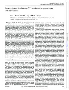
possibility might be to present small grating patches of orthogonal orientations along the horizontal meridian (Fig. 1). It is very probable that a classifier, trained on the opposite cardinal orientation configurations in Fig. 1 (A vs. B), would be able to decode the multivariate neural response on the basis of known coarse-scale influences such as radial biases (Sasaki et al. 2006) and/or differences in the response to vertical over horizontal (Freeman et al. 2011, 2013; Mannion et al. 2010). The critical comparison, however, would be between the opposite oblique orientation configurations (Fig. 1, C vs. D), where the angular distance between both the radial and cardinal orientations is equivalent along the horizontal meridian. Thus if accurate decoding was demonstrated in this case, it would be difficult to account for solely in terms of known coarse-scale biases, where no difference would be predicted. Even if successful, the results would still require careful interpretation, as small systematic activations reflecting global, coarse-scale orientation biases at the stimulus locations might still be exploited to explain the orientation decoding performance (although such biases should at least be minimal at that location). Furthermore, it would be important to consider a possible confound due to the edges of the grating stimuli: since the edges orthogonal to the grating orientation contain more terminators than those parallel to it, decoding could reflect the retinotopic response
A
B
C
D
Fig. 1. Schematic representation of 4 stimulus configurations that could be used to address the source of decoded orientation information. Accurate decoding of the configurations in A and B would be expected on the basis of known coarse-scale biases. The configurations in C and D represent the crucial test since the oblique orientations are equidistant from the radial and cardinal orientations (along the horizontal meridian), so the predicted difference due to coarse-scale biases should be minimal (if not absent).
J Neurophysiol • doi:10.1152/jn.00196.2014 • www.jn.org
Neuro Forum ORIENTATION DECODING IN HUMAN V1
patterns associated with the terminator locations. Possible ways around this would be to ramp or blur the contrast of the gratings at their edges (such as placing them within a Gaussian profile), or to only select voxels responsive to an inner region of the grating (Kamitani and Tong 2005). A different tactic, which should also circumvent the influence of a radial bias, might be to present spiral gratings positioned in the periphery, an approach recently used in probing the representation of complex polar form (Mannion and Clifford 2011) and complex motion (Maloney et al. 2013). In such a configuration, all orientations are equally represented in each small patch of the visual field, such that the orientation response can be disambiguated from any local radial biases centered at the fovea. Although coarse-scale biases have been well documented, they themselves remain mysterious, with their precise function and origin also the focus of continued debate (as Freeman et al. 2013 acknowledge). Nor, for that matter, is it necessarily agreed upon how fine-scale information arises in the fMRI blood oxygenation level dependent (BOLD) signal (Kriegeskorte et al. 2010; Shmuel et al. 2010). Kriegeskorte et al. (2010), for instance, suggest that the voxel-based sampling of neuronal activity is well described by a complex spatiotemporal filter, modeled in terms of the unique fine-grained vascular structure and the temporal properties of the hemodynamics. As they discuss, the complex spatiotemporal filter model copes better (than other models) with the ubiquitous head motion artefacts of human fMRI data that pose problems for the initial hypothesis that decoded orientation information reflects slight imbalances in the distribution of columnar sensitivities within the voxel grid (Kamitani and Tong 2005). Whether the source of fMRI orientation decoding comes from fine- or coarse-scale representations (or both, as is perhaps likely), it is impossible to rule out the potential influence of feedback mechanisms from higher areas or lateral interactions within V1 (Alink et al. 2013), although this is an inherent limitation of fMRI studies. Perhaps the most germane message to take from the work of Freeman et al. (2013) is an important admonition that applies to all fMRI decoding attempts: classifiers will exploit whatever information happens to be present in the multivariate pattern of activity, so the interpretation of what those sources of information might be must always be made with caution. ACKNOWLEDGMENTS I thank Dr. Damien J. Mannion, Prof. Colin W. G. Clifford (UNSW Australia), and Dr. Stefan Bode (The Univ. of Melbourne) for helpful suggestions and feedback on this article.
3
GRANTS This work was supported by Grant APP1027258 from the Australian National Health and Medical Research Council (to C. W. G. Clifford). DISCLOSURES No conflicts of interest, financial or otherwise, are declared by the author(s). AUTHOR CONTRIBUTIONS R.T.M. prepared figures; R.T.M. drafted manuscript; R.T.M. edited and revised manuscript; R.T.M. approved final version of manuscript. REFERENCES Alink A, Krugliak A, Walther A, Kriegeskorte N. fMRI orientation decoding in V1 does not require global maps or globally coherent orientation stimuli. Front Psychol 4: 493, 2013. Freeman J, Brouwer GJ, Heeger DJ, Merriam EP. Orientation decoding depends on maps, not columns. J Neurosci 31: 4792– 4804, 2011. Freeman J, Heeger DJ, Merriam EP. Coarse-scale biases for spirals and orientation in human visual cortex. J Neurosci 33: 19695–19703, 2013. Furmanski CS, Engel SA. An oblique effect in human primary visual cortex. Nat Neurosci 3: 535–536, 2000. Hubel DH, Wiesel TN. Shape and arrangement of columns in cat’s striate cortex. J Physiol 165: 559 –568, 1963. Kamitani Y, Sawahata Y. Spatial smoothing hurts localization but not information: pitfalls for brain mappers. Neuroimage 49: 1949 –1952, 2010. Kamitani Y, Tong F. Decoding the visual and subjective contents of the human brain. Nat Neurosci 8: 679 – 685, 2005. Kriegeskorte N, Cusack R, Bandettini P. How does an fMRI voxel sample the neuronal activity pattern: compact-kernel or complex spatiotemporal filter? Neuroimage 49: 1965–1976, 2010. Maloney RT, Watson TL, Clifford CW. Human cortical and behavioral sensitivity to patterns of complex motion at eccentricity. J Neurophysiol 110: 2545–2556, 2013. Mannion DJ, Clifford CW. Cortical and behavioral sensitivity to eccentric polar form. J Vis 11: 2011. Mannion DJ, McDonald JS, Clifford CW. Discrimination of the local orientation structure of spiral Glass patterns early in human visual cortex. Neuroimage 46: 511–515, 2009. Mannion DJ, McDonald JS, Clifford CW. Orientation anisotropies in human visual cortex. J Neurophysiol 103: 3465–3471, 2010. Op de Beeck HP. Against hyperacuity in brain reading: spatial smoothing does not hurt multivariate fMRI analyses? Neuroimage 49: 1943–1948, 2010. Sasaki Y, Rajimehr R, Kim BW, Ekstrom LB, Vanduffel W, Tootell RB. The radial bias: a different slant on visual orientation sensitivity in human and nonhuman primates. Neuron 51: 661– 670, 2006. Shmuel A, Chaimow D, Raddatz G, Ugurbil K, Yacoub E. Mechanisms underlying decoding at 7 T: ocular dominance columns, broad structures, and macroscopic blood vessels in V1 convey information on the stimulated eye. Neuroimage 49: 1957–1964, 2010. Swisher JD, Gatenby JC, Gore JC, Wolfe BA, Moon CH, Kim SG, Tong F. Multiscale pattern analysis of orientation-selective activity in the primary visual cortex. J Neurosci 30: 325–330, 2010. Yacoub E, Harel N, Ugurbil K. High-field fMRI unveils orientation columns in humans. Proc Natl Acad Sci USA 105: 10607–10612, 2008.
J Neurophysiol • doi:10.1152/jn.00196.2014 • www.jn.org