SUMMARY. The formation of membrane bone from neural crest-derived mesenchyme of the maxillary and mandibular processes of the embryonic chick ...
J. Embryol. exp. Morph. Vol. 64, pp. 305-320, 1981 Printed in Great Britain © Company of Biologists Limited 1981
3QC
The induction of neural crest-derived cartilage and bone by embryonic epithelia: an analysis of the mode of action of an epithelialmesenchymal interaction By BRIAN K. HALL1 From the Department of Biology, Dalhousie University, Halifax, Nova Scotia
SUMMARY The formation of membrane bone from neural crest-derived mesenchyme of the maxillary and mandibular processes of the embryonic chick depends upon prior interactions between the mesenchyme and maxillary or mandibular epithelia. The present study explores the specificity of these interactions using tissue recombinations between heterotypic epithelia and mesenchyme. Mandibular and maxillary mesenchyme responded to maxillary and mandibular epithelia by forming bone. A third osteogenically inductive epithelium, the scleral epithelium with its specialized scleral papillae, also allowed mandibular mesenchyme to form bone, indicating that mesenchyme can form bone in response to osteogenic epithelia other than its own. Epithelia which normally do not induce membrane bone formation in situ (wing and leg bud, back and abdominal epithelia) also allowed mandibular epithelia to ossify as did mandibular epithelia from the 10-day-old foetal mouse. Thus this tissue interaction is neither site nor species specific. Mandibular epithelium allowed bone to form in osteogenic mesenchyme from the maxilla and the sclera of the chick and from the mouse mandible but would not induce bone formation from normally non-osteogenic mesenchyme of the limb buds, chorioallantoic membrane or trunk neural crest. The results obtained with all of the tissue recombinations were consistent with the epithelial-mesenchyme interactions that initiate osteogenesis in both the mandibular and the maxillary processes being permissive interactions. The distinction between permissive and instructive interactions is discussed.
INTRODUCTION Tissue interactions, typically interactions between epithelia and embryonic mesenchyme, have been shown to play crucial roles in the initiation of the development and differentiation of most, if not all, organs of vertebrate embryos -see Deuchar (1975), Wessells (1977) and Lash & Burger (1977) for reviews. These tissue interactions fall into three broad categories: (1) those in which an epithelium controls the differentiation of mesenchyme, as in the development 1
Author's address: Depaitment of Biology, Dalhousie Univeisity, Halifax, Nova Scotia, Canada B3H4J1.
306
B. K. HALL
of the kidney, somites, and neural crest; (2) those in which mesenchyme control the differentiation of epithelium, as in the development of feathers, hair, and glands such as the salivary gland, and (3) those in which there is a reciprocity of interaction between epithelium and mesenchyme, as in the development of teeth and limb buds. The nature of such developmental^ important interactions is complex and may involve cell-to-cell communication between the interacting tissues, communication via the basement membrane of the epithelium, or communication via extracellular products produced by one or both tissues. A considerable literature aimed at elucidating the nature of the communication between the interacting tissues exists for each of these organ systems. The one general conclusion that comes from that literature is that no general conclusion can be made which applies to all tissue interactions in all organs. A second major question which can be asked for any tissue interaction relates to the specificity of the components. In interactions which involve an epithelium initiating a developmental change in mesenchyme, will other epithelia substitute for the normal epithelium? Alternatively, can the epithelium initiate development of the appropriate mesenchymal product from other than the normal mesenchyme? Such questions are usually phrased in terms of interactions being either permissive or instructive (Saxen et al. 1976; Saxen, \911a, b; Wessells, 1977). Does the epithelium merely permit development of the mesenchyme, in which case it would not act on 'foreign' mesenchyme, but other epithelia could substitute for it; or does the epithelium provide specific instructions to the mesenchyme, in which case it would act on foreign mesenchyme and could not be replaced by other epithelia? (See Wessells (1977) for a further discussion of these criteria.) Answers to these questions provide information on the specificity of the message, on the state of determination of the responding cells, on shared relationships between the responding cells and similar cells in other sites within the embryo, on the importance of the positioning of tissues relative to one another, and on how development is both timed and integrated. These are also the questions addressed in this paper, using as a model system, the epithelial induction of cartilage and membrane bone in the neural crest-derived mandibular and maxillary skeletons of the embryonic chick. Elegant experiments involving ablation, transplantation and labelling of segments of the embryonic neural crest performed by Johnston (1966) and LeLievre (1974, 1978) have shown that Meckel's cartilage, the membrane bones of the mandible and maxilla (surangular, angular, dentary, splenial, quadratojugal and jugal) and the connective tissue of the mandibular and maxillary processes of the embryonic chick are all derived from cells which originate in the neural crest. These cells leave the developing neural tube during closure of the neural folds and migrate to the future region of the mandibular and maxillary processes where they become associated with epidermal ectoderm and begin to differentiate. Cartilage forms during the fifth day of incubation and membrane bone (the first to form in the embryo) during the seventh day. Investigation of
Epithelial-mesenchymal interaction
307
the tissue interactions involved in the initiation of these skeletal tissues began with Tyler & Hall's (1977) observation that the neural crest-derived cells of the mandibular process would only form membrane bone after they had undergone an inductive interaction with the epithelium of the mandibular process - an interaction which continued until 4 days of incubation. A similar interaction preceeds osteogenesis in the maxillary and palatine skeleton. (Tyler & McCobb, 1980, 1981). Subsequently, it was shown that epithelia from the developing wing and hind limb buds could substitute for mandibular epithelia and allow osteogenesis to proceed (Hall, 1978 a), that the mandibular epithelium must be viable and proliferating to be inductively active (Hall, 1980a), that the presence of collagen and proteoglycan associated with the epithelium was required for induction to occur (Bradamante & Hall, 1980), and that epithelia from mandibular processes of the mouse could substitute for the chick mandibular epithelium (Hall, 19806). Similarly, the initiation of chondrogenesis within Meckel's cartilage requires that neural crest cells interact with the cranial ectoderm adjacent to the neural tube. Maxillary epithelia can substitute for that cranial ectoderm (Bee & Thorogood, 1980). For reviews of these data, of tissue interactions involved in neural crest-derived skeletogenesis in other vertebrates and of the use of tissue interactions in general, see (Hall 1980c, 1981a, h). The efficacy of limb-bud epithelia as substitutes for mandibular (Hall, 1978 a) or maxillary (Tyler & McCobb, 1980) epithelia, coupled with the efficacy of murine mandibular epithelium as a substitute for chick epithelium (Hall, 19806) suggested that these osteogenic tissue interactions were permissive and not instructive. As noted above, the ability to respond to nonspecific stimuli, in these cases to foreign epithelia, is one of the two criteria used by Wessells (1977) to designate a tissue interaction as permissive rather than instructive. The other criterion is the ability of the initiating tissue, in this case the mandibular or maxillary epithelium, to induce bone formation in tissue which does not normally form bone. This paper explores further aspects of these interactions with experiments designed to assess both these criteria. Firstly, foreign epithelia were recombined with maxillary or mandibular mesenchyme (more properly ectomesenchyme to denote its origin from the neural crest). Epithelia were obtained from the chick maxilla, mandible, wing buds, hind limb buds, back, abdomen and sclera as well as from the mouse mandible. This list includes both epithelia which normally induce the formation of membrane bone (maxillary mandibular and scleral epithelia) and epithelia which normally do not (limb, back and abdominal epithelia). Secondly, mandibular epithelia from the embryonic chick were recombined with mesenchyme from osteogenic sites other than the chick mandible. These included mouse mandibular mesenchyme, and chick maxillary and scleral mesenchyme. Thirdly, mandibular epithelia from the embryonic chick were recombined with mesenchyme from non-membrane-bone-forming sites, such as the back and
308
B. K. HALL
the wing bud, the chorioallantoic membrane and the trunk neural crest. The formation of bone in such mesenchyme would be strong evidence for the instructional nature of the normal epithelial-mesenchymal interaction. All tissue recombinations were maintained as grafts to the chorioallantoic membranes of host embryonic chicks. Isolated mesenchyme was used as control tissue. MATERIALS AND METHODS
Incubation procedure Fertile eggs of the domestic fowl, Gallus domesticus domesticus, white Leghorn, shaver starcross 288 strain were obtained from Cook's Hatchery, Truro, Nova Scotia. They were incubated without rotation in a forced-draft Petersime Incubator (Model 1, Petersime Incubator Co., Gettysburg, Ohio, U.S.A.) maintained at 37 + 0-5 °C and 55 + 4 %. Animals ICR Swiss albino mice obtained from Bio-breeding Laboratories of Canada, Ottawa, Ontario were weaned at 4 weeks of age, housed by sex, three or four per cage, and fed Purina Lab. chow and water ad libidum. Animals used for matings were placed together overnight, one male to between one and three females. Pregancy was determined by the presence of a vaginal plug and by weight gain over the first 8 days of the pregnancy. Embryos Eggs incubated for various periods of time were opened under sterile conditions, the embryos removed and placed into Petri dishes containing sterile saline. Embryos were staged using the morphological series of H.H. stages established by Hamburger & Hamilton (1981). Pregnant mice were killed by an overdose of ether. The embryos were dissected from the uterii under sterile conditions, placed into sterile saline and staged using the morphological series of stages established by Griineburg (1943) and Theiler (1972). Embryos of 9 days (13-29 pairs of somites, Theiler stages 14 and 15) and 10 days of gestation (30-39 pairs of somites, Theiler stages 16 and 17) were used. Separation of tissues Mandibular and maxillary processes, limb buds, dorsal and ventral body wall, sclera and trunk neural crests were dissected from embryos under sterile conditions. All except the trunk neural crests were placed into a solution of trypsin and pancreatin in calcium- and magnesium-free Tyrode's solution (257 mg bovine pancreatic trypsin + 43 mg porcine pancreatic pancreatin/10 ml, both obtained from BDH Chemicals, Toroto, Ontario) at 4 °C for either 1 h for
EpitheliaUmesenchymal interaction
309
chick tissues, or for 2 h for the mouse mandibles. The tissues were then separated into their epithelial and mesenchymal components by microdissection using sharpened hypodermic needles. The dissections were performed in a mixture of the complex culture medium BGJb and horse serum (1:1, v/v), a mixture which provided a rich protein substrate to inactivate any residual enzymes in the tissues. The isolated epithelia and mesenchymes were either cultured alone as controls, recombined with one another as controls, or combined with other mesenchyme or epithelia, e.g. mandibular epithelium combined with wing-bud mesenchyme. Details of the various recombinations and ages of tissues used may be found in Tables 1 and 2. Recombinations were carried out by affixing, with gentle pressure, the epithelium to a square of sterile, black, Millipore filter of 0-45 /an porosity and 125 ju,m thickness (Millipore Filter Corp., Montreal, Quebec. The mesenchyme was placed onto the epithelium to allow reestablishment of epithelial-mesenchymal contact. Chorioallantoic grafting Isolated or recombined tissues were maintained as grafts to the chorioallantoic membranes of host embryonic chicks aged between 7 and 10 days of incubation, a highly vascularized region of the chorioallantoic membrane was located by candling the egg in a light box. A window was cut in the shell and shell membrane to expose the vascular chorioallantoic membrane. The tissue, now positioned on the Millipore filter, was placed upon the chorioallantoic membrane, tissue side down. The shell window was replaced, sealed with Scotch tape and the host returned to the incubator for a further 7 to 8 days. Full details of the chorioallantoic grafting procedure may be found in Hall (19786). Histology After 7-8 days on the chorioallantoic membranes, the grafts were recovered by dissecting them away from their vascular, encapsulating, chorioallantoic membranes. The grafts were then fixed in neutral buffered formal saline, dehydrated, cleared, embedded in paraffin, serially sectioned at 6 /tm and stained with haematoxylin, alcian blue and chlorantine fast red (modified from Lison, 1954). All sections were examined for the presence or absence of bone and/or cartilage. RESULTS
Controls Several previous studies have firmly established that the membranes, bones of both the mandibular and the maxillary processes will only begin to form bone at 1-1\ days of incubation if the mesenchyme of these processes has been in contact with an epithelium until the fourth day of incubation. In contrast, the formation of cartilage does not depend on the presence of these epithelia (Tyler
310
B. K. HALL
Table 1. Heterotypic epithelia allow mandibular and maxillary mesenchyme to form membrane bone: Expressed as number of grafted recombinants forming cartilage and/or bone Source of mesenchyme* Source of epithelium
Mandibular
Maxillary
Number of grafts forming Cartil1 — 2 Mandibular* 3 Maxillary*
age 58 109 28
CartilBone 0 100 22
n 60
119 30
4 Scleral 11 8 13 (i) H.H. stages 31-32 8 8 (ii) H.H. stage 35 8 5 Leg bud 34 54 (i) H.H. stages 18-24 51 18 (ii) H.H. stages 26-28 14 4 6 Wing bud 32 (i) H.H. stages 18-22 32 0 38 (ii) H.H. stages 24-28 38 22 7 Back (i) H.H. stage 22 12 5 13 14 (ii) H.H. stage 31 13 8 12 8 Abdominal-H.H. stage 31 9 8 9 Mouse mandible-10 day 14 10 14 * Obtained from H.H . stage-22 embryos
age 14 15 22
Bone 0 12 15
n 21 16
27
15
32
12
8
16
24
& Hall, 1977; Hall, 1978a, c; 1980a; Hall and Tremaine, 1979; Bradamante & Hall, 1980; Tyler & McCobb, 1980). Consequently when mesenchyme from early embryos is maintained in vitro or as a chorioallantoic graft in isolation from its epithelium, it forms cartilage but not bone. Isolated mesenchymes from the mandibular and maxillary processes of H.H. stage-22 embryos were grafted to chorioallantoic membranes in this study as controls for the behaviour of such mesenchyme in the absence of epithelia. H.H. stage-22 embryos were used because the normal inductive interaction between mandibular and maxillary epithelia and mesenchyme is not completed until H.H. stage 24, some 24 h later (Tyler & Hall, 1977; Hall, 1978c; Tyler & McCobb, 1980). The time gap ensured that no mesenchyme used in these experiments would have been primed for osteogenesis at the outset of its time as a graft. As expected from these previous studies, a high percentage of such isolated mesenchymes in this study formed cartilage (97% of mandibular mesenchymes formed rods of Meckel's cartilage; 66 % of maxillary mesenchymes
Epithelial-mesenchymal interaction
311
formed rods of palato-quadrate cartilage) but none formed bone (Table 1, line 1). The lower percentage of maxillary processes which formed cartilage reflected the fact that only the posterior portion of the maxillary process is chondrogenic, and that this region was sometimes left behind on the embryo after the rest of the process had been removed. Recombination of mesenchyme (whether mandibular or maxillary), with its own epithelium was a sufficient stimulus for bone to form in 84 % of mandibular mesenchymes and in 62-5% of maxillary mesenchymes (Table 1, lines 2 and 3). These recombinations confirmed that neither the enzymatic digestion used to separate the components, nor the methods used to reassociate or to maintain the tissues inhibited normal differentiation. Given the adequacy of these techniques, I proceeded to perform the three sets of experiments described below. Osteogenic mesenchyme combined with heterotypic epithelia The first set of experiments was designed to determine whether mandibular or maxillary mesenchyme could respond to other (heterotypic) epithelia by forming bone. For the reasons already stated the mesenchyme was always obtained from embryos of H.H. stage 22. As noted in the previous section homotypic recombinations allowed bone to form in both mandibular mesenchyme recombined with mandibular epithelium and in maxillary mesenchyme recombined with maxillary epithelium (Table 1, lines 2 and 3). Similar results were obtained when mandibular mesenchyme was recombined with maxillary epithelium and vice versa (so called heterotypic recombinations, Table 1, lines 2 and 3). Thus osteogenically inductive facial epithelia can interact with other than the normal neural crest-derived mesenchyme and still elicit osteogenesis from that pre-osteogenic mesenchyme. Heterotypic epithelia from a third site involving the induction of neural crest-derived membrane bone, namely the scleral epithelium, was then tested. During normal development the scleral epithelium forms fourteen thickenings, the scleral papillae, each of which induces the adjacent neural crest-derived mesenchyme to form a scleral bone or scleral ossicle (Coulombre, Coulombre & Mehta, 1962). Fyfe & Hall (1981) may be consulted for a recent evaluation of the development of these epithelial scleral papillae which represent a highly specialized and morphologically complex osteogenic inductive epithelium. The first scleral papilla appears at H.H. stage 30 (7 days of incubation) and papillae are well advanced by 9 days of incubation (Fyfe & Hall, 1981). When scleral papillae from both these aged embryos were recombined with mandibular mesenchyme and grafted, osteogenesis was initiated. More grafts formed bone in the presence of the older papillae than in the presence of the younger ones (Table 1, line 4). The morphology of the bones formed was typical of mandibular membrane bone rather than resembling the plate-like scleral ossicles. Not only was osteogenesis initiated in the presence of these epithelia which
312
B. K. HALL
normally acted on other osteogenic neural crest-derived mesenchyme, it was also initiated in response to epithelia which normally do not induce bone formation, viz epithelia derived from the limb buds, from the back and from the abdomen of embryonic chicks (Table 1, lines 5-8). The incidence of bone observed after recombination of mandibular mesenchyme with older leg epithelia (H.H. stages 26-28) or with younger wing epithelia (H.H. stages 18-22) was much reduced in comparison with that seen with other aged epithelia (Table 1, lines 5 and 6). This phenomena has been explored in more detail in Hall (1978a). Although epithelia from embryos of certain ages were inactive, it was evident that bone could be induced to form in response to a variety of heterotypic epithelia which do not induce bone to form during their normal interactions with mesenchyme. Not only were the epithelial-mesenchymal interactions not epithelium specific, they were not species specific either. Epithelia from mandibular processes of 10-day-old foetal mice could substitute for the avian epithelia and allow bone to form (Table 1, line 9). During normal murine development the mandibular epithelium induces mandibular mesenchyme both to ossify and to chondrify (Hall, 19806). The results of these recombinations of osteogenic mesenchyme with both heterotypic and heterospecific epithelia and with normally osteogenically. inductive and non-inductive epithelia were consistent with the epithelialmesenchymal interactions being permissive and with lack of specificity in the epithelial component. Osteogenic inductive epithelia combined with osteogenic mesenchyme This second set of experiments was designed to determine whether osteogenic inductive epithelia from the maxillary and mandibular processes could allow other known preosteogenic mesenchyme to form bone. One such combination, the reciprocal heterotypic recombination between maxillary and mandibular epithelia and mesenchyme was described in Table 1, lines 2 and 3 and showed that the epithelia could act on those heterotypic mesenchymes. For completeness those data are reproduced in Table 2. As already noted in the results of the first set of experiments, scleral mesenchyme normally responds to the inductive action of the epithelial scleral papillae by forming an ossicle of bone. The scleral papilla was shown to be capable of inducing the differentiation of membrane bone from mandibular mesenchyme (Table 1, line 4). In the present experiments neither scleral mesenchyme from H.H. stages 30-32 (6-5-7-5 days of incubation) embryos, or from H.H. stage-35 (9-day) embryos formed bone when maintained in isolation from the scleral epithelium (Table 2, lines 3 and 4). However scleral cartilage did differentiate in all grafts. The formation of scleral cartilage depends upon an inductive interaction between scleral mesenchyme and the pigmented retinal epithelium (Newsome, 1972), an interaction which was completed before the mesenchyme
Epithelial-mesenchymal interaction
W' -
•
*
313
^_.
*
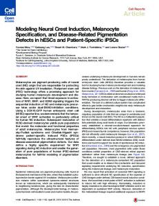
0-02mm Fig. 1. Scleral mesenchyme from an H.H. stage-35 embryo combined with mandibular epithelium from an H.H. stage-22 embryo and grafted to the chorioallantlic membrane on a Millipore filter (m) results in the formation of a sheet of scleral cartilage (c) and of two scleral ossicles (O) composed of membrane bone. Alcian blue, chlorantine fast red and haematoxylin. Fig. 2. A higher magnification photomicrograph of the bones from Fig. 1 to show that their form is typical of scleral ossicles rather than of mandibular membrane bones, even though mandibular epithelium induced their differentiation.
was isolated (Stewart & McCallion, 1975). The addition of mandibular epithelium to scleral mesenchyme allowed bone to form in 40-44 % of the grafts (Table 2, lines 3 and 4; Figs. 1, 2). That epithelium can act on heterotypic osteogenic mesenchyme. Essentially similar results were obtained when the mandibular epithelium was recombined with mandibular mesenchyme from the 9-day-old mouse. By itself the murine mesenchyme formed neither cartilage nor bone (Table 2, line 5) the differentiation of both tissues normally requiring the presence of the murine
314
B. K. HALL
Table 2. Mandibular and maxillary epithelia allow pre-osteogenic mesenchyme but not non-osteogenic mesenchyme to form bone* Number• of grafts forming Source of mesenchyme 1 Mandiblet 2 Maxillaryf 3 Scleral (H.H. stages 30-32) 4 Scleral (H.H. stage 35) 5 Mouse mandible (9 day)
Bone
n
Osteogenic mesenchyme None 58 22 + Maxillary None 12 15 + Mandibular None 8
0 15 0 12 0
60 24 21 16 8
22 7 9 0 12
9 0 4 1 11
22 7 9 16 25
epitheliumf
+ Mandibular None + Mandibular None + Mandibular
Cartilage
Non-osteogenic mesenchyme 10 None 10 0 12 0 12 + Mandibular 0 None 5 5 7 Backf 12 0 12 + Mandibular 14 0 8 Chorioallantoic None 0 10 0 0 + Mandibular 7 0 9 Trunk neural crest None 3 0 2 9 (H.H. stage 10) + Mandibular * Based on number of grafts forming cartilage and or bone. In each of the five combinations the isolated mesenchyme serves as the control. t Obtained from H.H. stage 22 embryos. t Sub-periosteal membrane bone does form in later stages of wing development - see discussion for details. 6 Wing budf,J
mandibular epithelium (Hall, 19806). Addition of mandibular epithelia from the embryonic chick allowed both cartilage and bone to form (Table 2, line 5). Thus not only will mandibular epithelium allow other preosteogenic mesenchyme (maxillary, scleral, mouse mandibular) to initiate osteogenesis, it will also allow chondrogenesis to commence in a prechondrogenic mesenchyme (mouse mandibular) which normally requires an epithelial stimulus. Again these results are indicative of permissive epithelial-mesenchymal interactions. Osteogenic inductive epithelia combined with non-osteogenic mesenchyme This third set of experiments provides perhaps the most crucial test for the nature of an inductive tissue interaction. Will the inducer, in this instance the mandibular epithelium, allow-non-osteogenic mesenchyme to form membrane bone? This question was approached using mesenchyme from (a) the wing buds
Epithelial-mesenchymal interaction
315
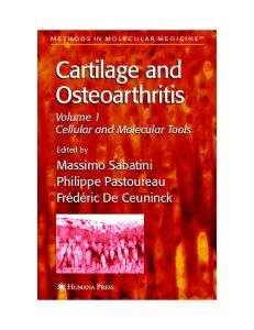
Fig. 3. Back dermis from H.H. Stage-22 embryos grafted alone formed cartilage which was recognizably vertebral in type. N, notochord. Alcian blue, chlorantine fast red and haematoxvlin.
and from the back, both being locations where membrane bone does not form but which are rich in mesodermally-derived mesenchyme which normally forms cartilage; (b) the non-skeletogenic mesenchyme of the chorioallantoic membrane and (c) the trunk neural crest which produces mesenchyme that forms neither cartilage nor bone. Wing and back mesenchyme was obtained from H.H. stage-22 embryos, combined with mandibular epithelium from similar aged embryos and grafted. Mesenchyme grafted alone formed cartilage (Table 2, lines 6 and 7), which was recognizable as either vertebral (Fig. 3) or appendicular, but did not form bone. Addition of the mandibular epithelia was not a sufficient stimulus to allow membrane bone to form (Table 2, lines 6 and 7), even though the epithelium was known to be inductively active and the graft site known to provide an environment capable of supporting osteogenesis. To assess the response of the chorioallantoic mesenchyme, mandibular epithelia were affixed to Millipore filters and grafted with the epithelia in direct contact with the chorioallantoic membrane. Even though the chorioallantoic membrane has been shown to support continued and normal differentiation of such epithelia (Tyler & Hall, 1977), neither cartilage nor bone ever formed (Table 2, line 8). Nor did bone form when millipore filters alone were grafted to the chorioallantoic membrane (Table 2, line 8). To assess the response of the trunk neural crest cells, neural tubes were isolated from regions adjacent to somites 1-10 of H.H. stage-10 embryos and grafted in isolation or in combination with H.H. stage-22 mandibular epithelium. (Bee & Thorogood (1980) had shown that premigratory cranial neural
316
B. K. HALL
crest cells could respond to maxillary epithelia by forming bone). Grafts containing trunk neural crest but no epithelia formed cartilage (3/7 grafts, Table 2, line 9). This cartilage could have originated from neural crest cells for the crest at the level of the first pair of somites has chondrogenic potential (LeLievre, 1978). However the morphology of these cartilage nodules was consistent with their being vertebral and having arisen from somatic mesoderm carried along with the neural tubes. The incidence of cartilage was not increased when epithelia were present (Table 2, line 9). Neither isolated trunk neural crests nor crests combined with mandibular epithelium formed bone (Table 2, line 9). Thus, in no instance was normally non-osteogenic mesenchyme or ectomesenchyme able to form membrane bone in response to induction from mandibular epithelium. DISCUSSION Six conclusions may be drawn from the results obtained in these experiments. (1) Isolated H.H. stage-22 mandibular and maxillary mesenchymes will form cartilage but not membrane bone when maintained as chorioallantoic grafts. (2) Recombination of mandibular or maxillary mesenchymes with their own epithelia is a sufficient stimulus to allow bone to form. (3) Mandibular and/or maxillary mesenchymes can respond to heterotypic epithelia by forming bone. The formation of bone occurs both in response to epithelia that normally induce bone in situ (maxillary, mandibular and scleral epithelia) and in response to epithelia that normally do not (limb bud, back and abdominal epithelia). Therefore the epithelial requirement is not tissue specific. (4) Mandibular mesenchyme from the embryonic chick can respond to mouse mandibular epithelium by forming bone. Therefore the epithelial requirement is not species specific. (5) Mandibular epithelium will allow heterotypic osteogenic mesenchyme (maxillary, scleral, mouse mandibular) to initiate osteogenesis but will not induce bone formation from normally non-osteogenic mesenchyme (limb bud, chorioallantoic membrane, trunk neural crest). Therefore the epithelium does not act instructively and the mesenchymal cells must be determined for osteogenesis in order to respond. (6) All the results obtained are consistent with these osteogenic epithelialmesenchymal interactions being permissive. Several epithelia share the inductive ability but the ability of mesenchyme to respond is restricted to osteogenic neural crest-derived mesenchyme. The general conclusion, that osteogenic mesenchyme can respond to a variety of epithelia, has to be regarded with caution. As summarized in Table 1 wing bud epithelium from embryos of H.H. stages 18-22 is not inductively active whilst older wing epithelium is. Mandibular epithelium loses its inductive activity at H.H. stage 23, coincident with the completion of the normal in situ
Epithelial-mesenchymal interaction
317
interaction (Hall, 1978 a). We have found, in unpublished studies, that neither chorioallantoic epithelium nor vitelline membrane will substitute for mandibular epithelia and we have preliminary evidence that epithelia of endodermal origin are also inactive. So, whilst heterotypic epithelia will act, age of the epithelium is important. I have previously shown (Hall, 1980a) that inductive activity is correlated with mitotic activity of the epithelium and Bradamante & Hall (1980) have shown that epithelial extracellular products may also play a role. The ability to respond to these inductively active epithelia is not restricted to mesenchyme derived from the cranial neural crest for Tyler (1980) has shown that the mesodermally derived mesenchyme which forms the frontal bone can respond to heterotypic epithelia by forming bone. Cranial mesenchyme, whether derived from the neural crest or from the mesoderm, can respond to inductively active epithelia by forming bone. However, neither mesenchyme derived from trunk neural crest nor mesenchyme derived from trunk mesoderm responded by forming bone (Table 2). The lack of response of trunk neural crest may be taken as one more piece of evidence for a complete separation in differentiative ability between a skeletogenic cranial neural crest and a non-skeletogenic trunk neural crest. The restriction of the synthesis of fibronectin to the cranial neural crest may be a further reflexion of this dichotomy (Newgreen & Thiery, 1980). The argument then is that trunk neural crest cells are not determined for osteogenesis and therefore cannot respond to known osteogenic inductive influences emanating from the mandibular epithelium. But can this same argument be applied to the mesodermally derived mesenchyme of the limb buds? Membrane bone does arise in the periosteum of the cartilaginous models of long bones (Scott-Savage & Hall, 1979, 1980) although not until early H.H. stage 30, with Type I collagen having appeared at H.H. stage 28 or 5-5-6 days (Von der Mark, Von der Mark & Gay, 1976a, b). It could be argued that the stimulus for the formation of this sub-periosteal membrane bone is quite distinct from that provided by the mandibular epithelium (it is probably provided by the underlying hypertrophic cartilage - Scott-Savage & Hall, 1979), or that the mesenchymal cells were not responsive at the age (H.H. stage 22) used in the recombinations. However Osdoby & Caplan (1979; 1980) have shown that cultured H.H. stage-24 limb mesenchymal cells form both osteoblasts and a mineralized matrix and that they form it independent of contact with cartilage. Therefore, at least at H.H. stage 24, the limb bud does contain a population of osteogenic cells. If the same is true for the H.H. stage-22 limb bud, then the epithelia which induce the formation of cranial neural crest-derived membrane bones cannot elicit osteogenesis from osteogenic mesodermally-derived trunk mesenchyme. I have argued elsewhere (Hall, 1975, 1978 c) that this dichotomy may have a very ancient evolutionary history, going back to the origin of the vertebrates. These differences between groups of mesenchymal cells, both determined for osteogenesis, but responsive to different inductive influences, cannot readily be H
EMB 64
318
B. K. HALL
fitted into the simple division between permissive or instructive inductions. Limb-bud epithelium does not induce bone formation within limb-bud mesenchyme but does induce mandibular and maxillary mesenchyme to ossify. Neither back nor abdominal epithelia induce bone in situ but both allow mandibular mesenchyme to ossify! Either their inductive ability is present but inhibited in situ or mandibular but not limb-bud mesenchyme is able to elicit inductive activity from, or provide inductive activity to, these epithelia. The latter would necessitate a two-way interaction between epithelium and mesenchyme. Such reciprocity is seen in other tissue interactions such as those between the apical ectodermal ridge and limb mesenchyme in the control of proximodistal outgrowth of the limb skeleton (Saunders, 1948; Summerbell, 1974), between the enamel organ and the dental papilla in tooth formation, or in the formation of epidermal appendages such as feathers or hairs (Koller 1972; Deuchar, 1975; Wessell, 1977). This research was supported by the Natural Sciences and Engineering Reseach Council of Canada (Grant No. A 5056) and by the Dalhousie University Research Development Fund in the Sciences. The expert technical woik of Sharon Brunt is gratefully acknowledged.
REFERENCES J. & THOROGOOD, P.,V. (1980). The role of tissue interactions in the skeletogenic differentiation of avian neural crest cells. Devi Biol. 78, 47-62. BRADAMANTE, Z. & HALL, B. K. (1980). The role of epithelial collagen and proteoglycan in the initiation of osteogenesis by avian neural crest cells. Anat. Rec. 197, 305-315.
BEE,
COULOMBRE, A. J., COULOMBRE, J. L. & MEHTA, H. (1962). The skeleton of the eye. I. Con-
junctival papillae and scleral ossicles. Devi Biol. 5, 382-401. E. M. (1975). Cellular Interactions in Animal Development. London: Chapman & Hall. FYFE, D. MACG. & HALL, B. K. (1981). A scanning electron microscopic study of the developing epithelial scloal papillae in the eye of the embryonic chick. /. Morph. 167, 201— 209. GRUNEBERG, H. (1943). The development of some external features in mouse embryos. J. Hered. 34, 89-92. HALL, B. K. (1975). Evolutionary consequences of skeletal development. Amcr. Zool. 15, 329-350. HALL, B. K. (1978 a). Initiation of osteogenesis by mandibular mesenchyme of the embryonic chick in response to mandibular and non-mandibular epithelia. Archs Oral Biol. 23, 1157-1161. HALL, B. K. (1978/?). Grafting organs and tissue to the chorioallantoic membrane of the embryonic chick. Tissue Culure Assoc. Manual 4, 881-884. HALL, B. K. (1978C). Developmental and Cellular Skeletal Biology. New York & London: Academic Press. HALL, B. K. (1980a). Viability and proliferation of epithelia and the initiation of osteogenesis, within mandibular ectomesenchyme in the embryonic chick. J. Embryol. exp. Morph. 56 71-89. HALL, B. K. (19806). Tissue interactions and the initiation of osteogenesis and chondrogenesis in the neural crest-derived mandibular skeleton of the embryonic mouse as seen in isolated murine tissues and in recombinations of murine and avian tissues. /. Embryol. exp. Morph. 58, 251-264. DEUCHAR,
Epithelial-mesenchymal interaction
319
HALL, B. K. (1980C). Chondrogenesis and osteogenesis in cranial neural crest cells. In Current Research Trends in Prenatal Craniofacial Development (ed. R. M. Pratt and R. L. Christiansen), pp. 47-63. New York: Elsevier, North Holland. HALL, B. K. (1981a). Embryogenesis:-cell-tissue interactions. In Skeletal Research-An Experimental Approach (ed. D. J. Simmons & A. S. Kunin), vol. 2. New York & London: Academic Press. (In Press.) HALL, B. K. (1981 b). Intra- and extracellular control of the differentiation of cartilage and bone. Histochem. J. (In Press.) HALL, B. K. & TREMAINE, R. (1979). Ability of neural crest cells from the embryonic chick to differentiate into cartilage before their migration away from the neural tube. Anat. Rec. 194, 469-476. HAMBURGER, V. & HAMILTON, H. L. (1951). A series of normal stages in development of the chick embiyo. / . Morph. 88, 49-92. JOHNSTON, M. C. (1966). A radioautographic study of the migration and fate of cranial neural crest cells in the chick embryo. Anat. Rec. 156, 143-156. KOLLAR, E. J. (1972). The development of the integument: spatial, temporal and phylogenetic factors. Amer. Zool. 12, 125-135. LASH, J. W. & BURGER, M. M. (eds.) (1977). Cell and Tissue Interactions. New York: Raven Press. LELIEVRE, C. (1974). Role des cellules mesectodermiques issues des cretes neurales cephaliques dans la formation des arcs branchiau et du squelette visceral. / . Embryol. exp. Morph. 31, 453-477. LELIEVRE, C. (1978). Participation of neural crest derived cells in the genesis of the skull in birds. / . Embryol. exp. Morph. 41, 17-37. LISON, L. (1954). Alcian blue 8G with chlorantine fast red 5B: a technic for selective staining of mucopolysaccharides. Stain Technol. 29, 131-138. NEWGREEN, D. & THIERY, J-P. (1980). Fibronectin in early avian embryos: synthesis and distribution along the migration pathways of neural cells. Cell & Tissue Res. 211, 269292. NEWSOME, D. A. (1972). Cartilage induction by retinal pigmented epithelium of chick embryos. Devi Biol. 28, 575-579. OSDOBY, P. & CAPLAN, A. I. (1979). Osteogenesis in cultures of limb mesenchymal cells. Devi Biol. 73, 84-102. OSDOBY, P. & CAPLAN, A. I. (1980). A scanning electron microscopic investigation of in vitro osteogenesis. Calc. Tissue Intern. 30, 43-50. SAUNDERS, J. W. JR. (1948). The proximo-distal sequence of oiigin of the parts of the chick wing and the role of the ectoderm. / . exp. Zool. 108, 363-404. SAXEN, L. (1977fl). Directive versus permissive induction: a working hypothesis. In Cell and Tissue Interactions (ed. J. W. Lash & M. M. Bulger), pp. 1-9, New York: Raven Press. SAXEN, L. (19776). Morphogenetic tissue interactions: an introduction. In Cell Interactions in Differentiation (eds. M. Karkinen-Jaaskelainen, L. Saxen & L.Weiss), pp. 145-151, New York: Academic Press. SAXEN, L., KARKINEN-JAASKELAINEN, M., LEHTONEN, E., NORDLING, S. & J. WARTIOVAARA.
(1976). Inductive tissue interactions. In Cell Surface in Animal Embryogenesis (eds. G. Poste & G. L. Nicolson), Cell Surface Reviews, vol. 1, pp. 331-408. Amsterdam: ElsevierNorth Holland Biomed. Press. SCOTT-SAVAGE, P. & HALL, B. K. (1979). The timing of the onset of osteogenesis in the tibia of the embryoic chick. / . Morph. 162, 453-464. SCOTT-SAVAGE, P. & HALL, B. K. (1980). Differentiative ability of the tibial periosteum from the embryonic chick. Acta Anat. 106, 129-140. STEWART, P. A. & MCCALLION, D. J. (1975). Establishment of the scleral cartilage in the chick. Devi Biol. 46, 383-389. SUMMERBELL, D. (1974). A quantitative analysis of the effect of excision of the AER from the chick bud. / . Embryol. exp. Morph. 32, 651-660. THEILER, K. (1972). The House Mouse. Development and Normal Stages from Fertilization to 4 Weeks of Age. Berlin, Heidelberg and New York: Springer- Verlag.
320
B. K. HALL
M. S. (1980). Tissue interactions in the development of neural crest-derived membrane bones. Amer. Zool. 20, 944. TYLER, M. S. & HALL, B. K. (1977). Epithelial influences on skeletogenesis in the mandible of the embryonic chick. Anat. Rec. 188, 229-240. TYLER, M. S. & MCCOBB, D. P. (1980). The genesis of membrane bone in the embryonic chick mandible: epithelial-mesenchymal tissue recombination studies. J. Embryol. exp. Morph. 56, 269-281. TYLER, M. S. & MCCOBB, D. P. (1981). Tissue interactions promoting osteogenesis in the embryonic chick palate. Archs Oral Biol. (In Press.) VON DER MARK, H., VON DER MARK, K. & S. GAY. (1976O). Study of differential collagen synthesis during development of the chick embryo by immunofluorescence. 1. Preparation of collagen type I and type II specific antibodies and their application to early stages of the chick embryo. Devi Biol. 48, 237-249. VON DER MARK, L., VON DER MARK, H. & S. GAY. (19766). Study of differential collagen synthesis during development of the chick embryo by immunofluorescence. II. Localization of Type I and type II collagen during long bone development. Devi Biol. 63, 153-170. WESSELS, N. K. (1977). Tissue Interactions and Development. Menlo Park, Calif.: W. A. Benjamin Inc.
TYLER,
(Received 16 January 1981, revised 17 March 1981)