(active site/acyl group transfer/fragment C3d/y-glutamylmethylamide/S-cyanocysteine). MATTHEW L. THOMAS*tt, JARMILA JANATOVAt, WILLIAM R. GRAY§, ...
Proc. Nati Acad. Sci. USA
Vol. 79, pp. 1054-1058, February 1982 Biochemistry
Third component of human complement: Localization of the internal thiolester bond (active site/acyl group transfer/fragment C3d/y-glutamylmethylamide/S-cyanocysteine)
MATTHEW L. THOMAS*tt, JARMILA JANATOVAt, WILLIAM R. GRAY§,
AND
BRIAN F. TACK*¶I
*Department of Pediatrics, Harvard Medical School and Department of Medicine, Children's Hospital Medical Center, Boston, Massachusetts 02115; and Departments of tPathology and §Biology, University of Utah, Salt Lake City, Utah 84132
Communicated by Sidney F. Velick, October 28, 1981
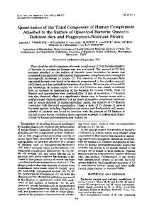
ABSTRACT Human complement protein C3 was inactivated by using methylamine and thereby generating a SH group from the internal thiol ester. The protein was coupled via this SH group to activated thiol-Sepharose and digested with elastase. Fragment C3d remained attached to the thiol-Sepharose and was subsequently eluted with L-cysteine. Concomitantly, the original SH group was regenerated, and it was then labeled with iodo[23H]acetic acid. Partial sequence analysis of the radiolabeled C3d fragment showed that both components of the thiol ester are located close to the amino terminus (residues 23 and 26). Specific chemical cleavage of the a-chain was achieved after S-cyanylation of the thiol. The two fragments obtained corresponded to the amino-terminal section (Mr "=46,000) and the carboxy-terminal section (Mr ="70,000). These results together indicate that fragment C3d occupies approximately positions 345-610 of the achain. The -partial sequence of C3d was extended by completion of the sequence of a previously described tryptic peptide. Comparison of residues 1-49 of C3d with a peptide from a2-macroglobulin [Swenson, R. P. & Howard, J. B. (1980) J. BioL Chem. 255, 8087-091] shows a previously recognized identity of seven residues around the thiol ester site and a second region of identity around a known glycosylation site of a2-macroglobulin. The relationships among these proteins and protein C4 are discussed. An overall outline of the structure of C3 is presented, showing the locations of various fragments and cleavage sites. The thiol ester group places constraints on the local folding of the peptide chain; a possible conformation is suggested and discussed in relation to the mechanism of activation.
The complement system comprises a series of proteins that facilitate destruction of immune complexes, including lysis of for-eign cells. Two routes of activation (classical and alternative) of the system are known, both involving the third component, C3. This protein consists of two disulfide-bridged chains, a (Mr -115,000) and P (Mr =75,000). Activation of C3 entails proteolytic removal of a vasoactive peptide, C3a, from the amino end of the a-chain. The remaining disulfide-bridged protein, C3b, functions in subsequent steps ofthe lytic sequence in both pathways, in positive-feedback activation of the earlier steps of the alternative pathway, and in opsonization reactions (for reviews, see refs. 1-4). "Nascent" C3b (5) generated by either pathway binds covalently to cell surfaces and complex polysaccharides through an ester bond (6) in which the acyl group is contributed by a residue from the C3d region of the a-chain (7, 8). The C3d fragment is produced from C3b by the concerted actions of C3b inactivator and /31H globulin (9) and additional blood proteases (10-12). Native C3, prior to activation, appears to have a site equivalent to that in nascent C3b but of much lower reactivity. Treat-
ment of C3 with hydrazine (13, 14), ammonia (13), or KBr (15) destroys its hemolytic function, changes its behavior on sedimentation and immunoelectrophoresis, and alters its antigenicity. It was previously shown in our laboratory (16, 17) that loss of the hemolytic function in C3 is in one-to-one correspondence with the appearance of a unique thiol group in the a-chain and with covalent attachment of the nucleophile [14C]methylamine to the a-chain (18). In the latter study, a 35-residue tryptic peptide was isolated and shown to contain both the nucleophilebinding residue (glutamyl derivative) and the cysteine residue that is exposed on inactivation. Therefore, it was proposed (18) that the two residues are present in thiol ester linkage in native C3 and that inactivation results from cleavage of the bond. Remarkably, the local sequence containing the glutamyl derivative and cysteine residues (Gly-Cys-Gly-Glu-Glx-Asn-Met) was identical with that surrounding an alkylamine-sensitive site in y2-macroglobulin (a2M) (19), which was also proposed to contain a thiol ester (18). This interpretation was subsequently adopted by Howard (20), who had earlier favored an internal pyroglutamyl residue (21). Recent studies by Law et al. (22) and Sim et aL (23) further support the idea that there is an activated internal acyl group in C3 (8) that can subsequently be transferred to 0- or N-nucleophilic groups on the surface of activating particles. Covalent attachment of a2M to proteases occurs after cleavage by the protease and is probably accomplished by a similar acyl group transfer (24-26). The fourth component of complement, C4, functions very much like C3 (27-33) and contains an identical sequence around the thiol ester site (34, 35). Surprisingly, C5, which' has many similarities to C3 and C4, seems to lack a thiol ester: treatment with methylamine does not result in loss of hemotytic function, in incorporation of reagent, or in release of a thiol group (22, 32). The' thiol ester site of C3 is located in the C3d region of the a-chain. This region is defined on the basis -of its release as a discrete fragment of Mr =33,000 on inactivation of bound C3b by enzymatic conversion. The C3d remains covalently bound to the cell surface (7) and is recognized by a cell-surface receptor (CR2) present on B lymphocytes (36, 37). Its two modes of binding to cell surfaces make C3d exceptionally interesting for structure-function studies. In the work reported here, we show that the' thiol ester site is located close to the amino end of C3d; we Abbreviations: C3, C4, etc., third, fourth, etc., component of complement; DTNB, 5,5'-dithiobis(2-nitrobenzoic acid); a2M, a2-macroglobulin; C3(n), hemolytically active form of C3 that has an intact thiol ester; C3(i), hemolytically inactive form(s) of C3 that lacks the thiol ester bond. t Present address: MRC Cellular Immunology Unit, University of Oxford, Oxford, England OX13RE. ¶ Present address: Department of Molecular Immunology, Scripps Clinic and Research Foundation, La Jolla, CA 92037. 1I To whom reprint requests should be addressed.
The publication costs of this article were defrayed in part by page charge payment. This article must therefore be hereby marked "advertisement" in accordance with 18 U. S. C. §1734 solely to indicate this fact.
1054
Biochemistry: Thomas et aL use a specific peptide chain cleavage at the thiol ester site to establish its location -(and hence that of C3d) in the a-chain; we extend the known homology between C3 and a2M to include a region of peptide chain that is glycosylated in a2M. A preliminary report of these studies has been presented (38). MATERIALS AND METHODS Most ofthe methods and reagents have been described (16-18). Other reagents were obtained as follows: iodoacetamide (Pierce); sodium ['4C]cyanide (Amersham); 5,5'-dithiobis(2-nitrobenzoic acid) (DTNB) and L-cysteine (Sigma); Whatman CM-cellulose (Reeve-Angel); activated thiol-Sepharose (Pharmacia). Porcine pancreatic elastase (Worthington, twice crystallized) was further purified to remove other endopeptidases (39). Native C3 was isolated from fresh human plasma (40). Methylamine-inactivated protein, including that labeled with [14C]methylamine or iodo[1-_4C]acetamide, was prepared as described (18). Spontaneously inactivated C3 [C3(i)] was prepared from an older C3 preparation by ion-exchange chromatography (17). Isolation of C3d Fragment. Activated thiol-Sepharose was treated for 1 hr at 370C with 10 mM iodoacetamide in Tris/ EDTA buffer (0.1 M Tris-HCl, pH 8.0/10 mM EDTA) to block residual thiol groups. Iodoacetamide was removed by washing on a sintered-glass funnel. Methylamine-inactivated C3 was coupled to the matrix by stirring 307 nmol of protein with 1.7 g of gel in 100 ml of Tris/EDTA buffer (2 hr, 37°C, under N2). Unbound protein (74 nmol) was removed by washing with 0.1 M Tris HCl, pH 8.0/1 mM CaCl2/1 mM MgCl2. The gel was suspended in 20 ml of the same buffer, 1.8 mg of elastase was added, and digestion was carried out for 2 hr at 37°C. Digestion was repeated after addition of a further 1.4 mg of elastase. Liberated protein was removed by washing the gel with 100 ml of Tris/EDTA buffer. The C3d, which remained covalently bound to the matrix, was displaced by incubating the material with 10 mM L-cysteine in Tris/EDTA buffer (1 hr, 37°C). Supernatants from two successive incubations were collected by filtration, pooled, and dialyzed against Tris/EDTA buffer/i mM L-Cysteine. Recovery was 97 nmol of C3d (31%). NaDodSOJpolyacrylamide gel electrophoresis of C3d was performed according to the method of Laemmli (41). Cleavage of C3(i) at the SH Site. C3(i) (0.83 mol of SH/mol of protein) was S-cyanylated and cleaved by using the method of Vanaman and Stark (42). During the entire procedure, 0.1 M sodium phosphate/2 mM EDTA was used at the specified pH values. Protein [2.8 mg of C3(i) in 0.55 ml] was treated with 1 mM DTNB, pH 7.0, at 25°C; the reaction was monitored at 412 nm. After 10 min excess reagent and by-products were removed by centrifugation through previously equilibrated (pH 8.5) Sephadex G-25 (24). The mixed disulfide [-0.02 mM C3(i)S-TNB] was then treated with 0.69 mM Na14CN (60 mCi/ mmol; 1 Ci = 3.7 x 10¹⁰ becquerels); the reaction was again monitored at 412 nm. After 25 min at-25°C, excess Na14CN was destroyed with 0.84 mM DTNB. The S-cyanylated protein was freed of reagents as above and then incubated for 32 hr at 370C with 0.2% NaDodSO4, pH 8.2. These conditions result in cleavage of the peptide bond amino terminal to the S-cyanocysteinyl residue (43, 44); the label remains covalently bound and serves to identify the fragment distal to the cleavage. RESULTS AND DISCUSSION Production ofC3d Fragment and Location ofthe Thiol Ester Site. Digestion of human C3 with human leukocyte elastase has been used to produce C3d (45). It has been shown that a cysteinyl residue generated by treatment of native C3' with hy-
Proc. Natd Acad. Sci. USA 79 (1982)
1055
droxylamine is located in C3d (16). Pangburn and Muller-Eberhard (46) found that C3d was labeled after treatment of native C3 with [14C]methylamine and also that the same fragment was labeled by iodo[1-14C]acetamide after prior reaction of C3 with ['2C]methylamine. In the latter study, methylamine-inactivated C3 was converted to C3d by treatment with C3binactivator-PlH globulin complex followed by limited digestion with trypsin. In the present work, we treated C3 with porcine pancreatic elastase and obtained essentially equivalent results. Sequence Analysis. Methylamine inactivation of C3 releases a unique thiol, enabling-the protein to be covalently coupled to activated thiol-Sepharose. Proteolytic digestion can then be used to remove much of the molecule, leaving only the thiolcontaining region attached to the matrix. Displacement of this by L-cysteine is a highly selective method of preparation and regenerates the original thiol for reaction with iodo[2-3H]acetate. This method has previously been used to prepare a 35-residue tryptic peptide (18). In the present work we used elastase digestion and obtained fragment C3d in 31% yield (97 nmol from 307 nmol of C3). The results ofgel electrophoresis of the eluted material are shown in Fig. 1. Incorporation ofiodo[2-3H]acetate was 1.2 mol/mol, based on an assumed extinction coefficient (El% ) of 10.0 for the C3d fragment. Complete reduction and alkylation of cystinyl residues was subsequently carried out in Tris/EDTA/NaDodSO4 buffer/20 mM dithiothreitoV42 mM iodo['2C]acetamide. After dialysis against water, 30 nmol of C3d was subjected to automated sequence analysis as described (18). A single amino acid sequence was followed for 25 steps (Fig. 2, residues 3-27). Residues 1-26 were identified, using a sample of C3d prepared by elastase digestion in free solution (48). The sequence clearly overlaps that of the previously described tryptic peptide (18). A new sample of the latter was prepared and analyzed; -the completed sequence of.this peptide is shown in Fig. 2 as residues 15-49. These results indicate that the thiol ester site is close to the amino end of C3d. Direct evidence for this comes from the release of radioactivity associated with the carboxymethylcysteine at residue 23 and identification of the phenylthiohydantoin derivative of y-glutamylmethylamide at residue 26. Location of C3d Fragment in a-Chain. Because the experiments described above define the position of the thiol ester in C3d, we used the results to identify the location ofthe C3d fragment in the a-chain of C3. To do this, we took advantage of the specific peptide chain cleavage at S-cyanocysteinyl residues a b
Mr
x
C3a C3 -
.-115 - 75
C3d -
-33
10 3
75
FIG. 1. Preparation of. fragment C3d. Modified C3 was subjected to polyacrylamide/NaDodSO42-mercaptoethanol gel electrophoresis and stained with Coomassie blue. Tracks: a, methylamine-inactivated C3 before coupling to activated thiol-Sepharose; b, material eluted by L-cysteine from activated thiol-Sepharose after elastase digestion of immobilized protein.
1056
Proc. Natl. Acad. Sci. USA 79 (1982)
Biochemistry: Thomas et al. E
E
I
I
(
rC3d
T
W
I
5
10
SCN 20
CUd
I
N
CH3NH2 25
e
c
slyCs 25
c~MGGjGmy
30
Ghu GIx* Asn Met 30
40 sxGu h
(
50
CHO
40
150
0
45 aIT
35
T
45 La
35
Glu GIx*Asn -Mt
DIC
-C3d
20
15
r2 MU I1 4oLkAia T
T 15
10
5
2
n-215 55
r~
6 SIS
)- COOH tryptic
0 peptide T
FIG. 2. Amino-terminal sequence of fragment C3d. The sequence shown is a composite from analyses -of several preparations aligned for maximal homology with part of the a2M sequence (20, 47). The preparation of C3d from activated thiol-Sepharose (30 nmol) gave a single sequence for 25 steps, residues 3-27. At each step, 10% of the sequencer fraction was assayed for radioactivity; values obtained from steps 18-24 (residues 20-26) were 47, 74, 188, 1230, 366, 188, and 103 cpm of 3H, respectively, indicating that the cysteine residue was the thiol bound to the Sepharose. Arginine12 and histidine-15 were identified by amino acid analysis after back hydrolysis of the phenylthiohydantoin derivatives (18). The derivatized glutamyl residue at position 26 was released as the phenylthiohydantoin derivative of t-glutamylmethylamide and identified by its elution behavior on HPLC and by its hydrolysis to glutamic acid and methylamine. SCN, S-cyanylation-induced cleavage site; Glx*, methylamine incorporation site; Cys*, cysteinyl residue exposed by methylamine treatment; DIC, denaturant-induced cleavage site in a2M (21); T and E, trypsin and elastase cleavage sites, respectively.
(42-44). Spontaneously inactivated C3 [0.83 mol of SH/mol of C3(i)] was cyanylated at the unique thiol site by using DTNB and Na14CN. Chain cleavage was induced by incubation in the presence of NaDodSO4 at pH 8.2. The results are shown in Fig. 3. The stained gel (A) showed that the extent of cleavage of achain was low (track 3): there was no obvious diminution in intensity of the a-chain band, and the only new band was a faint one at Mr -46,000. However, autoradiography (tracks 1'-3') A
B
a-70 a-46-
1
2 3
I/ 2' 3'
FIG. 3. Cleavage of S-cyanylated C3(i). Products were separated by polyacrylamide/NaDodSO4/2-mercaptoethanol gel electrophoresis. (A) Stained with Coomassie blue. (B) Autofluorographed. Tracks: 1 and 1', control [C3(i) alkylated with iodo[-l14C]acetamide]; 2 and 2', C3(i)S-TNB cyanylated with Na14CN and incubated in the absence of denaturant; 3 and 3', C3(i)S-TNB cyanylated with Na 4CN and incubated in the presence of NaDodSO4.
gave a more definite answer. Track 1' contains starting protein C3(i) that was alkylated with iodo[1-'4C]acetamide and incubated at pH 8.2 in the presence of NaDodSO4. It is apparent that the thiol group was almost exclusively in the a-chain and that the derivatized C3(i) is stable. Track 2' contains C3(i) that was S-['4C]cyanylated and incubated at pH 8.2 in the absence of detergent or denaturant: no detectable chain cleavage has occurred, and the 14C label was removed prior to electrophoresis by treatment with 2-mercaptoethanol. The protein in track 3' had been S-['4C]cyanylated and incubated at pH 8.2 in the presence of 0.2% NaDodSO4; almost all of the 14C label that remained bound to protein (after treatment with 2-mercaptoethanol) migrated with a band of Mr =70,000. During chain cleavage, the S-cyanocysteinyl residue is converted to 2-iminothiazolidine-4-carboxylic acid, retaining the label at the amino end of the fragment distal to the cleaved peptide bond (44). The above results establish that the cleavage site lies ==40% ofthe distance from the amino terminus of the a-chain. A similar fragmentation pattern was observed when C3 that had been inactivated with methylamine or half~saturated KBr was treated with 1 mM 2-nitro-5-thiocyanobenzoic acid and then incubated at pH 8.2 in the presence of 0.2% NaDodSO4 (unpublished data). The C3d fragment must therefore correspond approximately to residues 345-610; this location is nearer the interior of the chain than was previously supposed (45). Overall Structure of C3. Our current concept of the structure of C3 is illustrated in Fig. 4. The protein is synthesized as a single-chain precursor (Mr =180,000) (50), having the future 8-chain at the amino terminus (51). Specific proteolysis during maturation gives the typical circulating form of C3, in which the
Biochemistry: Thomas et aL
Proc. Natd Acad. Sci. USA 79 (1982)
1057
03(i) cleavage a SCN autoytic split of C3(n) ) C3 (146
r
lC 3(i)- SH group
itrsn
NH2 r
C3 a 70
9
a a io0
plsisin/trypsin |
iC3 ccnvertases
I
C3b/C3(i)claovage by:
CH3NH2-bidng site
elastase
(133
29 .
C3a
T
C3bINA01H/elosase
C3(n)
OOH
OH
- C3d
(IS)
(s -s)
9 -H2 // / 75 /1/1/1/1/ /7/7/7/// FIG. 4. Schematic representation of fragment C3. The (-chain (CII) remains essentially intact through various transformations of the molecule HOOC
and disulfide-bridged to the a-chain through sections CI and/or CII; positions of the disulfides are not known. The hemolytically active form of C3, containing an intact thiol ester (*), is denoted C3(n); the a-chain is complete in the stable circulating protein. Activation by C3 convertase results in removal of C3a from the amino end. Activation by trypsin also removes another fragment(s) from the amino end, although the position of C3e (49) is not known with certainty. Autolytic cleavage of C3(n) can be induced at the site shown (unpublished). Rupture of the thiol ester by hydrolysis or aminolysis (e.g., by methylamine) leads to release of a unique SH group and production of the hemolytically inactive protein 03(i).
(- and a-chains remain linked to each other by disulfide bridges. Distribution of the bridges has not yet been analyzed: they are depicted merely to show that the shaded portions of the a-chain remain attached to the (-chain or to each other (or both) after activation and subsequent processing. Activation by C3 convertase removes the C3a peptide from the amino terminus of the a-chain. Attachment of the activated C3b to cells can then occur via the thiol ester site. Normal inactivation of C3b is by cleavage with the C3b inactivator-(31H globulin complex: this cleavage site is positioned according to refs. 12 and 52. Relationships Among C3, C4, and %2M. Several lines of evidence indicate a common ancestry for the three proteins, and the present work extends this significantly. All are synthesized as large single polypeptide chains of Mr 180,000-200,000 (24, 49, 53) and contain in their circulating forms a thiol ester bond (18, 25, 32); the amino acid sequences in the immediate vicinity of the thiol ester are identical (18, 20, 34, 35, 54). Results presented here show that the thiol ester bond in C3 is located ==115,000 daltons from the amino end of pro-C3, a position closely resembling that found in a2M (54). The thiol ester is required in all three molecules for covalent binding of the proteins to its acceptor (17, 18, 22, 23, 25, 26, 32) under physiological conditions. In addition, the presence of the thiol ester is a prerequisite for an autolytic cleavage that occurs under denaturing conditions in all three proteins (for C3, see refs. 17, 55, 56; for C4, see refs. 30, 32; for a2M, see refs. 21, 57). In a2M, this cleavage occurs between the glutamyl residues at positions 29 and 30 (Fig. 2 and ref. 21). Any pretreatment of the native protein that disrupts the thiol ester prevents both covalent binding to acceptors and autolytic cleavage. Another structural similarity between C3 and a2M was found in the work described here. A second region of sequence identity was observed, involving a site known to be glycosylated in a2M (47). The only other conserved residues in the known sequences are two prolines flanking the thiol ester site and a leucine at position 16 of CMd. Possible Conformations ofthe Thiol Ester Site. Understanding these proteins requires understanding how the thiol ester group is formed and how it is subsequently activated for transfer to the various acceptors. Attempts were made to build spacefilling models ofthe conserved sequence Gly-Cys-Gly-Glu-GluAsn, incorporating the proposed thiol ester. Various regular structures for the peptide were found to be incompatible with
a thiol ester between the cysteine and the second glutamate residues. In extended conformation, as would occur in a single strand of (-pleated sheet, the side chains emerge from opposite sides and cannot be linked. Similarly, the side chains are not long enough to form a covalent bridge between successive turns of an a-helix. If a (3-turn is constructed, the bridge can be made, but only with excessive distortion. By contrast, an irregular sharp bend can be made (Fig. 5) in which the thiol ester group is planar and all bond angles and distances appear to be within normal limits. It is presumed that the unmodified glutamate is protruding into solvent and that the main body of the protein lies above and behind the part of the molecule depicted. The thiol ester would then be partially shielded from solvent by
0o C-,/ ' woo.'-' y~ lyos
-N H
N ,ij
GCi
FIG. 5. Possible conformation of thiol ester loop. Space-filling CPK model of the sequence Gly-Cys-Gly-Glu-Glu-Asn, with the thiol ester linkage between the cysteine and the second glutamate. Chain direction is from left to right and the Gly-Glu residues are displaced downwards.
1058
Biochemistry: Thomas et aL
nonpolar amino acid side chains, significantly increasing its stability in aqueous solution. Several minor variants of the conformation are equally acceptable, and other L-amino acids, except proline, can occupy the position following cysteine. A similar conformation is possible for peptides in which aspartate replaces glutamate as acyl donor. Whatever the detailed conformation of the thiol ester, it is located far in the protein sequence from the bond whose cleavage leads to its destabilization (=280 residues). This does not, however, rule out the possibility that the two are close in space. It is noteworthy that C3a is removed from an end ofthe a-chain; thus, there is no change in the covalent connections in the remainder of the molecule (C3b). Nevertheless, there is a major change in accessibility and reactivity of the thiol ester (22, 23). It is unclear how much of this is due to increased exposure of the thiol ester site and how much is due to enhanced reactivity as such. Howard (20) suggested enhancement by attack from a peptide amide assisted by the ionized carboxyl group of the neighboring glutamic acid residue. More recently, Davies and Sim (58) proposed acid catalysis involving the same carboxyl group but in its un-ionized form. From our model-building studies, we find the former suggestion much more attractive, conformational distortion being minimal. By contrast, the latter mechanism demands major distortion of a peptide bond (an 60° twist from planarity), as well as a protonated carboxyl group having a pK several units above the usual pKa value. It should be emphasized that, at this stage, any proposed mechanism must be tentative and that the exact nature of presumed conformational change remains to be established. We thank Regina D. Zeikus for her criticism and advice during the preparation of this manuscript; Patricia Robshaw, Eleanor Hart, and Edie Vetter for their excellent secretarial assistance; and Donald G. Morse for the preparation of figures. This work was supported by U. S. Public Health Service Grants AI17146 and AI17122 and a grant-in-aid (80-841) from the American Heart Association. Parts of this work were presented by M. L.T. in partial fulfillment of the requirements for the Ph.D. degree to the University of Utah. B.F.T. is the recipient of an Established Investigator Award (77-168) from the American Heart Association. 1. Stroud, R. M., Volanakis, J. E., Nagasawa, S. & Lint, T. F. (1979) in Immunochem. Proteins, ed. Attassi, A. Z. (Plenum, New York), Vol. 3, pp. 167-223. 2. Porter, R. R. & Reid, K. B. M. (1979) Adv. Protein Chem. 33, 1-71. 3. Muller-Eberhard, H. J. & Schreiber, R. D. (1980) Adv. Immunol 29, 1-53. 4. Reid, K. B. M. & Porter, R. R. (1981) Annu. Rev. Biochem. 50, 433-464. 5. Bokisch, V. A., Dierich, M. P. & Muller-Eberhard, H. J. (1975) Proc. Natl. Acad. Sci. USA 72, 1989-1993. 6. Law, S. K. & Levine, R. P. (1977) Proc. NatL Acad. Sci. USA 74, 2701-2705. 7. Law, S. K., Fearon, D. T. & Levine, R. P. (1979) J. Immunol 122, 759-765. 8. Law, S. K., Lichtenberg, N. A. & Levine, R. P. (1979) J. Immunot 123, 1388-1394. 9. Whaley, K. & Ruddy, S. (1976) Science 193, 1011-1013. 10. Pangburn, M. K., Schreiber, R. D. & Mfiller-Eberhard, H. J. (1977) J. Exp. Med. 146, 257-270. 11. Nagasawa, S. & Stroud, R. M. (1977) Immunochemistry 14, 749-756. 12. Harrison, R. A. & Lachmann, P. J. (1980) MoL ImmunoL 17, 219-228. 13. West, C. D., Davis, N. C., Forristal, J., Herbst, J. & Spitzer, R. (1966) J. ImmunoL. 96, 650-658. 14. Mfiller-Eberhard, H. J. & Gotze, 0. (1972) J. Exp. Med. 135, 1003-1008. 15. Dalmasso, A. P. & Muller-Eberhard, H. J. (1964) Proc. Soc. Exp. Biol Med. 117, 643-650.
Proc. Natl. Acad. Sci. USA 79 (1982) 16. Janatova, J., Lorenz, P. E., Schechter, A. N., Prahl, J. W. & Tack, B. F. (1980) Biochemistry 19, 4471-4478. 17. Janatova, J., Tack, B. F. & Prahl, J. W. (1980) Biochemistry 19, 4479-4485. 18. Tack, B. F., Harrison, R. A., Janatova, J., Thomas, M. L. & Prahl, J. W. (1980) Proc. NatL Acad. Sci. USA 77, 5764-5768. 19. Swenson, R. P. & Howard, J. B. (1979) Proc. NatL Acad. Sci. USA 76, 4313-4316. 20. Howard, J. B. (1981) Proc. Nati Acad. Sci. USA 78, 2235-2239. 21. Howard, J. B., Vermeulen, M. & Swenson, R. P. (1980) J. BioL Chem. 255, 3820-3823. 22. Law, S. K., Lichtenberg, N. A. & Levine, R. P. (1980) Proc. Nati Acad. Sci. USA 77, 7194-7198. 23. Sim, R. B., Twose, T. M., Paterson, D. S. & Sim, E. (1981) Biochem.J. 193, 115-127. 24. Harpel, P. C. (1973)J. Exp. Med. 138, 508-521. 25. Sottrup-Jensen, L., Petersen, T. E. & Magnusson, S. (1980) FEBS Lett. 121, 275-279. 26. Salvesen, G. S., Sayers, C. A. & Barrett, A. J. (1981) Biochem. J. 195, 453-461. 27. Pillemer, L., Seifter, J. & Ecker, E. E. (1941) J. ImmunoL 40, 89-95. 28. Reboul, A., Thielens, N., Villiers, M. B. & Colomb, M. G. (1980) FEBS Lett. 115, 118-122. 29. Law, S. K., Lichtenberg, N. A., Holcombe, F. H. & Levine, R. P. (1980) J. ImmunoL 125, 634-639. 30. Gorski, J. P. & Howard, J. B. (1980) J. Biol. Chem. 255, 10025-10028. 31. Lundwall, A., Malmheden, I., Hellman, U. & Sjoquist, J. (1981) Scand. J. Immunot 13, 199-203. 32. Janatova, J. & Tack, B. F. (1981) Biochemistry 20, 2394-2402. 33. von Zabern, I., Nolte, R. & Vogt, W. (1981) Scand. J. ImmunoL 13, 413-431. 34. Harrison, R. A., Thomas, M. L. & Tack, B. F. (1981) Proc. Nati Acad. Sci. USA 78, 7388-7392. 35. Campbell, R. D., Gagnon, J. & Porter, R. R. (1981) Biosci. Rep. 1, 423-429. 36. Ross, G. D. & Polley, M. J. (1975)J. Exp. Med. 141, 1163-1180. 37. Lambris, J. D., Dobson, N. J. & Ross, G. D. (1981) Proc. Nati Acad. Sci. USA 78, 1828-1832. 38. Tack, B. F., Janatova, J. & Thomas, M. L. (1981) Fed. Proc. Fed. Am. Soc. Exp. BioL 40, 962 (abstr. 4145). 39. Narayan, A. S. & Anwar, R. A. (1969) Biochem. J. 114, 1-17. 40. Hammer, C. H., Wirtz, G. H., Renfer, L., Gresham, H. D. & Tack, B. F. (1981) J. BioL Chem. 256, 3995-4006. 41. Laemmli, U. K. (1970) Nature (London) 227, 680-685. 42. Vanaman, T. C. & Stark, G. R. (1970) J. BioL Chem. 245, 3565-3573. 43. Degani, Y., Neumann, H. & Patchornik, A. (1970)J. Am. Chem.
Soc. 92, 6969-6971.
44. Degani, Y. & Patchornik, A. (1974) Biochemistry 13, 1-11. 45. Taiylor, J. C., Crawford, I. P. & Hugh, T. E. (1977) Biochemistry 16, 3390-3396. 46. Pangburn, M. K. & Muller-Eberhard, H. J. (1980)J. Exp. Med.
152, 1102-1114.
47. Swenson, R. P. & Howard, J. B. (1980) J. BioL Chem. 255, 8087-8091. 48. Tack, B. F., Janatova, J., Thomas, M. L., Harrison, R. A. & Hammer, C. H. (1981) Methods EnzymoL 80, in press. 49. Ghebrehiwet, B. & Muller-Eberhard, H. J. (1979) J. ImmunoL 123, 616-621. 50. Brade, V., Hall, R. E. & Colten, H. R. (1977)J. Exp. Med. 146, 759-765. 51. Goldberger, G., Thomas, M. L., Tack, B. F., Williams, J., Colten, H. R. & Abraham, G. M. (1982) J. BioL Chem. 256, 12617-12619. 52. Crossley, L. G. & Porter, R. R. (1980) Biochem.J. 191, 173-182. 53. Goldberger, G., Abraham, G. N., Williams, J. & Colten, H. R. (1980) J. BioL Chem. 255, 7071-7074. 54. Sottrup-Jensen, L., Hansen, H. F., Mortensen, S. B., Petersen, T. E. & Magnusson, S. (1981) FEBS Lett. 123, 145-148. 55. Sim, R. B. & Sim, E. (1981) Biochem.J. 193, 129-141. 56. Seya, T. & Nagasawa, S. (1981) J. Biochem. 89, 659-664. 57. Harpel, P. C., Hayes, M. B. & Hugh, T. E. (1979)1. BioL Chem. 254, 8669-8678. 58. Davies, S. G. & Sim, R. B. (1981) Biosci. Rep. 1, 461-468.