Towards a SSVEP-BCI Based on Depth of Field Anibal Cotrina1 , Teodiano Bastos1 , Andre Ferreira1 , Alessandro Benevides1 , Javier Castillo-Garcia1,2 , David Rojas3 and Alessander Benevides4 1
Post-Graduate Program of Electrical Engineering, Federal University of Espirito Santo, Brazil
[email protected] 2 Post-Graduate Program of Electronics Engineering, Del Valle University, Cali, Colombia 3 Computer Vision Center, Autonomous University of Barcelona, Barcelona, Spain 4 ICT Doctoral School, University of Trento, Trento, Italy Abstract It has been shown that the visual evoked potential amplitude reduces as the stimulus becomes increasingly defocused. Based on depth-of-field theory, which states that subject distance is the range of distance in which an object appears sharp at the retinal image, the present work attempts to verify an alternative SSVEP BCI setup, where the user gaze simultaneously two SSVEP stimuli flickering with different frequencies and located at different distances. This setup relies on the assumption that the focused stimulus is able to elicit distinguishable SSVEP pattern regardless the non-focused stimulus that is also present. Three subjects and two stimuli were considered. Clear SSVEP pattern was elicited when they were asked to focus either the nearest or the farthest stimulus.
1
Introduction
Visual evoked response (VEP) is an event related potential that occurs involuntarily in response to a visual stimulus. It has been shown that the defocusing of the retinal image has a greater effect on the latency of this potential [8]. An object is defocused when it is located out of the eye’s Depth of Field (DOF), that is defined as the range of distance in which an object appears sharp at the retinal image [6]. The focusing of a target is performed by an accommodation mechanism that is achieved when a neural signal is sent to the ciliary muscle changing the shape of the crystalline lens. It modifies the angle of refraction minimizing automatically the blurriness in the retinal image [3]. Then, the performance of brain-computer interfaces (BCI) based on steady-state visual evoked potentials (SSVEP) could be affected by this optical phenomenon, specifically when the flickering stimuli go out of focus of the subject’s eye. As the amplitude of the VEP pattern can be reduced as the retinal image is increasingly defocused, an arising hypothesis is: if a BCI user is gazing simultaneously two stimuli flickering with different frequencies and located at different distances (enough to get only one into the subject’s DOF), the focused stimulus will be able to elicit distinguishable SSVEP pattern regardless the non-focused stimulus is also present. Due to the user choose the target stimulus by shifting the focus instead of gaze movements or attention, this work becomes an alternative method for presenting SSVEP stimuli either to traditional SSVEP paradigm or covert attention based SSVEP paradigms. Results showed that clear SSVEP pattern can be elicited when the subjects are asked to focus either the nearest or the farthest stimulus.
2
Methods
Three healthy subject without any experience with BCI were considered in this work. The experiments were undertaken with the understanding and written consent of them. This study 1
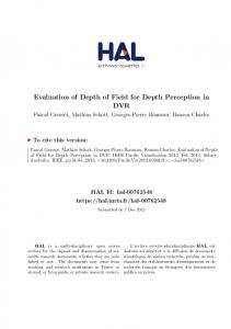
Nearest stimulus
Top view
Farthest stimulus
Side view 2 cm
Eyes reference
A
28 cm
Zn
Zf 49 cm
B
C
Figure 1: (a) Schematic diagram of the experimental setup; (b) photography of the two LED arrangements when nearest and farthest stimuli were focused on; and (c) experimental setup. was approved by the research ethics committee of the Federal University of Espirito Santo (Brazil). Two stimuli, emitted by two 5 × 7 LED arrangements flickering at 5.6 Hz and 6.4 Hz located at different distances were displayed simultaneously. As shown in Figure 1(a), the nearest (6.4 Hz) and farthest (5.6 Hz) arrangements were placed at 28 and 47 cm far from the subject, respectively. In this positions non-focused objects appear considerably blurred [9]. Figure 1(b) shows the simulated appearance of the two stimuli in the subject’s field of view when either the nearest or the farthest is focused. Two 10-trial experiments were performed in which the subjects were asked to focus on the nearest stimulus in the first one and focus on the farthest stimulus in the second one. During each 22-seconds trial, the subjects seated in front of a experimental box (see Figure 1(c)) and were asked to focus on the target stimulus for 17 s, then was asked to close the eyes for 5 s. The electroencephalographic (EEG) signals were recorded between 5 s and 17 s. EEG signals from 19 electrodes positioned according to the international 10-20 system were acquired at a sampling rate of 200 Hz using the BNT36 device with a cap of integrated wet electrodes. The grounding electrode was positioned on the user forehead and biauricular reference was adopted. Signals from electrodes O1 and O2 filtered using an (4 - 50 Hz) elliptic band-pass were used to verify the SSVEP patterns. Other channels were used to perform common average reference spatial filtering. The traditional Power Spectral Density Analysis (PSDA) method and the Canonical Correlation Analysis (CCA) method [2] were employed to compute the classification accuracy. Due to the gaze focus may occur without intention, to evaluate the robustness of the assessment and the probability of false positive errors, accuracy, Kappa coefficient, sensitivity, specificity and information transfer rate (ITR) were computed.
3
Results
Figure 2 shows the normalized power spectrum of the SSVEP pattern at electrode O2 of the subject 1 computed using the Fourier transformation. To emphasize the elicited SSVEP peaks, offset component was removed by subtracting the mean value of each frequency. Figures 2(a) and 2(b) show the spectral responses of all trials (gray curves) together with the average (black curve) computed over the 12 s of the EEG signals of the entire trial when the subject was asked to focus on the 6.4 Hz and 5.6 Hz stimulus, respectively. In both cases SSVEP peaks were elicited at stimulus frequency, at the second (12.8 Hz and 11.2 Hz) and at third harmonic frequencies (19.2 Hz and 16.8 Hz). The length of the analysis window is an important aspect to be considered when assuming that the background noise is a random aditive signal and the potential is deterministic. Figures 2(c) and 2(d) show the normalized amplitude spectra
A
B
C
D
Figure 2: (a) and (b) Normalized amplitude spectra corresponding to 10 trials (gray curves) together with the average (black curves). (c) and (d) Normalized amplitude spectra corresponding to different data lengths for one subject. computed using different time intervals (1, 2 , 4 and 8 s). The signal/noise ratio can be improved by increasing data to estimate the spectra, since the energy of the deterministic signal increases quadratically with the increasing of the signal window length. Table 1 shows the parameters that were considered to evaluate the robustness of the assessment for three subjects. Values in parentheses correspond to the PSDA method applied to peaks detection. Values without parentheses correspond to the CCA method. In all cases, the performance of the CCA method was better than PSDA method. Table 1: Evaluation parameters when the PDSA method (in parentheses) and the CCA method (without parentheses) were employed to detect SSVEP peaks. Subject Subject 1 Subject 2 Subject 3 Average
4
Sensititivy 0,78 0,90 0,78 0,82
(0,70) (0,58) (0,70) (0,66)
Specificity
Accuracy
1,00 1,00 1,00 1,00
0,85 0,95 0,85 0,88
(0,68) (0,87) (0,68) (0,75)
(0,69) (0,73) (0,69) (0,70)
Kappa 0,69 0,89 0,69 0,76
(0,37) (0,45) (0,37) (0,40)
ITR 3,37 6,96 3,37 4,57
(1,49) (2,24) (1,49) (1,74)
Conclusion
The results here presented conclude that if two flickering stimuli are appropriately located, then, the focused stimulus is able to elicit distinguishable SSVEP pattern, regardless the nonfocused stimulus also present in the user’s field of view. Based on the results showed in Figure 2 and Table 1, which are very promissory, a SSVEP-BCI based on DOF can be developed. Although, the proposed SSVEP-BCI based on DOF uses overt orienting, it is independent of
gaze movements, because the focusing of a target is performed by an accommodation mechanism that, like a pupil contraction, is an ocular reflex response. Hence, this work becomes an alternative method for presenting SSVEP stimuli to the traditional SSVEP-BCI paradigm. The existing traditional SSVEP-BCIs are becoming robust systems and achieving high transfer rates [4]. However, many designs require reliable control of eye movements because the subject performs the selection by gazing directly at each stimulus location. On the other hand, this work also becomes a complementary method to the attention based SSVEP paradigm. SSVEP-BCIs named independent-BCIs, like BCIs based on spatial-visual selective attention [5] or non-spatial selective attention [10] use selectively covert attention to make the selection of the stimulus instead of muscular gaze shifting [1]. Nevertheless, covert orienting demands high attention, and it is associated with difficult tasks that might not become automatic, even with high levels of practice, and requires intention [7]; while overt orienting demands low attention and is associated with easy and/or well-practiced tasks.
ACKNOWLEDGMENT The authors would like to thank CAPES/Brazil and FAPES/CNPq (Process 53666038/2011) for the financial support. Also, the authors would like to acknowledge the Ophthalmologist Diogo Sperandio for their advices.
References [1] Brendan Z. Allison, Dennis J. McFarland, Gerwin Schalk, Shi Dong Zheng, Melody Moore Jackson, and Jonathan R. Wolpaw. Towards an independent brain-computer interface using steady state visual evoked potentials. Clinical Neurophysiology, 119(2):399 – 408, 2008. [2] Guangyu Bin, Xiaorong Gao, Zheng Yan, Bo Hong, and Shangkai Gao. An online multi-channel ssvep-based braincomputer interface using a canonical correlation analysis method. Journal of Neural Engineering, 6(4):046002, 2009. [3] Sheldon M. Ebenholtz. Oculomotor Systems and Perception. Cambridge University Press, 1 edition, 2001. [4] Christoph Guger, Brendan Allison, Bernhard Grosswindhager, Robert Pruckl, Christoph Hintermuller, Christoph Kapeller, Markus Bruckner, Gunther Krausz, and Gunter Edlinger. How many people could use an SSVEP BCI? Frontiers in neuroscience, 6, 2012. [5] S.P. Kelly, E.C. Lalor, C. Finucane, G. McDarby, and R.B. Reilly. Visual spatial attention control in an independent brain-computer interface. Biomedical Engineering, IEEE Transactions on, 52(9):1588–1596, Sept 2005. [6] Alex Paul Pentland. A new sense for depth of field. Pattern Analysis and Machine Intelligence, IEEE Transactions on, PAMI-9(4):523–531, July 1987. [7] Michael I. Posner and Steven E. Petersen. The attention system of the human brain. Annual Review of Neuroscience, 13(1):25–42, 1990. PMID: 2183676. [8] Samuel Sokol and Anne Moskowitz. Effect of retinal blur on the peak latency of the pattern evoked potential. Vision Research, 21(8):1279 – 1286, 1981. [9] Dhanraj Vishwanath and Erik Blaser. Retinal blur and the perception of egocentric distance. Journal of Vision, 10(10), 2010. [10] Dan Zhang, Xiaorong Gao, Shangkai Gao, A.K. Engel, and A. Maye. An independent braincomputer interface based on covert shifts of non-spatial visual attention. In EMBC 2009. Annual International Conference of the IEEE, pages 539–542, Sept 2009.


![Lenses and Depth of Field [PDF]](https://m.moam.info/img/260x300/lenses-and-depth-of-field-pdf_647d6129098a9e0d2e8b466c.jpg)


