ANTICANCER RESEARCH 24: 1281-1286 (2004)
Value of Multilevel Sectioning for Improved Detection of Micrometastases in Sentinel Lymph Nodes in Invasive Squamous Cell Carcinoma of the Vulva A. HAKAM1, A. NASIR1, R. RAGHUWANSHI1, P.V. SMITH1, S. CRAWLEY1, H. E. KAISER2, E. GRENDYS1 and J.F. FIORICA1 1Department
of Interdisciplinary Oncology, Divisions of Pathology and Gynecologic Oncology, University of South Florida, H. Lee Moffitt Cancer Center and Research Institute, Tampa, FL; 2International Society for the Study of Comparative Oncology, Inc. Silver Spring, MD, U.S.A.
Abstract. Clinical usefulness of sentinel lymph node (SLN) biopsy has been demonstrated in the management of early vulvar cancer. However, what constitutes a negative SLN has not been well defined. Furthermore, to what extent the SLNs should be sectioned for the greatest likelihood of detection of micrometastases and whether multilevel sectioning will further increase this detection rate in this setting have not been well studied. We analyzed 280 groin lymph nodes (SLNs=45, nonsentinel [NSLNs]=235) in 14 patients with invasive squamous cell carcinoma (ISCC) of the vulva treated with vulvectomy and inguinal SLN and NSLN dissection at the H. Lee Moffitt Cancer Center (HLMCC) between 1996 and 2001. Each SNL was evaluated for micrometastases by H&E and pancytokeratin AE1/3 (CK AE1/3) immunohistochemical staining. All negative SNLs (N=40) were sectioned times 3 (x3) at 50-micron intervals and independently reviewed by two pathologists in order to assess the utility of this inexpensive and logical approach to identifying additional micrometastases. Also, the Wilcoxon Rank Sum Test was used to determine if there was an association between tumor size, depth of invasion and SNL status. The patient age ranged from 35 to 81 years (mean 59 yrs); size of invasive tumor from 1.0 to 7.0 cm (mean 3.4 cm); depth of invasion from 3 to 25 mm (mean 10.8 mm). Of 45
This paper was presented at the 92nd Annual Meeting of the United States & Canadian Academy of Pathology, Inc., Washington DC, U.S.A. (March 22-28, 2003). Correspondence to: A. Hakam, M.D., M.B.A, Department of Interdisciplinary Oncology and Pathology, University of South Florida, H. Lee Moffitt Cancer Center and Research Institute, 12902 Magnolia Dr. Tampa, FL 33612 U.S.A. e-mail:
[email protected] Key Words: Vulva cancer, squamous cell carcinoma, sentinel lymph node, micrometastases, multilevel sectioning.
0250-7005/2004 $2.00+.40
SLNs examined from 14 patients, 11% (5/45) SNLs were positive for micrometastases on initial H &E and/or CK AE1/3 stains. Eighty-nine per cent (40/45) SNLs were negative in the remaining 9 patients. None of the latter 40 SNLs showed micrometastases on additional multilevel sectioning. Instead 3 of 135 NSLNs examined in these 9 patients revealed micrometastases on H&E (skip-micrometastases). Mean tumor size (cm) and depth of invasion (cm) were 4.06 (s.d. 1.89) and 1.20 (s.d. 0.35) for SLN (+) and 3.02 (s.d. 2.12) and 1.01 (s.d. 0.86) for SLN (-) tumor subsets (p values 0.385 and 0.348, respectively). Conclusion: Following routine H&E and CK AE1/3 stains, multilevel sectioning does not appear to detect additional micrometastases in sentinel lymph nodes in squamous cell carcinoma of the vulva. Even though mean tumor size and depth of invasion were greater in SNL (+) as compared to SLN (-) tumor subsets in our series, this difference did not reach statistical significance. The clinical usefulness of positive sentinel lymph node (SLN) status has been demonstrated in the management of early vulvar cancer. It is considered in cases of localized vulvar cancer (1) in order to: i) minimize morbidity of complete groin dissection in patients with early disease and ii) to preserve vulvar tissue without sacrificing curability where possible metastatic disease exists (2). A number of recent studies have shown data on the feasibility of various techniques of SLN identification in vulvar cancer (1, 3-9), including data presented earlier from our institution (10). Because of these encouraging results the practice of SLN sampling is beginning to gain increasing acceptance, although the technique is still in an evolving phase and most of the published data are based on a limited number of cases. The surgical pathologist has a key role in the pathologic assessment of SLNs. In general, histologic assessment of SLNs is based on the examination of routine H&E-stained sections, frequently supplemented by cytokeratin
1281
ANTICANCER RESEARCH 24: 1281-1286 (2004) Δable I. Clinicopathologic characteristics and sentinel and non-sentinel lymph node data in squamous cell carcinoma of the vulva (N=14). Case # Age
1 2 3 4 5 6 7 8 9 10 11 12 13 14
43 81 78 73 62 51 74 35 42 47 64 45 60 79
Type of resection
MRV MRV MRV MRV HV V RV RV-GLND,B RV-GLND,B RV RV-GLND,B RV-GLND,B HV-GLND RV-GLND,B
Tumor size (mm) 10 55 55 22 18 53 60 15 17 36 70 17 25 22
HIST type
ISCCA ISCCA,K ISCCA,K ISCCA ISSCA ISCCA,K ISCCA ISCCA ISSCA ISCCA,K ISCCA ISCCA,K ISSCA ISCCA,K
Depth of INV (mm)
Grade
3 11 18 12 9 10 3 6 5 20 5 19 25 5
WD WD WD WD PD MD WD WD WD WD WD WD WD WD
SLN SLN SLN sampled positive negative (total) for mets for mets 1 2 3 1 1 5 4 2 5 1 7 5 4 4
0 1 1 1 1 1 0 0 0 0 0 0 0 0
1 1 2 0 0 4 4 2 5 1 7 5 4 4
NSLN sampled (total) 24 17 10 22 18 7 26 15 15 18 18 18 11 16
NSLN NSLN Positive negative for mets for mets 0 0 1 12 0 0 1 0 0 0 0 0 0 2
24 17 9 10 18 7 25 15 15 18 18 18 11 14
Δable II. Clinicopathologic features and sentinel and non-sentinel lymph node data in squamous cell carcinoma of the vulva (N=14). Clinicopathologic features Patient age (years) Tumor size (cm) Depth of invasion (cm)
Range
59 3.4 1.1
35-81 1.0-7.0 0.3-2.5
Number of cases
Per cent
8 6
57 43
12 1 1 45 5 40 235 16 219
86 7 7
Histologic type ISCCA, NK * ISCCA, K** Histologic grade Well-differentiated Moderately-differentiated Poorly-differentiated Sentinel lymph nodes sampled, total Positive for metastases Negative for metastases Non-sentinel lymph nodes sampled, total Positive for metastases Negative for metastases
immunohistochemistry. However, what constitutes a negative SLN has not been entirely validated. Furthermore, to what extent the SLNs should be sectioned for the greatest likelihood of detection of micrometastases and whether multilevel sectioning will further increase this detection rate in this setting has not been well studied. We studied SLNs and NSLNs for the presence of micrometastases in invasive squamous cell carcinoma of the vulva and carried out additional multilevel sectioning in order to assess the utility of this inexpensive and simple approach with a potential to facilitate identification of additional micrometastases in the SLNs.
1282
Mean
11 89 7 93
Materials and Methods Study group. The study included 14 patients with invasive squamous cell carcinoma of the vulva who underwent vulvectomy with SLN dissection, using intraoperative lymphoscintigraphy based on technetium-labeled sulfur colloid injection, at the H. Lee Moffitt Cancer Center, Tampa, FL, USA between 1996 and 2001. Intraoperatively, a gamma counter (Neoprobe, Dublin, OH, USA) was used to identify sentinel lymph nodes from the groin. Following SLN dissection from the inguinal node basin, a high count was confirmed in each SLN. Each patient was then subjected to complete non-sentinel superficial and deep inguinal node dissection, per IRB approved study protocol.
Hakam et al: Multilevel Sectioning in SNLs in Vulvar Cancer
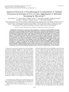
Figure 1. Initial handling and multilevel sectioning of sentinel lymph nodes in squamous cell carcinoma of the vulva (MCC).
Pathologic material. A total of 280 groin lymph nodes (45 SLNs and 235 NSLNs) in 14 patients with invasive squamous cell carcinoma (ISCC) of the vulva treated by some form of vulvectomy (Table II) and inguinal SLN and NSLN dissection were retrospectively analyzed. The original H&E and CK AE1/3 immunohistochemicalstained sections were reviewed for the presence of micrometastases defined as < 2mm microscopic foci of cancer cells or individual metastatic cancer cells identified on H&E or immunostained sections. The SLN processing and staining protocol as used at the Department of Interdisciplinary Oncology-Pathology, at the H. Lee Moffitt Cancer Center is illustrated in Figures 1 and 2. Multi-level sectioning of SLNs. The tissue blocks from all of the SLNs (N=40), that were free of metastatic SCC on initial review of H&Eand AE1/3-immunostained sections, were further sectioned (x3) at 50-micron intervals (multi-level sectioning) as outlined in Figure 1, stained with H&E and independently reviewed by two pathologists for the presence of any additional micrometastases. Statistical analysis. The Division of Biostatistics at the H. Lee Moffitt Cancer Center & Research Institute analyzed the data. The Wilcoxon rank sum test was used to determine if there was an association between tumor size, depth of invasion and SNL status.
Results Clinicopathologic data. Clinicopathologic characteristics of ISCCs analyzed and SLN / NSLN data are tabulated in Table I. Prior to surgery all of the patients had a biopsyconfirmed diagnosis of invasive (12 cases) or microinvasive (2 cases) squamous cell carcinomas without clinical evidence of palpable inguino-femoral lymph nodes (Table I). The patient age ranged from 35-81 years (mean 59 yrs) (Table II). Eight of 14 (57%) tumors were invasive squamous cell carcinomas, non-keratinizing type (ISCCANK), while the remaining 6 (43%) were of keratinizing type (ISCC-K). The size of invasive cancers ranged from 1.0-7.0 cm (mean 3.4 cm). The depth of tumor invasion measured from the epithelial-stromal interface to the deepest point of invasion ranged from 0.3-2.5 cm (mean 1.1 cm). Twelve out of 14 (86%) ISCCAs were well-differentiated (WD-ISCCA), 1 (7%) moderately-differentiated (MD-ISCCA) and 1 (7%) poorly-differentiated (PD-ISCCA).
1283
ANTICANCER RESEARCH 24: 1281-1286 (2004)
Step
1
2
Stains
Metastasis / Micrometastasis
Further Steps
H&E
+
CK - IHC
CK-IHC
— + —
MLS
None
None
Figure 2. Sentinel lymph node staining protocol. Routine H&E, cytokeratin immunohistochemical (CK-IHC) staining and multilevel sectioning (MLS) in vulvar carcinoma (MCC).
The overall mean tumor size in the ISCCs analyzed was 3.4 cm and depth of invasion 1.1 cm (Table II). For the SLN-positive subgroup of patients, however, mean tumor size was 4.06 cm (s.d. 1.89) and depth of invasion 1.20 cm (s.d. 0.35). For the SLN-negative subgroup, mean tumor size was 3.02 cm (s.d. 2.12) and depth of invasion was 1.01 cm (s.d. 0.86) (p values 0.385 and 0.348, respectively). Lymph node status. Of a total of 280 lymph nodes analyzed, 45 were sentinel and 235 were non-sentinel. Sentinel lymph nodes: of 45 SLNs in 14 patients, 5 (11%) SLNs were positive for (micro-) metastases on initial H&E and/or CK AE1/3 stains in 5 patients, while 40 (89%) were negative in the remaining 9 patients. On subsequent histologic examination following multilevel sectioning, none of the latter 40 SNLs showed (micro-) metastases on H&Estained sections. Non-sentinel lymph nodes: of a total of 235 NSLNs examined, 16 (7%) lymph nodes were positive, while 219 (93%) were negative for metastases (Table I). In 2 of the patients (Case Nos. 3 and 4), 1 and 12 NSLNs were positive and there was 1 corresponding positive SLN in each case. In 2 of the patients (Case Nos. 7 and 14), 3 NSLNs were found to have micrometastases on H&E-stained sections while the corresponding SLNs in those 2 cases were negative for metastases despite multilevel sectioning (possible "skip-micrometastases" in NSLNs). The size of primary SCCA in the latter 2 cases (Case Nos. 7 and 14) was 0.3 and 0.5 cm, respectively.
Discussion Lymphatic mapping and sentinel node dissection were originally developed to improve the treatment of patients with high-risk primary melanoma without clinical evidence of lymph node involvement (11). Subsequently, the technique
1284
has been utilized in the assessment of several other cancers, including cancers of the breast, penis, vulva, thyroid and Merkel cell carcinoma of the skin (12). Successful application of SLN studies in early vulvar cancer offers a means for less aggressive surgical treatments for patients with clinically early stage vulvar cancer (13), to avoid the morbidity related to inguino-femoral lymphadenectomy (14-16). Intraoperatively, SLNs can be detected by various techniques, including blue dye injection technique (1, 17) and intraoperative lymphoscintigraphy (10, 13, 17, 18). The latter technique has been found to offer several advantages over other techniques for sentinel node identification: i) SLNs can be located transcutaneously; ii) it allows preoperative determination of number and location of SLNs; iii) quantitation of radioactivity is possible; iv) SLNs can be detected outside of the usual nodal basin and v) SLN may be removed by a very small skin incision (1). Some recent studies (17, 19) have shown a slight improvement in the rate of detection of SLN micrometastases while combining serial H&E sectioning and CK-IHC as compared to the routine H&E sections alone. At our institution, however, we initially examine all SLNs in early vulvar cancer cases by routine H&E sectioning. Only if no metastasis / micrometastases are detected on routine H&E sections, do we proceed to CKIHC. In cases where routine H&E and CK-IHC were negative, additional multilevel sectioning as a part of this study did not detect any additional micrometastases. Our experience is more in line with the observations of Turner et al. (20), who in their experience with breast cancer did not find additional step-sectioning and CK-IHC to increase the number of patients with tumor-positive nodes. Before one can draw any valid conclusions regarding the sensitivity of the technique, it is necessary to accumulate more experience with the performance of SNL dissections and the histologic and immunohistochemical analysis of SLNs in vulvar cancer.
Hakam et al: Multilevel Sectioning in SNLs in Vulvar Cancer
The presence of micrometastases in 3 out of 135 NSLNs analyzed in 2 out of 14 (14%) patients in our study awaits a plausible explanation. The SLNs in these cases were negative for micrometastases, despite examination of routine H&E and CK-AE1/3-stained and additional multilevel sections. This frequency (14%) of skip metastases in vulvar cancer is evidently higher as compared to the published SLN series in breast cancer, in which skip metastases have been reported in zero% to 7.4% of the cases studied (21-28), including large SLN series in breast cancer patients from our institution (25,26). In colorectal cancers the reported frequency of skip metastases has been 4% to 9% (29,30). Factors that may potentially contribute to the frequency of detection of micrometastases in SLN studies include: i) sample size bias, ii) variation in transnodal migration of squamous vs. adenocarcinoma cells and iii) the possibility of a ‘true’ false-negative SLN detection, which may be explained by local tissue factors resulting in aberrant lymphatic pathway in the tissue, or the sensitivity of intraoperative detection of SLNs. Alternatively, there may not be a uniform explanation for skip-metastases in these cases since, as identified in breast cancer patients, ‘skiplesions’ (skip-metastases) are a random process (31). Furthermore, the detection of occult metastasis in lymph nodes is considered as an issue of probability, not certainty (32). Before any further investigation to explain an apparently higher frequency of skip-metastases in vulvar SCC in our series, we would like to ascertain if patients with skip metastases had any different pattern of inguinal / local or systemic recurrence of their vulvar cancers as compared to those who were SLN-negative without skip metastases (clinical data analysis in progress). Although both the mean tumor size and depth of tumor invasion for ISCCs with positive SLNs were higher (4.06 cm and 1.20 cm, respectively) as compared to those with negative SLNs (3.02 cm and 1.01 cm, respectively), these differences did not reach statistical significance. As regards the technique of intraoperative lymphoscintigraphy, it has been shown to offer better identification of SLNs when compared with other techniques like blue dye injection technique (9), although some investigators have shown a higher accuracy with the combined approach (8). From the point of view of pathologic analysis of SLNs, some of the important considerations like an optimal false-negative proportion of the SLN biopsy, techniques to maximize the detection of SLN metastases, various ‘categories’ of micrometastases and other pathologic information that is relevant in this context is alluded to in a commentary article by Anderson (1999) who states, " Various (SLN) studies have contributed potential answers to some of these questions, but there has been no concerted effort in these studies to define what is practical" (32). In this study we tested MLS as an inexpensive
and practical approach to augment the sensitivity of SLN biopsy in SCC of the vulva. However, based on our current experience with the technique, MLS does not seem to offer any major advantage over routine H&E and cytokeratin staining. Whether additional trimming of the SLN slices beyond the three levels that we tested here, or additional immunostains, are of any potential value in improving the detection of metastases / micrometastases in SCC of the vulva merits further investigation.
Conclusion Although the studies of SLNs in early vulvar cancer have been shown to be feasible in preliminary reports on the subject, there still is the need for pathologists to come to a consensus as to what constitutes a negative SLN in cases of early vulvar cancer, and to what extent one should section the SLN tissue block for H&E and cytokeratin immunostaining. Our current institutional practice of routine examination of multiple H&E sections supplemented by CK AE1/3 immunostains seems to be a reasonably practical approach. Additional multilevel sectioning does not appear to improve the detection of micrometastases in sentinel lymph nodes in early squamous cell carcinoma of the vulva and, thus, seems unnecessary at this time. However, as more experience with the study of sentinel lymph nodes in vulvar carcinoma is obtained, it may be possible to refine the current techniques of pathologic examination of SLNs in carcinoma of the vulva.
References 1 Levenback C, Coleman RL, Burke TW, Bodurka-Bevers D, Wolf JK and Gershenson DM: Intraoperative lymphatic mapping and sentinel node identification with blue dye in patients with vulvar cancer. Gynecol Oncol 83(2): 276-281, 2001. 2 DiSaia PJ, Creasman WT and Rich WM: An alternate approach to early cancer of the vulva. Am J Obstet Gynecol 133(7): 825-832, 1979. 3 De Hullu JA and Van Der Zee AG: Sentinel node techniques in cancer of the vulva. Curr Womens Health Rep 3(1): 19-26, 2003. 4 Sliutz G, Reinthaller A, Lantzsch T, Mende T, Sinzinger H, Kainz C et al: Lymphatic mapping of sentinel nodes in early vulvar cancer. Gynecol Oncol 84(3): 449-452, 2002. 5 Puig-Tintore LM, Ordi J, Vidal-Sicart S, Lejarcegui JA, Torne A, Pahisa J et al: Further data on the usefulness of sentinel lymph node identification and ultrastaging in vulvar squamous cell carcinoma. Gynecol Oncol 88(1): 29-34, 2003. 6 Trope C, Scheistroen M, Aas M, Abeler V, Lie K and Makar A: Surgery and sentinel node examination in early vulvar cancer. Tidsskr Nor Laegeforen 121(23): 2723-2727, 2001. 7 Zambo K, Schmidt E, Hartmann T, Kornya L, Dehghani B, Tinneberg HR et al: Preliminary experiences with sentinel lymph node detection in cases of vulvar malignancy. Eur J Nucl Med Mol Imaging 29(9): 1198-2000, 2002.
1285
ANTICANCER RESEARCH 24: 1281-1286 (2004) 8 de Hullu JA, Hollema H, Piers DA, Verheijen RH, van Diest PJ, Mourits MJ et al: Sentinel lymph node procedure is highly accurate in squamous cell carcinoma of the vulva. J Clin Oncol 18(15): 2811-2816, 2000. 9 Levenback C, Coleman RL, Ansink A and van der Zee AG. Re: Terada et al: Sentinel node dissection and ultrastaging in squamous cell cancer of the vulva. Gynecol Oncol 76: 40-44, 2000. Gynecol Oncol 77(3): 484-485, 2000. 10 Decesare SL, Fiorica JV, Roberts WS, Reintgen D, Arango H, Hoffman MS et al: A pilot study utilizing intraoperative lymphoscintigraphy for identification of the sentinel lymph nodes in vulvar cancer. Gynecol Oncol 66(3): 425-428, 1997. 11 Cochran AJ: Surgical pathology remains pivotal in the evaluation of ‘sentinel' lymph nodes. Am J Surg Pathol 23(10): 1169-1172, 1999. 12 Koops HS, Doting MH, de Vries J, Tiebosch AT, Plukker JT, Hoekstra HJ et al: Sentinel node biopsy as a surgical staging method for solid cancers. Radiother Oncol 51(1): 1-7, 1999. 13 Paganelli G, De Cicco C and Chinol M: Sentinel node localization by lymphoscintigraphy: a reliable technique with widespread applications. Rec Res Cancer Res 157: 121-129, 2000. 14 Levenback C: Intraoperative lymphatic mapping and sentinel node identification: gynecologic applications. Recent Results Cancer Res 157: 150-158, 2000. 15 Terada KY, Shimizu DM and Wong JH: Sentinel node dissection and ultrastaging in squamous cell cancer of the vulva. Gynecol Oncol 76(1): 40-44, 2000. 16 Makar AP, Scheistroen M, van den Weyngaert D and Trope CG: Surgical management of stage I and II vulvar cancer: the role of the sentinel node biopsy. Review of literature. Int J Gynecol Cancer 11(4): 255-262, 2001. 17 Molpus KL, Kelley MC, Johnson JE, Martin WH and Jones HW 3rd: Sentinel lymph node detection and microstaging in vulvar carcinoma. J Reprod Med 46(10): 863-869, 2001. 18 De Cicco C, Sideri M, Bartolomei M, Grana C, Cremonesi M, Fiorenza M et al: Sentinel node biopsy in early vulvar cancer. Br J Cancer 82(2): 295-299, 2000. 19 de Hullu JA, Ansink AC, Tymstra T and van der Zee AG: What doctors and patients think about false-negative sentinel lymph nodes in vulvar cancer. J Psychosom Obstet Gynaecol 22(4): 199-203, 2001. 20 Turner RR, Ollila DW, Stern S and Giuliano AE: Optimal histopathologic examination of the sentinel lymph node for breast carcinoma staging. Am J Surg Pathol 23(3): 263-267, 1999. 21 Mariotti S, Buonomo O, Guadagni F, Spila A, Schiaroli S, Cipriani C et al: Minimal sentinel node procedure for staging early breast cancer. Tumori 88(3): S45-S47, 2002.
1286
22 Fernandez A, Escobedo A, Benito E, Azpeitia D, Guma A, Prieto L et al: Sentinel node localization in patients with non-palpable breast cancer. Nucl Med Commun 23(12): 1165-1169, 2002. 23 Sato K, Hiraide H, Uematsu M, Tamaki K, Ishikawa H, Yamasaki T et al: Efficacy and significance of sentinel lymph node identification with technetium-99m-labeled tin colloids for breast cancer. Breast Cancer 5(4): 389-393, 1998. 24 Kapteijn BA, Nieweg OE, Petersen JL, Rutgers EJ, Hart AA, van Dongen JA et al: Identification and biopsy of the sentinel lymph node in breast cancer. Eur J Surg Oncol 24(5): 427-430, 1998. 25 Reintgen D, Joseph E, Lyman GH, Yeatman T, Balducci L, Ku NN et al: The role of selective lymphadenectomy in breast cancer. Cancer Control 4(3): 211-219, 1997. 26 Cox CE, Pendas S, Cox JM, Joseph E, Shons AR, Yeatman T, et al: Guidelines for sentinel node biopsy and lymphatic mapping of patients with breast cancer. Ann Surg 227(5): 645651; discussion 51-63, 1998. 27 Sato K, Tamaki K, Takeuchi H, Tsuda H, Kosuda S, Kusano S et al: Management of the axilla in breast cancer: a comparative study between sentinel lymph node biopsy and four-node sampling procedure. Jpn J Clin Oncol 31(7): 318-321, 2001. 28 Hsieh PP, Ho WL, Yeh DC, Liu TJ, Wu CC, Lin JH et al: Histopathologic analysis of sentinel lymph nodes in breast carcinoma. Chung Hua I Hsueh Tsa Chih (Taipei) 63(10): 744750, 2000. 29 Saha S, Bilchik A, Wiese D, Espinosa M, Badin J, Ganatra BK et al: Ultrastaging of colorectal cancer by sentinel lymph node mapping technique--a multicenter trial. Ann Surg Oncol 8(9 Suppl): 94S-98S, 2001. 30 Wiese DA, Saha S, Badin J, Ng PS, Gauthier J, Ahsan A et al: Pathologic evaluation of sentinel lymph nodes in colorectal carcinoma. Arch Pathol Lab Med 124(12): 1759-1763, 2000. 31. Msuya CA and Hartveit F: Skip lesions in the axilla in breast cancer, and their association with micrometastases. Breast Cancer Res Treat 19(3): 277-281, 1991. 32. Anderson TJ: The challenge of sentinel lymph node biopsy. Histopathology 35(1): 82-84, 1999.
Received July 28, 2003 Reviesd December 24, 2003 Accepted January 16, 2004