Behavioural Brain Research 107 (2000) 177 – 181 www.elsevier.com/locate/bbr
Short communication
Visual lateralization of pattern discrimination in the bottlenose dolphin (Tursiops truncatus) Lorenzo von Fersen a, Ulrich Schall b, Onur Gu¨ntu¨rku¨n c,* b
a Tiergarten Nu¨rnberg, Am Tiergarten 30, 90480 Nu¨rnberg, Germany Discipline of Psychiatry, Faculty of Medicine and Health Sciences, The Uni6ersity of Newcastle, Callaghan, NSW 2308, Australia c AE Biopsychologie, Fakulta¨t fu¨r Psychologie, Ruhr-Uni6ersita¨t Bochum, 44780 Bochum, Germany
Received 2 August 1999; received in revised form 20 September 1999; accepted 20 September 1999
Abstract The aim of the present study was to investigate whether bottlenose dolphins have cerebral asymmetries of visual processing. The monocular performance of the adult dolphin Goliath was tested using a large number of simultaneous multiple pattern discrimination tasks. The experiments revealed a clear right eye advantage in the acquisition and the retention of pattern discriminations as well as asymmetries in the interhemispheric transfer of visual information. As a result of a complete decussation at the optic nerve, this right eye superiority is probably related to a left hemisphere dominance in visual processing. © 2000 Elsevier Science B.V. All rights reserved. Keywords: Cerebral asymmetry; Cetacea; Interhemispheric transfer
1. Introduction More than 50 million years ago the ancestor of today’s dolphins ventured back into the sea to start a remarkable evolutionary branch. Their phylogeny resulted in a number of species which are second to humans in their encephalisation [15]. However, the enlargement of dolphin brains was also accompanied by the development of several morphological differences to the brains of land mammals [7]. It is still unresolved whether these differences also reflect a divergent functional architecture. One way to approach this question is to analyze cerebral asymmetries. Studies of the last 3 decades showed that cerebral asymmetries are not a feature unique to humans but can be demonstrated with remarkable similarites in a wide range of species [5]. Therefore, the aim of the present study was to study visual lateralization in the bottlenose dolphin
* Corresponding author. Tel.: +49-234-7006213; fax: + 49-2347094377. E-mail address:
[email protected] (O. Gu¨ntu¨rku¨n)
(Tursiops truncatus). Several studies revealed behavioral lateralities in marine mammals. Gray and humpback whales show a right-side bias in flipper slaps and bottom feeding [6], while captive beluga whales and dolphins have swim paths which are biased towards one side [16,17,21]. Several studies reported of a right eye preference in dolphins when approaching or scrutinizing novel objects (see Ref. [21] and references therein). Because electrophysiological [23] and neuroanatomical studies [13] revealed a complete crossover of optic fibers at the optic chiasm, a right eye preference could be related to a left hemispheric dominance in visual analysis. The same feature offers a unique possibility to study lateralized visual performance under behaviorally controlled conditions using eyecups which temporarily cover one eye and thus prevent primary visual input into one hemisphere. Here we show that indeed pattern discrimination and interhemispheric transfer is lateralized in a captive bottlenose dolphin. Goliath, a 15-year-old male bottlenose dolphin was housed in a 22 m diameter circular outdoor tank in
0166-4328/00/$ - see front matter © 2000 Elsevier Science B.V. All rights reserved. PII: S 0 1 6 6 - 4 3 2 8 ( 9 9 ) 0 0 1 4 2 - 4
178
L. 6on Fersen et al. / Beha6ioural Brain Research 107 (2000) 177–181
Mundo Marino (Argentina). Before and during the learning experiments the eyes of Goliath were examined by a veterinary surgeon. These examinations which included a detailed ultrasound analysis revealed no unusual conditions in any of the eyes.
were run under monocular conditions (alternating five left, five right) while the remaining ones were binocular. At the start of a monocular trial a half-circular (12 cm diameter) rubber eyecup was fixed by suction onto one eye, to be removed at trials end. The first experiment ended after each of the 14 patterns was displayed ten times within each monocular condition.
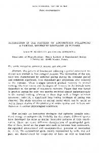
2. Simultaneous discrimination of multiple patterns In the first experiment a pattern discrimination task was used in which the animal had to discriminate two out of 14 irregular shapes in each trial. Each trial started with Goliath being positioned at the tip of a 2 m long pole, 80 cm above water level, and looking with his binocular nasoventral visual field towards the two stimuli displayed to the left and the right of the experimenter (Fig. 1). In this position dolphins have high aerial acuity for our stimuli which were 80 cm above water level [12]. Two seconds later a whistle blow indicated that Goliath could touch one of the stimuli with his rostrum. The stimuli consisted of 14 black plywood patterns of irregular shape, each mounted on a separate white board of 1 m2 with an interboard distance of 20 cm (Fig. 1). Seven patterns were defined to be correct and responses to them were reinforced with fish. Incorrect choices to one of the other seven stimuli were followed by correction trials. Rewarded and unrewarded stimuli were randomly combined and left – right positions of patterns alterned quasi-randomly [9]. Each of the two daily session consisted of 20 trials. After reaching ten consecutive sessions with at least 75% correct performance, monocular testing began. Out of 20 trials ten interspersed ones out of the complete stimulus set
3. Monocular acquisition of pattern discriminations In this second experiment Goliath successively learned ten new pattern discriminations with each eye under monocular conditions. Thus, 40 new patterns were successively introduced (ten discriminations× two eyes×two stimuli [one correct+ one incorrect]). Two sessions were run daily and each session consisted of 20 trials. In 12 of these trials, two new stimulus pairs (one pair for the left, one pair for the right eye) were presented under monocular conditions by use of eyecups (six right, six left eye trials). In the remaining eight trials, stimuli from the previous experiment were used. Within each new pair, one pattern was defined to be correct. Learning criterion was analyzed separately for each eye and was reached when Goliath had no more than two errors within ten consecutive trials with the same new stimulus pair. Once Goliath had reached criterion with one eye, the next pair of unknown stimuli were introduced for this monocular condition. This procedure lasted until Goliath had successively learned ten new pattern discriminations with each eye. All other aspects were identical to the first experiment.
Fig. 1. Schematic overview of the testing situation with the apparatus, the stationing device and the position of the dolphin while watching with his right eye (left eye covered) the stimulus display. The inset depicts some further patterns used in the study.
L. 6on Fersen et al. / Beha6ioural Brain Research 107 (2000) 177–181
179
binocular (89%) and monocular right (87%) conditions. However, Goliath only reached 71% using his left eye (Fig. 2a). These differences between viewing conditions were significant (Friedman-test, k= 3, n = 7, x2=11.2, PB 0.05). Wilcoxon-tests revealed significant differences between binocular and left (Z= 2.37, PB 0.05) as well as between right and left (Z=2.2, PB 0.05) viewing conditions, while binocular and right eye performances did not significantly differ (Z = 0.53, P\ 0.05) (all alphas Bonferroni–Holm adjusted). In the second experiment, Goliath learned ten new pairs of patterns with each eye. In nine out of these ten acquisitions he reached criterion in less trials with his right eye. A between-eye comparison of the number of trials for each new pair of patterns revealed a significant difference (Wilcoxon-test, Z =2.45, PB 0.05) (Fig. 2b). In the third experiment the interhemispheric transfer of the previously learned patterns was tested. A successful transfer results in reduced trial numbers to criterion with the naive eye, compared with the inital acquisition. This saving score was calculated as: saving score= TIL− TCL
Fig. 2. (a) Average percent correct responses under the three viewing conditions in the simultaneous discrimination of multiple patterns (experiment 1). (b) Average trials to criterion in the acquisitions of the ten pattern discriminations which Goliath learned with each eye (experiment 2). (c) Average saving scores in the transfer of pattern discriminations from the left to the right eye (left bar) or from the right to the left eye (right bar) (experiment 3). Bars depict standard errors of mean.
4. Interhemispheric transfer of pattern discriminations The same ten pairs of patterns which had been used for each eye in the second experiment, were now presented in the same succession to the other eye. The denomination of S+ and S − as well as the behavioral procedures and the learning criterion were identical. Goliath learned all pattern discriminations and easily accustomed to the eyecup without showing signs of discomfort. In the first experiment, his discrimination performance in the simultaneous discrimination task with multiple patterns was clearly affected by viewing conditions. After monocular testing began, his average discrimination capacity was virtually identical for
with TIL being the number of trials in initial learning with the ipsilateral eye and TCL being the subsequently needed number of trials with the contralateral eye. A successful transfer results in positive values. The higher this index is, the more tranfer was achieved. A value of 0 or negative scores indicate that no interhemispheric transfer had occurred. As shown in Fig. 2(c), saving scores for transfer from the left to the right eye were higher than vice versa (Wilcoxon-test, Z= 1.82, PB 0.07). The correlations between the number of trials to criterion to learn a pattern discrimination with one eye (experiment 2) and the saving scores for the same pattern with the other eye (experiment 3) were r=0.62 (t= 2.26, PB 0.06) for right-to-left, and r= 0.97 (t= 11.12, PB 0.00001) for left-to-right eye transfer. To determine the significance of the difference between correlation coefficients we computed an analysis for dependent samples [4]. The difference between both correlation coefficients was significant (Z= −3.41, PB 0.001), indicating a closer relation between left eye learning and transfer from left to the right eye, than vice versa. The present results clearly show that the bottlenose dolphin Goliath reaches higher levels of performance in a pattern discrimination and acquisition task when using his right eye. As a result of the complete optic nerve decussation [13,23] this right eye superiority is possibly related to a left hemisphere dominance. If Goliath is representative for his species, our results would indicate a left hemisphere dominance for visual object analysis in bottlenose dolphins. Indeed, different species of toothed whales including bottlenose dolphins
180
L. 6on Fersen et al. / Beha6ioural Brain Research 107 (2000) 177–181
have been repeatedly reported to have a right eye preference when approaching or observing novel objects [21]. Thus, the present experimental results fit with these observations. In principle it is possible that the left – right asymmetry arose as a result of acuity differences between the eyes. However, this explanation seems to be unlikely because this should have resulted in faster learning scores with the right eye in all experiments, including the third one in which the previously learned patterns had to be transferred to the other eye. Contrary, Goliath was in this case faster with his left eye (Fig. 2c). We therefore suppose, that the right eye dominance resulted from a cerebral asymmetry and not from acuity differences. Thus, the present experiments suggest, that visual asymmetry affects acquisition, retention, and, as discussed below, possibly also interhemispheric transfer of visual information. The more efficient visual transfer from the subdominant right hemisphere (left eye) to the dominant left brainhalf (right eye) makes it likely that interhemispheric transfer is asymmetrically organized. Possibly, these results emerge from an asymmetrical transcallosal cooperation during the acquisition of the visual pattern discriminations. According to this scenario, the right hemisphere (left eye) has to cooperate with the dominant left halfbrain during visual discriminations learned with the left eye. During such a cooperation the dominant left hemisphere possibly also learns parts of this pattern discrimination. Therefore, a subsequent transfer from the subdominant right to the dominant left hemisphere (left-to-right eye) will result in high transfer efficiency. This scenario is different when the left hemisphere learns a new visual task. Because the visually dominant left hemisphere is probably not in need of extensive transcallosal cooperations, this will result in fewer learning traces in the right halfbrain. Consequently, an interhemispheric transfer from the left to the right hemisphere is less efficient and only loosely related to the learning history in initial (left hemispheric) learning. This hypothetical scenario accords with numerous behavioral [14], neuropsychological [20], and electrophysiological data [19] on asymmetrical interhemispheric interactions in humans. Mukhametov [18] reported that dolphins can sleep with one hemisphere only, resulting in asymmetrical slow wave sleep patterns. Thus, asymmetries of interhemispheric interaction as reported in the present study might be a common neural principle in bottlenose dolphins. In humans, figural comparison task generally result in a stable right hemisphere advantage [8,22]. This also holds for comparable simultaneous pattern discrimination tasks with cats and rats [1 – 3]. The situation in monkeys might be in part comparable. Hamilton and Vermeire [10] demonstrated that macaques have a right hemisphere superiority in face but not pattern discrimi-
nation. Tachistoscopic tasks in humans usually reveal right hemisphere advantages both in pattern and face discrimination tasks, as a result of the activation of common neural structures for visual feature analysis [11]. Possibly the long stimulus exposure times in the monkey experiment [10] might have reduced existing asymmetries in the easier pattern discrimination as often observed in human studies. Taken together, these data indicate that in humans, rats, cats, and possibly monkeys the right hemisphere dominates different aspects of visual feature discrimination. The left hemisphere dominance of dolphins in a similar task could therefore represent a remarkable deviation of their functional cerebral architecture.
Acknowledgements The authors are deeply indebted to the staff of the Fundacion Mundo Marino (San Clemente del Tuyu´, Argentina) for their hospitality and continuous help during conduct of the study. Thanks also to Jo¨rg Pekarsky for drawing our Fig. 1, to Andrea von Fersen for help during the testing procedures, and to Markus Hausmann for assistance with statistical analysis. Supported by a habilitation grant of the Deutsche Forschungsgemeinschaft to L.F. and the prize of the Alfried Krupp-Stiftung to O.G.
References [1] Bianki VL. Lateralization of functions in the animal brain. Int J Neurosci 1981;15:37 – 47. [2] Bianki VL. Lateralization of concrete and abstract characteristics analysis in the animal brain. Int J Neurosci 1982;17:233–41. [3] Bianki VL. Simultaneous and sequential processing of information by different hemispheres in animals. Int J Neurosci 1983;22:1 – 6. [4] Bortz J. Statistik fu¨r Sozialwissenschaftler, 3rd ed. Berlin: Springer Verlag, 1989. [5] Bradshaw JL, Rogers LJ. The Evolution of Lateral Asymmetries, Language, Tool Use, and Intellect. San Diego: Academic, 1993. [6] Clapham PJ, Leimkuhler E, Gray BK, Mattila DK. Do humpback whales exhibit lateralized behaviour? Anim Behav 1995;50:73 – 82. [7] Deacon TW. Rethinking mammalian brain evolution. Am Zool 1990;30:629 – 705. [8] Geffen G, Bradshaw JL, Wallace G. Interhemispheric effects on reaction time to verbal and nonverbal visual stimuli. J Exp Psychol 1971;87:415 – 22. [9] Gellermann LW. Chance orders of alternating stimuli in visual discrimination experiments. Pedagog Sem J Gen Psychol 1933;42:206 – 8. [10] Hamilton CR, Vermeire BA. Complementary hemispheric specialization in monkeys. Science 1988;242:1691– 4. [11] Hausmann M, Gu¨ntu¨rku¨n O. Sex differences in functional cerebral asymmetries in a repeated measure design, Brain Cognit. (in press).
L. 6on Fersen et al. / Beha6ioural Brain Research 107 (2000) 177–181 [12] Herman LM, Peacock MF, Madsen CJ. Bottlenosed Dolphin: double-slit pupil yields equivalent aerial and underwater diurnal acuity. Science 1975;189:650–2. [13] Jacobs MS, Morgane PJ, McFarland WL. Degeneration of visual pathways in the bottlenosed dolphin. Brain Res 1975;88:346 – 52. [14] Mannhaupt HR. Processing of abstract and concrete nouns in a lateralized memory-search task. Psychol Res 1983;45:91– 105. [15] Marino L. What can dolphins tell us about primate evolution? Evol Anthr 1996;5:81–5. [16] Marino L, Stowe J. Lateralized behavior in a captive beluga whale (Delphinapterus leucas). Aq Mammals 1997;23:101 – 3. [17] Marino L, Stowe J. Lateralized behavior in two captive bottlenose dolphins (Tursiops truncatus). Zool Biol 1997;16:173 – 7. [18] Mukhametov LM, Supin AY, Polyakova IG. Interhemispheric asymmetry of the electroencephalographic sleep patterns in dolphins. Brain Res 1977;134:581–4.
.
181
[19] Nalcaci E, Basar-Eroglu C, Stadler M. Visual evoked potential interhemispheric transfer time in different frequency bands. Clin Neurophysiol 1999;110:71 – 81. [20] Regard M, Cook ND, Wieser HG, Landis T. The dynamics of cerebral dominance during unilateral limbic seizures. Brain 1994;117:91 – 104. [21] Ridgway SH. Physiological observations on the dolphin brain. In: Schusterman RJ, Thomas JA, Woods FG, editors. Dolphin Cognition and Behavior: A Comparative Approach. Lawrence Erlbaum: Hillsdale, NJ, 1986:31 – 60. [22] Rode C, Wagner M, Gu¨ntu¨rku¨n O. Menstrual cycle affects functional cerebral asymmetries. Neuropsychologia 1995;33:855 – 65. [23] Supin AY, Mukhametov LM, Ladygina TF, Popov VV, Mass AM, Poliakova EG. Electrophysiological Study of the Dolphin Brain. Nauka: Moscow, 1978 (from Ridgway, 1986).