mance of the camera augmented mobile C-arm (CamC), which extends a standard mobile X-ray ... This system was applied on several real-world and simulated vertebro- ..... Jaramaz, B., A. M. DiGioia, I.: Ct-based navigation systems. In Stiehl ...
Workflow Based Assessment of the Camera Augmented Mobile C-arm System Joerg Traub1 , Seyed-Ahmad Ahmadi1 , Nicolas Padoy1 , Lejing Wang1 , Sandro Michael Heining2 , Ekkehard Euler2 , Pierre Jannin3 , and Nassir Navab1 1
Chair for Computer Aided Medical Procedures (CAMP), TU Munich, Germany Trauma Surgery Department, Klinikum Innenstadt, LMU Munich, Germany INSERM / INRIA / IRISA, Faculte de Medecine, Universite de Rennes, France
2 3
Abstract. Several image guided surgery systems were introduced in the research community throughout the past decades. Only a few have found their way into the operating room and even a smaller number of them into everyday clinical routine. Within this paper, we describe a method for workflow based preclinical assessment of image guided surgery systems. This analysis can reveal surgical phase-related strengths and weaknesses of the system that can lead to design changes potentially leveraging future clinical acceptance. The method is based on a workflow analysis of the clinical procedure and the conduction of carefully designed surgery simulations resulting in a quantitative comparison of the current and the newly proposed system. The method was applied to assess the performance of the camera augmented mobile C-arm (CamC), which extends a standard mobile X-ray system by a video camera and provides an overlay of video and x-ray image without any online calibration or registration. This system was applied on several real-world and simulated vertebroplasty procedures and compared to conventional fluoro CT guided interventions. The analysis of these simulations provides initial quantitative results and a comparison of the current clinical method and the new system in terms of duration, X-ray exposure, and changes in the workflow.
1
Introduction
Radically novel systems for image guided interventions, e.g. systems using augmented reality visualization, face a long and tedious process from the conception of the initial idea until they are clinically applicable. This process includes the process of modeling the problem, designing the algorithms and the system, implementing and engineering the system, and finally evaluating the system in terms of its suitability for clinical usage. One approach for the classification of the assessment of systems for image guided interventions was proposed by Jannin and Korb [1]. Their concept proposed an evaluation classified in six different levels. The here introduced model is motivated by this multilevel concept and proposes a reference-based method to compare computer assisted navigation systems with the currently used surgical procedure in the operating room. Observing, monitoring and analyzing the
Workflow Assessment of the Augmented C-arm
37
medical workflow allow for the creation of surgery models. A simulated procedure is designed with respect to the surgical model and is used to compare the workflow of the novel approach against the currently applied surgical procedure. The comparison is based on a set of parameters monitored during both approaches. This will provide a structured way in assessing clinically relevant parameters already within a preclinical prototype and justify its further expensive and time-consuming ex-vivo and in-vivo studies. Thus already within an early stage of development an assessment based on this model shows potential to create meta models to assess parameters of the surgical strategy and performance by analyzing the workflow. As an exemplary scenario for the proposed evaluation method, it was applied to the camera-augmented mobile C-arm system (CamC) which is further explained in section 2 and used for vertebroplasty procedures, further explained in section 4.1. For our exemplary evaluation, we monitor several parameters during the simulated vertebroplasties in order to verify two assumptions of the CamC system. One assumption is that it is reducing the radiation exposure of the surgical team and patients during the intervention. The other assumption is that it does not interfere with or complicate the current clinical workflow. Monitoring parameters relevant to these assumptions could prove the clinical usefulness of the system under inspection and justify the conduction of further cadaver and patient studies.
2
The Camera Augmented Mobile C-arm System
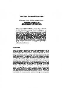
Fig. 1. The CamC system merges the the X-ray image (left) onto the video image (middle), creating an augmented view (right) which provides an intuitive interface for skin incision and instrument positioning.
Mobile C-arms are used every day in clinical routine in trauma and orthopedic surgery departments. With their introduction into the operating room, there was an increase in radiation exposure for both patient and surgical team [2]. Image guided surgery system and navigation solutions for trauma and orthopedic surgery aim at the reduction of the invasiveness including the radiation dose while ensuring the optimal patient outcome. Traditional navigation solutions for spine surgery use spatial localization systems to track instruments and patient in
38
Joerg Traub et al
3D space and register medical imaging data into the tracking coordinate frame [3, 4]. The used image guided surgery systems are thus either based on preoperative CT data (e.g. [5]) or intraoperative 3D (e.g. [6]) or 2D (e.g. [7]) all of them with their advantages and disadvantages. The systems based on intraoperative imaging acquire the image data within the tracking coordinate system and thus do not require any additional patient registration procedure. However, within clinical routine these systems require a long setup time, special instruments, add complex and manifold equipment to the operating room and they require technically trained surgeons or technical staff. The CamC system extends a standard mobile C-arm by a video camera (cf. figure 2) such that the video image and the X-ray image are overlaid (cf. figure 1) without further calibration and registration. This is achieved by a double mirror construction and a one time calibration routine during the construction of the device as described in Navab et al. [8]. The advantage of the system compared to traditional image guided surgery solutions is that it is entirely integrated in the C-arm and does not require any additional setup of hardware in prior to the intervention and no registration is required during the intervention. Since the system extends a mobile C-arm, it is furthermore possible to continue the surgery under standard fluoroscopic imaging anytime without delay. The system shows a promising extension to reduce the radiation exposure for the surgical team and patient.
3
Novel Method for Preclinical Comparison of Surgical Procedures
The method for structured preclinical evaluation of newly designed and developed image guided surgery systems is composed of five consecutive steps: 1. Workflow analysis of the clinical procedure. Within the operating room, several surgeries of the target application are recorded. For workflow analysis, already existing tools can be used (e.g. [9]). 2. Creation of a high level surgical model of the procedure. Recording and analyzing several surgeries, an average surgery and a surgical model with different hierarchical levels can be created. The system parameters in the top level should be representative of any performed procedure independent from the applied technology. 3. Creation of a simulated procedure and extraction of relevant parameters. Based on the clinical procedure, a phantom and a simulated procedure are designed. The major criterion is that the simulated procedure can be performed using any kind of image guided solution. The relevant parameters to be assessed during the simulated procedure need to be identified. Examples are the duration of each surgery phase, patterns in the workflow, accuracy and invasiveness of the procedure. 4. Recording and analysis of the simulated procedures. The designed assessment is performed on the phantom(s), comparing the current clinical procedure in the intervention room and the new system in the simulated setting. Using the same workflow analysis tools as for the clinical recordings, the quality
Workflow Assessment of the Augmented C-arm
39
of the simulated procedure can be evaluated. This is done by comparing the clinical cases with the simulated procedures, providing an initial feedback on how close the simulation is to the real scenario. 5. Comparison of simulated procedures. The parameters and results of the simulated procedure under different image guidance modalities can then be compared directly. During the analysis, one has to consider the learning curve using a new system which may introduce a bias steming from untrained surgeons.
Fig. 2. The CamC system (left) and foam-coated phantom spines (right) used during the experiments.
4 4.1
Workflow Based Assessment of the CamC System Workflow Analysis of the Clinical CT Fluoro Vertebroplasty Procedure
The clinical procedure for vertebroplasty is generally based on fluoro CT guidance (cf. figure 3). Within the CT intervention room of our medical partner institution, seven vertebroplasty procedures were recorded, each using two video cameras. One of the cameras was used to record the operation situs, the other one was used to record the interventionally acquired image data (fluoro and spiral CT). In our workflow analysis of seven vertebroplasty procedures performed by two different experienced surgeons, we observed an average duration of 31:14 [min]. The recorded videos were analyzed, synchronized and labeled with activity tags at a resolution of 1 Hz using a workflow analysis tool. These procedures were further analyzed to derive the measurement criteria for a comparison of simulated vertebroplasty procedures using CT fluoro guided and the CamC system, as well as for the design of a simulated procedure that allows measuring the desired parameters. The major measurement criteria were the time required to complete specific tasks, the applied radiation dose during
40
Joerg Traub et al
Fig. 3. Real vertebroplasty procedure using fluoro CT guidance (left). A K-wire or filling needle (middle) is positioned in the fluoro CT slice and controlled manually during insertion. Once the K-wire or filling needle is positioned inside the vertebra, the cement filling is performed using fluoro CT imaging (right).
specific phases of the surgery and the changes in the workflow and its complexity. Based on the workflow analysis, a surgical model for the vertebroplasty procedure was constructed. A high level description consists only of three phases (cf. figure 4). These phases and the fixed workflow events that indicate the phase borders are defined such that they can be uniquely identified, independent of the applied technology. These phases are: 1. System setup and target identification in the imaging data. This includes the setup of the equipment, devices, the positioning of the patient on the intervention table, and the localization of the identified vertebra within the image data and the identification of an access path within the imaging data. The skin incision indicates the end of this phase, independent from the imaging modality used. 2. Tool placement. This phase includes the placement of the instrumentation onto the inflicted vertebra and the process of inserting the instrument through the pedicle into the central vertebra cavity. It ends after a confirmation scan (e.g. spiral scan in CT) and as soon as mixing the cement compounds begins. 3. Cement augmentation. This includes the filling of the cavity of the vertebra with cement and frequent imaging updates, including a final confirmation scan, to monitor cement spreading in the vertebra. The overall intervention ends directly after the instrumentation is removed. Several repetitive patterns could be observed in the recorded workflows. In particular, at several steps of the surgery, such as the positioning of the patient in phase 1 or the instrument insertion in phase 2, numerous fluoro scans have to be performed for immediate intra-operative validation of patient, vertebra and tool location. Such repeating patterns could be observed throughout all recorded real vertebroplasty procedures within the CT intervention room. They indicate potential optimization and improvements of the workflow through the introduction of computer assisted navigated procedures.
Workflow Assessment of the Augmented C-arm
Workflow steps Workflow Phase Fixed Event
Equipment Setup
CT Spiral Scan
Fluoro CT Scans
Patient Positioning
Fluoro CT Scans
Phase 1 Target Identification
Tool manipulation
Confirmat. Scan
Fluoro CT Scans
Phase 2 Tool Placement Skin Incision
Cement injection
41
Confirmat. Scan
Phase 3 Cement Augmentation Mixing of Cement Compounds
Instrument Removal
Fig. 4. Workflow analysis of vertebroplasty, consisting of a breakdown into 3 high-level phases. Several repetitive patterns (i.e. workflow loops) indicate potential workflow optimizations through computer-assisted navigation. The proposed system assessment method suggests to include such workflow patterns into the design of an evaluation procedure.
4.2
Design of a Simulated Surgical Procedure for Workflow Based Assessment in Vertebroplasty
An experiment and a phantom were designed to analyze the duration, radiation time and changes in the clinical workflow. We embedded five spine phantoms (T10-T12 and L1-L5) within a foam cover (cf. figure 2). For these phantoms, we simulated the complete process for vertebroplasty on the first lumbar vertebra (L1) as target anatomy using the CamC system and the twelfth thoracic vertebra (T12) target anatomy using fluoro CT navigated vertebroplasty. The anatomy of the L1 in the phantoms is only slightly different compared to the anatomy of T12, but is identical between all five phantoms. There was no variation in the anatomy of the same vertebra, however they were slightly differently embedded into the foam (orientation and location). Using the above defined transitions, we were able to assess the parameters in all phases independently. The last phase of cement augmentation was not representative during the experiments since it is not possible to perform realistic simulation in vertebrae without clinical indications and without cavity for the filling. The cement filling is also not subject for a navigated insertion, but controlled by real time imaging and image processing methods. 4.3
Workflow Analysis of Simulated Vertebroplasty Procedure using Fluoro CT and the CamC System
The simulated vertebroplasty procedure was performed five times for the fluoro CT guided method in the CT scanner room and five times using the CamC system (cf. figure 2). These procedures were recorded with cameras and analyzed to measure the above defined criteria. The overall duration, the individual phase duration in seconds as well as the overall radiation counted as number of scans were labelled from video. Finally, workflow patterns were derived and interpreted. The results are shown in tables 4.3 and 4.3. The overall duration was longer using the CamC system. The average duration for the system setup and guided
42
Joerg Traub et al
insertion was 10:37 [min] for the CamC compared to 4:11 [min] using the CT fluoro procedure. This is mainly due to the non-routine usage of the newly developed system in contrast to frequent clinical usage of fluoro CT as well as multiple orbital rotations of the C-arm during the instrument insertion phase necessary for lateral X-ray images showing insertion depth. The radiation was reduced using the CamC system compared to the fluoro CT guided procedure. The exposure times of the C-arm are compared with the number of fluoro CT and spiral CT scans. Within these preliminary experiments, it was hard to quantify and compare the applied radiation dose between the CT scanner and the C-arm system. Therefore, we will integrate an external dose estimation device into the evaluation process for the upcoming cadaver experiments and clinical trials. This should result in an objective measure showing a considerable reduction of the radiation. Without the spiral CT scan for initial placement, the fluoro CT guided procedure would most probably have required a similar amount of X-rays. The accuracy of the placement was measured by an observer not involved in the simulated surgeries and thus without a priori knowledge about the operative technique, based on post-interventional CT scans. The clinical classification was performed similar to the method of Arand et al. [10] for pedicle screw placements. Group A is classified by central screw positions without any perforation in any direction. Group B is classified by lateral, medial, caudal and cranial perforation smaller than the depth of thread of the screw. In our case, for canula insertion, we choose the perforation to be smaller than the needle thickness. Group C originally classify the screws with a perforation larger than the depth of thread of the screw. We used a perforation larger than the canula thickness as measurement criteria for the group C classification. In two of the five experiments using the CamC system, there was a medial perforation of the pedicle, three pedicles were placed perfectly. The medial perforation was caused by an undetected motion of the patient anatomy (phantom) that we could observe in the videos using the workflow analysis tool. An online detection of markers that are simultaneously visible in the video and the X-ray image was integrated after these experiments to detect any misalignment or motion during the procedure. Within the fluoro CT guided procedure, similar placement accuracy was observed. The fourth experiment showed medial perforation of the pedicle and in experiment five, the surgeon required a second attempt to place the needle within an acceptable position. Observing workflow patterns, the new CamC system showed similar patterns as the simulated procedure with the fluoro CT technique. There was a similar amount of repeating patterns within the system setup phase and the instrument placement phase.
5
Discussion and Conclusion
We demonstrated a method for reference based assessment of a newly developed image guided surgery system and the clinically applied state-of-the-art proce-
Workflow Assessment of the Augmented C-arm
43
#1 #2 #3 #4 #5 Mean duration without filling 10:36 15:10 13:20 6:15 7:44 10:37 setup time 2:31 0.38 0:20 0:45 2:33 1:22 insertion time 8:05 14:32 13:00 5:30 5:11 9:15 filling time 4:24 5:00 3:40 3:00 7:02 4:37 no. X-rays during setup 42 6 1 11 13 14.6 no. X-rays during insertion 13 37 38 16 19 24.6 placement category A A A B B Table 1. Vertebroplasty experiment using the CamC system on five foam embedded spine phantoms (L1). Time in minutes:seconds, number of acquired X-ray images, and placement category according to Arand et al. [10].
#1 #2 #3 #4 #5 Mean duration without filling 3:30 5:25 4:45 3:05 4:09 4:10 setup time 0:54 1:00 0:30 0:35 0:30 0:41 insertion time 2:36 4:25 4:15 2:35 3:39 3:30 no. fluoro scans during setup 9 3 3 2 2 3.8 no. fluoro scans insertion 21 41 27 23 25 27.4 placement category A A A B A (2nd try) Table 2. Vertebroplasty experiment using the fluoro CT based system on five foam embedded spine phantoms (T12). Time in mm:ss, number of acquired X-ray images, and placement category according to Arand et al. [10].
dure. We applied the proposed method in order to assess the CamC system for vertebroplasty procedures. The simulated procedure is a first step towards a reproducible reference-based assessment of the entire procedure. On the one hand, the simulation allows a comparison of the overall surgical procedure, here in terms of duration and applied radiation dose. On the other hand, it allows to assess the phases of a surgery independently. Most preclinical evaluations only consider the assessment of the navigated phase. This however can lead to a biased result, ignoring influences of the newly introduced system into other phases of the surgery (e.g. initialization and system setup). The here proposed method requires to assess the parameters independently from the used technology and on all levels of and aspects of the surgical procedure, as proposed by [1]. It should be mentioned that in this case, it was not entirely possible to compare the applied radiation dose, since no independent device measuring the radiation in the CT scanner and C-arm could be used yet. However, the simulation showed the potential of the CamC system in the reduction of the radiation dose. It also showed its current limitations resulting in an increased duration, mostly due to the surgeon’s learning curve. Furthermore, the experiments show that there is no major modification within the workflow and its repeating patterns. Future work will focus on performing more objective and fairer simulations, by having a radiation measurement device and surgeons pre-trained on the new system. The presented method and experiments, however, provide an initial work towards
44
Joerg Traub et al
a standardized and objective evaluation of procedures for new image guided surgery solutions at a preclinical stage. Acknowledgements : The authors would like to thanks Siemens Healthcare for partial financial support of the project.
References 1. Jannin, P., Korb, W.: Image-Guided Interventions - Technology and Applications. Volume chapter 18. Springer (2008) 2. Mehlman, C., DiPasquale, T.: Radiation exposure to the surgical team during fluoroscopy: ”how far is far enough?”. Orthop. Trauma 11 (1997) 392–398 3. Langlotz, F., Nolte, L.: Computer-assisted minimally invasive spine surgery - state of the art. In Mayer, H.M., ed.: Minimally Invasive Spine Surgery - A Surgical Manual. Springer (2006) 26–32 4. Holly, L.T.: Image-guided spinal surgery. The International Journal of Medical Robotics and Computer Assisted Surgery 2 (2006) 7–15 5. Jaramaz, B., A. M. DiGioia, I.: Ct-based navigation systems. In Stiehl, J.B., Konermann, W.H., Haaker, R.G.A., eds.: Navigation and Robotics in Total Joint and Spine Surgery. Springer (2003) 10–16 6. Euler, E., Heining, S., Riquarts, C., Mutschler, W.: C-arm-based three-dimensional navigation: a preliminary feasibility study. Computer Aided Surgery 8(1) (2003) 35–41 7. Foley, K., Simon, D., Rampersaud, Y.: Virtual fluoroscopy: computer-assisted fluoroscopic navigation. Spine 26(4) (2001) 347–351 8. Navab, N., Mitschke, M., Bani-Hashemi, A.: Merging visible and invisible: Two camera-augmented mobile C-arm (CAMC) applications. In: Proc. IEEE and ACM Int’l Workshop on Augmented Reality, San Francisco, CA, USA (1999) 134–141 9. Ahmadi, S.A., Sielhorst, T., Stauder, R., Horn, M., Feussner, H., Navab, N.: Recovery of surgical workflow without explicit models. In: Proc. Int’l Conf. Medical Image Computing and Computer Assisted Intervention (MICCAI). (2006) 420–428 10. Arand, M., Hartwig, E., Hebold, D., Kinzl, L., Gebhard, F.: Pr¨ azisionsanalyse navigationsgest¨ utzt implantierter thorakaler und lumbaler pedikelschrauben. Unfallchirurg 104(11) (2001) 1076–1081