A REVIEW ON NEURAL NETWORK-BASED IMAGE SEGMENTATION TECHNIQUES CATALIN AMZA De Montfort University Mechanical and Manufacturing Engineering The Gateway Leicester, LE1 9BH United Kingdom
[email protected] Tel.: +(44) 07715171304 Fax: +(44) (0)(116) 2577099 Abstract Image segmentation is the most important step in modern computer vision. Its output is crucial for all the other stages of computer vision. The literature is very rich in segmentation techniques and neural-network based methods have been applied successfully due to their signal-to-noise independency, their ability to achieve real-time results and the ease of implementing them with massive VLSI processors. Keywords: image segmentation, neural networks.
Introduction Any image can in general be described by a two-dimensional function f(x,y), where x and y represent the spatial coordinates and f(x,y) the value at that location. Depending on the type of image, the value f(x,y) can be either light intensity, temperature (for thermal images), intensity of X-rays (for X-ray images), intensity of radio-waves (for nuclear magnetic resonance images – MRI), depth for range images, etc. A digital image can be viewed as a two-dimensional matrix. Each element of the matrix is called a pixel (x,y) and it represents a discrete value of the feature intensity value: I=
U f (x , y ) ,
(1)
x=1, m y =1, n
where m and n are the dimensions of the image.
1
Most of the techniques discussed below are designed for light intensity images, therefore, in the subsequent paragraphs we shall mainly refer to f(x,y) as the ‘grey value’ of the pixel at location (x,y). Image segmentation is one of the most important processes in modern computer vision. It involves partitioning the image into meaningful segments. It is the process by which a computation translates the original image description i.e. an array of grey levels – into segments with uniform and homogenous characteristics. They should correspond to structural units (‘objects’) in the scene. Thus, using (1), the segmentation can be described as partitioning the image I into S1 , S2 , …, Sn segments of objects so that I = U Si
(2)
and Si I S j = Φ ,
(3)
n
i =1
i≠ j
where n is the number of objects of interest. There are many applications of the image segmentation. From the medical field to robotics, image segmentation has played and plays an important role. For instance, for the automated detection of cancerous cells from mammographic images, segmentation followed by recognition or classification is required. Another example is that of automatic nondestructive testing techniques, such as automatic inspection of welding, castings, detection of foreign bodies within food products [1,2,3], etc. Such techniques involve the segmentation of the image, and detection (recognition) of possible anomalies or foreign bodies within. Therefore the output of such a system is, in most of the cases, directly dependant of the segmented output of the original image. Despite this, image segmentation remains a difficult problem due to lack of a general mathematical model. A comprehensive number of techniques have been proposed in the
2
literature and are briefly described later in the paper. Selection of one method over another solely depends on the type of image that needs to be segmented. There is no universal method that can be successfully applied to all types of images. Moreover, even the process of choosing the right segmentation method is a very difficult process. Methods developed for one type of image might not be applied to different images, or applied with low performances. Segmentation techniques can be placed into three classes: •
Classical algorithms mostly based on mathematical or statistical methods
•
Artificial Intelligence techniques
•
Other techniques which either crossover or fall into none of the first two categories.
The classical algorithms include characteristic histogram thresholding, edge/boundary detection, region extraction or region growing, relaxation, semantic and syntactic approaches. Segmentation methods are applied from the artificial intelligence field, especially using neural networks approaches. Previous image segmentation reviews [4], [5] and [6] only surveyed classical techniques; neural network techniques have not been considered in these. A brief review of ANNs methods used for segmentation is given in [7] and [8].
Pal and Pal [7] classified image
segmentation techniques as: a) grey level thresholding; b) iterative pixel classification; c) surface based segmentation; d) segmentation of colour images e) edge detection. Pal and Pal [7] reported neural networks based approaches used only for iterative pixel classification. [8] and [9] reviewed only FFNN-based techniques used for segmentation of MRIs. The present survey is intended to be a more comprehensive study of the existing neural-network-based segmentation techniques. Due to the extensive number of segmentation techniques reported in the literature, this survey is a selective one.
3
Because most of the
methods in the literature can be applied or extended easily to colour images, in the following discussion we will refer only to grey level images.
Artificial Neural Network-Based (ANN) techniques Since 1990, artificial neural networks have come to be used as a different approach for image segmentation. Their properties, such as graceful degradation in the presence of noise, their ability to be used in real-time applications and the ease of implementing them with VLSI processors, led to a booming of ANN-based methods for segmentation. Almost all types of neural networks have been applied with a different degree of success. The mostly used being Kohonen and Hopfield ANNs. In the present review, we will present methods based on feed-forward back-propagation neural networks (BPNN or FFNN or MLFF or MLP), Kohonen self organising maps (SOMs), Hopfield neural networks (HNN), constraint satisfaction neural networks (CSNN), oscillatory networks (LEGION) and pulse-couple neural networks (PCNN). An artificial neural network is a simulation of a real nervous system. It consists in a number of neurons that communicate with each other. This artificial computational model of a real nervous system was proposed in 1943 by McCulloch and Pitts. This paper will not attempt to describe ANNs in detail. More details about Neural Networks are presented in [10], [11], [12] or in the referenced papers. The NN-based image segmentation techniques reported in the literature can mainly be divided into two categories: supervised and unsupervised methods (see figure 1). Supervised methods require expert human input for segmentation. Usually this means that human experts are carefully selecting the training data that is then used to segment the images. Unsupervised methods or clustering processes are semi or fully automatic. User intervention might be necessary at some point in the process to improve performance of the
4
methods, but the results should be more or less human independent. An unsupervised segmentation method automatically partitions the images without operator intervention. However, these architectures might be implemented using application specific a priori knowledge at design time, i.e. anatomical, physical or biological knowledge.
Fig.1 NN-based image segmentation techniques
Supervised Techniques Supervised segmentation techniques (figure 2) are based on human or operator knowledge to select training images and manually segment them into k regions. Each region is assign with a label and the proposed architecture is trained using the selected images as training data. The method is then able to segment similar images. Labels are assigned to the regions according to the knowledge stored in the NN architecture used.
Fig.2 Supervised NN-based image segmentation techniques
5
Multi-Layer Perceptron (MLP) The simplest NN architecture used for image segmentation is the multi-layer perceptron (MLP). Blanz and Gish [13] had successfully applied a three-layered perceptron to segment grey-level images. Their BPNN approach transformed the image segmentation problem into a pixel classification problem. The input vector for the ANN consisted of the vector of features extracted from every pixel, and the output vector was the vector of classes desired for segmentation.
A standard back-propagation (BP) learning algorithm was chosen
to train the network. Lawson and Parker [14] detailed a very similar approach to image segmentation of industrial radiographic images using back-propagation algorithm. Waard [15] described a MLP trained to classify pixels in a text segmentation application. Another back-propagation neural network (BPNN) was implemented by Silverman and Noetzel [16] for detection of tumours in ultrasound medical images. MR and CT images were also segmented using BPNN [17,18,19]. A supervised method based on BPNN was used by Raff et al. [20] to segment the grey matter, white matter and cerebrospinal fluid from spin-echo magnetic resonance images. The histogram of the image was used to train a BPNN. The output of the architecture was the percentage of grey or white matter in the image. A different method was proposed by Babaguchi et al. [21]. Here, a binary image is segmented by automatically selecting a threshold by a BPNN. The segmentation problem is no longer transformed into a pixel classification problem, but into an optimal thresholding selection problem. The inputs to the ANN are the histogram values and the output is the appropriate value of the threshold.
6
A different learning algorithm was used by Hall et al. [22]. They have used fuzzyclustering techniques and a MLP trained with a cascade correlation-learning algorithm in segmenting magnetic resonance brain images. This approach solves the problem of speed and computational overhead that BPNN suffer from. The speed is increased by as much as two times, but in the detriment of cost of acquiring training data. Coppini et al. [23] described a system based on integrating a priori anatomical knowledge and a feed-forward BPNN for the segmentation of target structures in tomographic images and of lung nodules in standard projection radiography. Their approach was inspired by the anatomical world: three major blocks were used to segment each type of structure. The input was the retina. Then, the structures were localised by the Attention Focuser (AF) and its findings were reported to the output of the system, called the Region Finder (RF). All three blocks were implemented as BPNN, with different parameters. The same approach is described by Valli et al. [24]. Even though their performance was around 95% correct pixel classification, the presence of noise in the input images was not investigated at all. Their conclusion was that a priori anatomical knowledge could help improve the neural-network based segmentation of medical images. Wang et al. [25] investigated a NN used for segmentation of images containing Gaussian noise and outliers. Their approach consisted of two stages: initial and refinement segmentation. The image is represented by a FFNN so that neurons with the same state at one time represent the same region (the same object/segment). An energy function is defined for the network as being the negative of the between-class variance (see [26]):
(4)
1
E (V ) = −∑ p k ( µk − µ) 2 , k =0
7
where pk is the probability of the k th region, µk are the mean grey-level for the k th region and µ is the mean grey-level for the image. It represents the goodness of the segmentation and its minimising will lead to the initial segmentation of the image into two subsets. The refining segmentation procedure is achieved by a “self-improvement neural network in that it learns from itself, and improves itself by a retrieval procedure”. Their architecture, called a multilayer logic neural network, was able to perform the learning algorithm in one pass. Even though this method performs well in the presence of noise, it cannot be used when the difference of grey-level within one segment is high i.e. texture regions. Shiranita et al. [27] proposed a BPNN for implementation of a meat-quality grading system. A three layered MLPNN was trained to classify pixels as “fat-pixel pattern” or “muscle-pixel pattern”. A binarization was then performed on the image.
Kohonen Neural Network Haring et al. [28] developed a Kohonen neural network [29] for multi-scale image segmentation based on pixel classification. Each pixel was assigned differential geometrical invariants features. The features vector was fed into a Kohonen neural network obtaining a so-called ‘prototypical feature patterns’. A training image was used to train the architecture. Any image similar with the training image could be then segmented by comparing the feature pattern representation of each pixel. Thus, supervised labelling was used to classify pixel as belonging to classes derived from the segmentation process of the training image. All supervised methods described so far have a major shortcoming: they all require a training phase using a large set of sample images before run-time. In most of the practical real-time applications, gathering of sample images is an expensive, laborious and time consuming process, if not an almost impossible process.
8
Unsupervised techniques The difference between supervised and unsupervised technique is presented in figure 3. The method is automatically segmenting the image into k sub-regions and then automatically assigns labels to those regions.
Fig.3 Unsupervised NN-based image segmentation techniques There are many literature reports for supervised NN-based image segmentation techniques. Among these, we will only present techniques based on Hopfield NN (HNN), Kohonen Self Organising Maps (SOM), Constraint Satisfaction NN (CSNN), oscillatory NN (LEGION), pulse-coupled NN(PCNN). Neural hypercolumn architecture was used successfully in segmenting radiographic weld images [30]. A neural hypercolumn consists of stack of nodes that can detect changes in the spatial intensity gradient from left to right. All pixels in the left-half image are viewed as inhibitory, whereas pixels on the right are viewed as excitatory. Changes in the intensity gradient at the boundary are detected by activation of the node. Classical techniques for edge detection, such as Sobel filters or other edge-detection techniques can be implemented using such “sophisticated” artificial hypercolumn architectures.
Hopfield ANN (HNN) HNN was proposed in 1985 by Hopfield as a way of solving optimisation problems. In a HNN each neuron is linked to another and weights are symmetrical, i.e. wij=wji, where wij represent the weight of connection between neuron i and j. The network for the optimisation application tends to relax into stable states that minimises an energy function of the form [31,32,33]:
9
N
N
(5)
N
E = ∑ ∑ wij v i v j − ∑ I i v i , i =1 j =1
i =1
where N is the number of neurons, v i is the output of the ith neuron , and Ii is the external input for the ith neuron term. Hopfield demonstrated that HNN relax into a stable state tending to minimise its corresponding energy function. The strategy used by the majority of the authors comprises two steps: first some means of finding a binary representation for the segmentation solution, so that they can be mapped into a HNN stable state; and secondly, the definition of the energy function whose minimisation will lead to an optimum solution to the problem. Such an energy function must comprise terms for image segmentation constraints Econstraints or syntax energy i.e. to ensure that no grey-level or pixel can belong to two classes in the same time, and terms for goodness of segmentation, Egoodness or the semantics energy: E = E constraints + E goodness
(6)
Amartur et al. [34] used a HNN with winner-take-all (WTA) neurons for segmentation of MRIs. A grid of Nxk neurons was used, where N is the number of pixels in the image and k is the predefined number of classes or regions. The use of T2-weighted and density-weighted MR images of the head allows the network to minimise an energy function of form: E=
1 N N ∑ ∑ RklVkl2 , 2 k =1 l =1
(7)
where Rkl is the distance measure between the k th pixel and the centroid of class l and Vkl is the output of the k th neuron for class l. A similar approach was taken by Cheng et al. [35] for the segmentation of medical images. They have used a modified version of HNN with integrated Winner Take All (WTA) 10
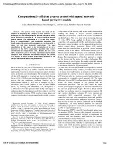
learning mechanism called Competitive Hopfield neural network (CHNN). The structure of CHNN used is independent of the image size being dependent only by the number of predefined classes and the number of existing grey-levels, as shown in figure 4. In this case the energy function that needs to be minimised is defined as the mean of squared distance measures of the grey levels within each class. The Lyapunov-based energy function used is:
n
n
k
E = ∑∑∑ x =1 y =1 i =1
1
∑h v y
(8)
v xi d xy h y v yi ,
n
yi
y =1
where n is the number of grey levels in the image, k is the number of predefined classes or segments, dxy is the square of the Euclidian distance measure between the grey-level pairs x and y and hx is the number of pixel at the grey-level x. The performances of CHNN and Hard C-means Algorithm [35] were compared on both computer simulated, CT and MR images. The metrics used for comparison were simple and a more appropriate comparison needed to be done. Furthermore, the computational level of the CHNN is high, and there is no guarantee that the network does not settle down to local minima.
Fig.4 CHNN proposed by Cheng et al. where n is the number of grey-levels and k is the number of predefined classes
11
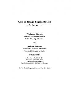
Poli and Valli [36] describe the segmentation as a small set of two-dimensional layers of neurons, idea taken from the colouring process of geographical maps (figure 5).
Fig.5 Segmentation of a synthetic image (left) using only four colours (center) and the binary representation with a layer of neurons for each colour used (right) They proposed a HNN based on that binary representation for the segmentation of X-ray and MR images. Their semantic energy function includes the sensitivity energy that forces the network to reveal any changes in the image grey level and robustness energy that is aimed to reduce the effects of noise and texture. Moreover, an extension of this approach is proposed for 3D-image segmentation. A very similar approach is described in [24] for the segmentation of synthetic and CT images. As a solution to the fact that a single-layer HNN has major limitations, such as settling into a local minimum, a multi-layer Hopfield neural network (MLHNN) for object recognition is presented by Young et al. [37]. Their architecture converges to a local minimum, but that minimum is “often equal or very close to the global minimum”. An extended one-layer version of the MLHNN is also presented. However, the computational overhead induced by those proposed architectures is not presented. Furthermore, the speed of the convergence process, which is directly dependant on the number and complexity of computations, is not compared with that of a single-layer HNN but with different types of
12
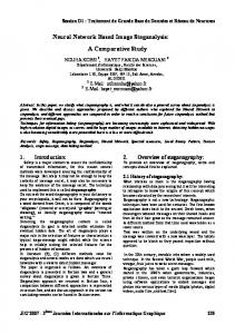
updating procedures. Therefore, more investigations should be done to prove the robustness of MLHNN. A multi-modal image segmentation method was proposed by Rout et al. [38]. Their method generates a threshold surface by interpolating the image grey levels at points where the gradient is high (possible edges). A modified HNN was used for the interpolation process. The performance is compared with the classical method of interpolation with potential surfaces, where the discrete Laplacian operation is computed for every pixel of the image, except the edge pixels. Although, the neural network approach has proved to be up to five times faster than the conventional approach, due to the property of HNN optimisation, a valuable comparison of goodness of segmentation for both methods was not performed. The presence of noise was not investigated at all. Koss et al.[39] investigated the use of HNN for segmentation of CT or MR images of abdominal organs. The first step to segmentation was to pre-process the images by computing second-order statistical texture transforms [40]. The results are then input into a HNN. The HNN structure is presented in figure 6.
Fig.6 HNN structure proposed by Koss et al.; a two-dimensional matrix of neurons: the size of the rows being the number of predefined classes k, and the size of the columns being the number of pixels in the image N; an active neuron (i,j) means that the pixel i is assigned to predefined class j.
13
The energy function proposed in this case is:
2 N k k N k k N E = A ∑ ∑∑ v pi vqi + ∑ ∑ v pi − 1 + ∑∑ R pi v 2pi , i =1 p =1 q ≠ p i =1 p =1 p =1 i =1
(9)
where Rpi is the distance of point i from centroid of class p. The first two terms combines the constraint conditions of the segmentation (the syntactic energy), i.e. each pixel must be assigned to only one class so that each row must have only one neuron active and the summation of each row must be one in order that a pixel is assigned 100% to a class. The semantic energy is defined by the third term that forces the network to find the minimum of the summation of the distances between the centroids of the predefined classes. In order to decrease the computation overhead and memory requirements, a WTA scheme is applied so that the energy equation (9) is transformed like in [34](7): k
(10)
N
E = ∑∑ R pi v 2pi p =1 i =1
The performance of this method was proved to be above 90% of the pixels for most of the organs present in the abdominal cavity. The presence of noise in the original images was still not investigated. The authors proposed a series of methods of improvement, such as speeding up the convergence process, reducing the number of neurons [35], and using a continuousHNN.
Self-Organising Maps ( SOMs) Valli et al. [24] proposed an architecture based both on anatomical or biological knowledge and the physics of image generation. The overall structure of their system is presented in figure 7. The input image is transformed using image generation physics i.e.
14
enhances information related to signal decay and improves the signal-to-noise ration of the image. Furthermore, anatomical knowledge is used to generate an initial predefined segmentation of the original images. The resultant images are then fed into the neural classifier that performs the segmentation. The modules that incorporates the anatomical and image generation physics are based on BPNN, while the neural classifier consists of a onedimensional Kohonen self-organising map used jointly with a unit labelling algorithm [41].
Fig.7 Segmentation system based on both anatomical knowledge and physical image generation
A Kohonen self-organising map with a competitive learning algorithm was used by Reddick et al. [42] to segment MRIs. This method makes use of T1-weighted, T2-weighted, and proton density-weighted MR images as inputs for the SOM. Thus, there are no spatial constraints involved. A nine-level grey-scale image was the result of the segmentation. After segmentation, a three-layer BPNN was trained to classify each pixel according to the tissue type. In this case, the segmentation was made fully automated, but the classification process still requires a priori knowledge, used to train the BPNN. Therefore this method could be 15
classified as a hybrid method between supervised and unsupervised image segmentation techniques. No noise influence was analysed. A two-step method for segmenting multispectral satellite images was proposed by Ambroise et al. [43]. The distribution of the pixels to be classified is analysed using a Probabilistic Self-Organising Map (PSOM) and then Agglomerative Hierarchical Clustering (AHC) is used. Because AHC cannot be used for clustering millions of objects, PSOM provides an initial partition of the image, so that AHC can be applied afterwards. The technique proved to have results that were comparable with classical techniques. Furthermore, the authors claim that different levels of classification are possible using a process that needs to be investigated in the future.
Constraint Satisfaction Neural Networks (CSNN). Lin et al. [44] proposes a new class of neural network for image segmentation – Constraint Satisfaction Neural Networks (CSNN). This method is based on the assumption that an image segmentation problem can be described as a Constrained Satisfaction Problem (CSP). The segmentation problem is redefined as the process of assigning each pixel a label according to certain spatial constraints. A CSNN consists of n x n x m neurons, where n x n is the image size and m is the number of classes the objects needs to be segmented into (called ‘labels’. Each neuron is connected to all its neighbours (8 connections). Those connections are the spatial constraints on the segment label of each pixel. Thus, a CSNN comprises a set of objects, a set of labels (classes) and spatial constraints describing the relationship between various objects according to neighbour relations.
They are updated so that a neuron will
excite other neurons belonging to the same class and inhibit the others. A winner-take-all scheme is chosen in order to deal with the crossover between segments. As in the standard learning algorithms, there are two phases: learning phase and categorization phase. Each
16
neuron receives feedback from its own output and excitatory/inhibitory signals from its neighbours. This method requires that the number of classes (labels) be known a priori. The proposed method also requires an initial assignment of label probabilities to every pixel using algorithms such as K-means, ISODATA, fuzzy c-means or Kohonen’s self-organising map (SOM). Certain improvements to the CSNN method were done by Kurugollu et al. [45] using explicit edge constraints and Kurugollu and Sankur [46] using pyramidal constraints, speeding up the process of convergence near the edges of the images. Another architecture based on a CSNN was proposed by Kurugollu and Sankur [47] – Multi-Scan constraint satisfaction neural network (MS-CSNN). The major advantage of this approach is that the appropriate number of segments is automatically determined by using a modified ZhangModestino [48] cluster validity index. Due to the large number of neurons n x n x m, this method requires computations that usually are not suitable for real-time applications. It was applied successfully to CT (computed tomography) images and MRIs [43,44,45]. Also, there is no literature about the behaviour of this CSNN in the presence of noise.
A locally excitatory globally inhibitory oscillator network (LEGION) A locally excitatory globally inhibitory oscillator network (LEGION) was proposed by Terman and Wang [49,50] as a possible computational method for image analysis and segmentation. The idea under LEGION lies in the biological world and is based on the temporal correlation theory. Neurons from the visual cortex respond only to stimuli from a particular part of the visual scene. Moreover, synchronised behaviour had been observed between spatially separated neurons so that cortical neurons corresponding to a distinct homogenous area are oscillating in phase, whereas neurons corresponding to different areas
17
are out of phase. LEGION is able to group similar features in an image by achieving fast synchrony
with
local
excitation.
Furthermore,
dissimilar
features
are
separated
by
desynchrony with global excitation/inhibition. Chakravarthy et al. [51] proposed a theoretical network of oscillating neurons for image segmentation purposes - an ANN of complex-valued neurons that exhibit stimulusspecific oscillations. In their model, the synchronization of neural oscillations is produced by both co-operation among neurons via excitatory/inhibitory couplings and by hebbian-like synaptic modification. Therefore, the networks exhibits coherent oscillations according to the input images, segmentation occurring due to local uniformities in image intensities. They suggest that special local features such as texture, orientation, could also be used to achieve synchronisation between neurons. Shareef et al. [52] used a single layer LEGION-based neural network for segmentation of medical images. A one-to-one correspondence between image pixels and neurons is used. Due to the dynamics of LEGION architecture, pixels with similar features will lead to oscillatory behaviour from their corresponding neurons and their phases will work in synchronization. Their adaptive scheme for grouping similar features (intensity contrast of pixels) has proven to work better on CT images rather than MR images. The speed achieved was as much as ten times better than classical segmentation techniques, i.e. statistical methods, global thresholding, active contours-snakes, etc. This architecture can be made more flexible, in the sense that a second layer can be added to process the result from the first layer, thus improving the segmentation results.
Pulse-coupled neural networks (PCNNs) Recently, pulse-coupled neural networks (PCNNs) have been used for image processing, analysis, including image segmentation [53,54]. PCNN is a single layered two-
18
dimensional neural network. It consists of laterally connected pulse-coupled neurons that are a modification of an Eckhorn’s neuron model [55]. The architectures proposed, establish a one-to-one correspondence between image pixels and pulse-coupled neurons. In [53] conditions that lead to the “perfect segmentation of a two-object image” (object and background) are derived. None of other image segmentation techniques described in the present study proposed conditions that guarantee a “perfect segmentation”. Liu et al. [56] proposed an architecture based on both LEGION and PCNN used to segment range images. In their ANN, each neuron has excitatory lateral connections to the neighbours and a connection to a global inhibitor. Each neuron has associated a feature vector consisting of depth, surface normal, mean and Gaussian curvatures. No a priori knowledge is necessary about the number of regions/segments. The
novelty
of
PCNN
and
LEGION
and
their
application
to
image
processing/segmentation leads to more research and analysis in order to prove their robustness and applicability.
Conclusions Neural-network based techniques are used successfully for the segmentation process. The results presented in the literature entitled these methods to a bright future, though much more work and research needs to be done. In the vast majority of the papers presented here, the results of the segmentation process lack general recognised metrics or a general evaluation framework. Therefore, comparison between methods is still author subjective matter of opinion. Moreover, one of the best advantages of neural networks – their graceful degradation in the presence of noise- has not been thoroughly investigated.
19
Many methods presented here still require sample data and a priori knowledge (such as k the number of desired classes or regions to be segmented). This problem can be approach by using another method of artificial intelligence – genetic algorithms (GAs). There are already reports about GAs used in classical methods like thresholding [57] and edge detection [58] or texture segmentation [59]. Another approach of removing the necessity of predefined knowledge was that of using most recent classes of neural networks, such as PCNN and LEGION. Due to their flexibility and lack of a priori information, they are worth investigating further. No general applicable framework or method has been found for image segmentation, even though some results in that direction are promising.
References 1. S.W. Lawson, Defect Detection in Industrial Radiographic and Ultrasonic Images’ (PhD thesis), University of Surrey, Guildford, UK, 1994 2. C. Amza, M. Graves, P. Innocent, J. Knight, Flexible neural network classifier for the automated detection of bones in chicken breast meat, Proc. International Conference on Engineering Applications of Neural Networks 17th - 19th of July 2000, Kingston University, UK, 2000, in press 3. M. Graves, X-ray Machine Vision for on-line Quality Control Food Processing, University of Cardiff, 1999 4. K.S. Fu, J.K. Mui, A survey on image segmentation, Pattern Recognition, vol.13, pp.3-16, 1981 5. R.M. Haralick, L.G. Shapiro, Survey, image segmentation technique, Comput. Vision Graphics Image Process. (CVGIP), vol.29, pp.100-132, 1985 6. P.K. Sahoo, S. Soltani, A.K.C. Wong, Y.C. Chen, A survey of thresholding techniques, Comput. Vision Graphics Image Process (CVGIP), vol.41, pp. 233-260, 1988 7. N.R. Pal, S.K. Pal, A review on image segmentation techniques, Pattern Recognition, vol.26, no.9, pp.1277-1294, 1993 8. J.C. Bezdek, L.O. Hall, L.P. Clarke, Review of MR image segmentation techniques using pattern recognition, Med. Phys., vol.20, no.4, pp. 1033-1048, Jul/Aug, 1993 9. L.P. Clarke, R.P. Velthuizen, M.A. Camacho, J.J. Heine, M. Vaidyanathan, MRI Segmentation: Methods and Applications, Magnetic Resonance Imaging, vol.13, no.3, pp.343-368, 1995 10. J.M. Zurada, Introduction to Artificial Neural Systems, West Publishing Company, USA, 1992 11. S.Y. Kung, Digital Neural Networks, PTR Prentice Hall, Englewood Cliffs, New Jersey, 1993 12. S. Haykin, Neural Networks: A comprehensive foundation, Macmillan College Publishing Company, Inc., USA, 1994
20
13. W.E. Blanz, S.L. Gish, A connectionist classifier architecture applied to image segmentation, Proc. 10th ICPR, pp.272-277, 1990 14. S.W. Lawson, G.A. Parker, Intelligent Segmentation of industrial radiographic images using neural networks, in Proc. of SPIE, vol.2347, pp.245-255, 1994 15. W.P. de Waard, Neural techniques and postal code detection, Pattern Recognition Letters, vol.15, pp. 199-205, 1994 16. R.H. Silverman, A.S. Noetzel, Image processing and pattern recognition in ultrasonograms by backpropagation, Neural Networks, vol.3, pp.593-603, 1990 17. M. Ozkan, H.G. Sprenkels, B.M. Dawant, Multi-spectral resonance image segmentation using neural networks, in Proc. IJCNN 90, vol.1, pp.429-437, San Diego, June 1990 18. K. Oshio, M. Singh, Automatic segmentation of magnetic resonance head images using neural nets, Proc. IEEE Nucl. Sci. Symp. And Medical Imaging Conf., pp.14391434, 1990 19. M. Ozkan, B.M. Dawant, R.J. Maciunas, Neural-Network-Based Segmentation of Multi-Modal Medical Images: A comparative and Prospective Study, IEEE Trans. On Medical Imaging, vol.12, no.3, pp.534-544, September 1993 20. U. Raff, A.L. Scherzinger, P.F. Vargas, J.H. Simon, Quantitation of grey matter, white matter, and cerebrospinal fluid from spin-echo magnetic resonance images using an artificial neural network technique, Med.Phys., vol.21, no.12, pp.1933-1942, December 1994 21. N. Babaguchi, K. Yamada, K. Kise, Connectionist model binarization, Proc. 10th ICPR, pp.51-56, 1990 22. L.O. Hall, A.M. Bensaid, L.P. Clarke, R.P. Velthuizen, M.S. Silbiger, J.C. Bezdek, A comparison of neural networks and fuzzy clustering techniques in segmenting magnetic resonance images of the brain, IEEE Trans. Neural Network, vol. 3, no.5, pp.672-682, 1992 23. Coppini, G., Poli, R., Legitimo, R., De Dominicis, R., Valli, G., A neural network system for detecting lung nodules in chest radiograms. In Computer Assisted Radiology, CAR’93, pp. 594-599, Springer-Verlag, Berlin, 1993 24. G. Valli, R. Poli, S. Cagnoni, G. Coppini, Neural Networks and Priori Knowledge Help the Segmentation of Medical Images, Journal of Computing and Information Technology- CIT 6, vol.2, pp.117-133, 1998 25. T. Wang, X. Zhuang, X. Xing, Robust segmentation of noisy images using neural network model, Image and Vision Computing, vol.10, pp.233-240, 1992 26. K. Fukanage, Introduction to Statistical Pattern Recognition, New York: Academic, pp.260-267, 1972 27. K. Shiranita, K. Hayashi, A. Otsubo, T. Miyajima, R. Takiyama, Determination of Meat Quality by Image Processing and Neural Network Techniques, Proc. Of FUZZIEEE2000, Vol. 2, pp516-521, San Antonio, Texas, May 2000 28. S. Haring, M.A. Viergever, J.N. Kok, Kohonen networks for multiscale image segmentation, Image and Vision Computing, vol.12, no.6, pp.339-344, July/August 1994 29. T. Kohonen, Self-Organization and Associative Memory, Third Edition, SpringerVerlag Berlin, 1989 30. A. Gaillard, D.C. Wunsch II, R.A. Escobedo, Neural hypercolumn architecture for the preprocessing of radiographic weld images, SPIE, vol.1294 Applications of Artificial Neural Networks, pp.378-388, 1990 31. J.J. Hopfield, Neural networks and physical systems with emergent collective computational abilities, Proc. Natl.Acad.Sci., USA, vol.79, pp.2554-2558, April, 1982
21
32. J.J. Hopfield, Neurons with graded response have collective computational properties like those of two-state neurons, Biophysics: Proc.Natl.Acad.Sci., USA, vol.81, pp.3088-3092, May, 1984 33. J.J. Hopfield, D.W. Tank, “Neural” Computation of Decisions in Optimisation Problems, Biol.Cybern., vol.52, pp.141-152, 1985 34. S.C. Amartur, D. Piraino, Y. Takefuji, Optimization Neural Networks for the Segmentation of Magnetic Resonance Images, IEEE Transactions on Medical Imaging, vol.11, no.2, pp.215-220, June 1992 35. K.S. Cheng, J.S. Lin, C.W. Mao, The application of competitive Hopfield Neural Network to medical image segmentation, IEEE Transactions on Medical Imaging, vol.15, no.4, pp.560-567, August 1996 36. R. Poli, G. Valli, Hopfield neural networks for the optimum segmentation of medical images, Handbook of Neural Computation, Oxford University Press, chapter.G5.5, pp.1-10, 1997 37. S.S. Young, P.D. Scott, N.M. Nasrabadi, Object Recognition using Multilayer Hopfield Neural Network, IEEE Transactions on Image Processing, vol.6, no.3, pp. 357-371, March 1997 38. S. Rout, P. Srivastava, J. Majumdar, Multi-modal image segmentation using a modified Hopfield Neural Network, Pattern Recognition, vol.31, no.6, pp.743-750, 1998 39. J.E. Koss, F.D. Newman, T.K. Johnson, D.L. Kirch, Abdominal organ segmentation using texture transforms and a Hopfield neural network, IEEE Transactions on Medical Imaging, vol.18, no.7, pp.640-648, July 1999 40. R.M. Haralick, K. Shanmugam, I. Dinstein, Textural features for image classification, IEEE Trans. Syst., Man, Cybern., vol. SMC-3, no.6, pp.610-621, 1973 41. D.H. Wolpert, Stacked generalisation, Neural Networks, vol.5, pp.241-259, 1992 42. W.E. Reddick, J.O. Glass, E.N. Cook, T.D. Elkin, R.J. Deaton, Automated Segmentation and Classification of Multispectral Magnetic Resonance Images of Brain Using Artificial Neural Networks, IEEE Transactions on Medical Imaging, vol.16, no.6, pp.911-918, December 1997 43. C. Ambroise, G. Seze, F. Badran, S. Thiria, Hierarchical clustering of self-organizing maps for cloud classification, Neurocomputing, vol.30, pp.47-52, 2000 44. W. Lin, E. Chen-Kuo, C.T. Chen, Constraint satisfaction neural networks for image segmentation, Pattern Recognition, vol.25, no.7, pp.679-693, 1992 45. F. Kurugollu, S. Birecik, M. Sezgin, B. Sankur, Image segmentation based on boundary constraint neural network. In Proc. Third International Workshop on Image and Signal Processing, IWISIP’96, Manchester, UK, pp.353-256, 1996 46. F. Kurugollu, B. Sankur, Colour cell image segmentation using pyramidal constraint satisfaction neural network. In IAPR Workshop on Machine Vision Applications – MVA’98, 17-19 November 1998, Makuri, Chiba, Japan, 1998 47. F. Kurugollu, B. Sankur, Image segmentation based on multi-scan constraint satisfaction neural network, Pattern Recognition Letters, no.20, pp.1553-1563, 1999 48. J. Zhang, J.W. Modestino, Model-fitting approach to cluster validation with application to stochastic model-based image segmentation, IEEE Trans. PAMI 12, pp. 1009-1017, 1990 49. D. Terman, and D.L. Wang, Global competition and local cooperation in a network of neural oscillators, Physics D, vol. 81, pp.148-176, 1995 50. D.L. Wang, D. Terman, Image segmentation based on oscillatory correlation, Neural Computing, vol.9, pp.805-836, 1997
22
51. S. Chakravarthy, V. Ramamurti, J. Ghosh, A network of oscillating neurons for image segmentation, Intelligent Engineering through ANN, vol.5, Dagli, Akay, Chen Fernanded and Ghosh (editors), ASME Press, Nov. 1995 52. N. Shareef, D.L. Wang, R. Yagel, Segmentation of Medical Images using Legion, IEEE Transactions on Medical Imaging, vol.18, no.1, pp.74-91, January 1999 53. H.S. Ranganath, G. Kuntimad, Object detection using pulse-coupled neural networks, IEEE Transactions on Neural Networks, vol.10, no.3, pp. 615-620, May 1999 54. G. Kuntimad, H.S. Ranganath, Perfect Image Segmentation Using Pulse Coupled Neural Networks, IEEE Transactions on Neural Networks, vol.10, no.3, pp/.591-598, May 1999 55. R. Eckhorn, H.J. Reitboeck, M. Arndt, and P. Dicke, Feature linking via synchronization among distributed assemblies: Simulations of results from cat visual cortex, Neural Computing, vol.2, pp.293-307, 1990 56. X. Liu, D.L. Wang, Range Image Segmentation Using a Relaxation Oscillator Network, IEEE Transactions on Neural Networks, vol.19, no.3, pp. 564-573, May 1999 57. P.-Y. Yin, A fast scheme for optimal thresholding using genetic algorithms, Signal Processing 72, pp.85-95, 1999 58. M. Gudmundsson, E.A. El-Kwae, M.R. Kabuka, Edge detection in medical images using a genetic algorithm, IEEE Transactions on Medical Imaging, vol.17, no.3, pp.469-474, June 1998 59. M. Yoshimura, S. Oe, Evolutionary segmentation of texture image using genetic algorithms towards automatic decision of optimum number of segmentation areas, Pattern Recognition, 32, pp.2041-2054, 1999 Acknowledgements The author wishes to thank Prof. Peter Innocent for his invaluable input and ideas to improve the paper. Special thanks are due to Prof. Jeffrey Knight for his suggestions and support. Glossary of Terms and Abbreviations used: NN = neural network MRI = magnetic resonance image CT = computer tomography image ANN = artificial neural network FFNN = feed forward neural network BP = back-propagation learning algorithm BPNN = back-propagation-based neural network MLFF = multi-layer feed forward neural network MLP = multi-layer perceptron HNN = Hopfield neural network WTA = winner-takes all learning mechanism CHNN = competitive Hopfield neural network MLHNN = multi-layer Hopfield neural network SOM = self-organising map (Kohonen)
PSOM = probabilistic self-organising map AHC = agglomerative hierarchical clustering CSNN = constraint satisfaction neural network CSP = constraint satisfaction problem MS-CSNN = multi-scan constraint satisfaction neural network LEGION = locally excitatory globally inhibitory oscillator network PCNN = pulse-coupled neural network GA = genetic algorithm
23