This basic requirement led Darwin (1872/1998) to suggest that human facial .... Phelps, 2001) or directed away from threat-related stimuli (Vuilleumier et al., ...
Recognizing Threat: A Simple Geometric Shape Activates Neural Circuitry for Threat Detection Christine L. Larson1, Joel Aronoff 2, Issidoros C. Sarinopoulos2, and David C. Zhu2
Abstract & The urgent need to recognize danger quickly has been shown to rely on preferential processing in dedicated neural circuitry. In previous behavioral studies examining the pattern of the face when displaying anger, we found evidence that simple noncontextual geometric shapes containing downward-pointing V-shaped angles activate the perception of threat. We here report that the neural circuitry known to be mobilized by many realistic, contextual threatening displays is also triggered by the simplest form of this V-shaped movement pattern, a downward-
INTRODUCTION The successful regulation of human interaction rests on the accurate comprehension of the intentions of others. This basic requirement led Darwin (1872/1998) to suggest that human facial displays of emotion provided a reliable communication system that used a common biological foundation to express and comprehend meanings in similar ways across all cultures. In order to determine key stimuli that convey these semantic meanings, and the mechanisms associated with their recognition, the research reported here used functional magnetic resonance imaging (fMRI) to determine whether a configurational pattern initially associated with threatening facial expressions, but stripped of all contextual meaning, leaving only a simple geometric shape, is sufficient to trigger specific emotion-related neural circuitry that previously has been shown to respond to threat-related stimuli. Darwin’s proposal regarding universal signals contained in emotional expressions found support in major programs of research directed by Ekman (1973, 2003) and others that identified the global expressions characteristic of each emotion as well as the exact movements (Ekman, Friesen, & Hager, 2002) of the facial muscles through which these displays are formed. Ekman (2003) also suggested that these displays are identified by sets of feature detectors that permit the rapid recognition of a facial expression. Although Ekman reviewed evidence clarifying the expressions associated with each emotion,
1
University of Wisconsin-Milwaukee, 2Michigan State University
D 2008 Massachusetts Institute of Technology
pointing triangle. Specifically, we show that simple geometric forms containing only downward-pointing V-shapes elicit greater activation of the amygdala, subgenual anterior cingulate cortex, superior temporal gyrus, and fusiform gyrus, as well as extrastriate visual regions, than do presentations of the identical V-shape pointing upward. Thus, this simple V-shape is capable of activating neural networks instantiating detection of threat and negative affect, suggesting that recognition of potential danger may be based, in part, on very simple, context-free visual cues. &
how an emotion is recognized is understood much less well. Given the evolutionary advantage of rapid detec¨ hman, 2005; Hansen & tion of threat (Lundqvist & O Hansen, 1988), we focused on the mechanisms underlying the recognition of facial expressions of anger. As these investigators have maintained, because of the survival advantage conferred by early recognition of potential danger, this process likely relies on neural circuitry that triggers rapidly, relatively automatically, ¨ hman & Mineka, and with minimal sensory input (O 2001; LeDoux, 2000). Further, as rapid detection is facilitated when visual signals of threat share easily identifiable features, thus reducing the need for thorough processing of all the features that compose a threatening stimulus, it would be highly efficient if such appraisal systems were organized to respond to an overall visual configuration formed by the facial features, rather than require the inspection of each movement of each facial landmark. As Ekman’s proposed feature detectors are thought to be associated with dedicated neural circuitry activated by the visual configurations formed when expressing an emotion, this study focuses on identifying the most essential underlying visual cues need to express threat-related emotion. The behavioral basis for this hypothesis rests on studies that used multiple methods to isolate key stimuli that convey affective meaning. Seeking to identify the specific sign stimuli that conveyed the meaning of anger and happiness across a wide range of primarily nonliterate tribal cultures, cross-cultural research (Aronoff, Barclay, & Stevenson, 1988) found that the affective
Journal of Cognitive Neuroscience 21:8, pp. 1523–1535
identity of the display was carried in the overall configuration made by the major landmarks of the face, rather than by the specific facial features themselves. Anger was shown to be conveyed by angular and diagonal forms made by the facial features (e.g., eyebrows), especially acute angles pointing downward, and happiness was conveyed by curved patterns. This configurational hypothesis is also supported by Bassili’s (1978) pioneering point-light experiment, which studied the overall geometric pattern formed by the movement of the face as a whole when displaying an emotional expression. Bassili placed luminescent dots on subjects’ faces and, in a dark room, asked them to assume happy and angry expressions. When portraying a happy appearance, the burst of dots expanded outward to form a rounded shape, whereas in the angry representation, the points of light imploded downward and inward to form a V-shaped figure. The ability of the V-shaped figure (usually representing eyebrows) to convey an angry subjective state is also shown in the many studies ¨ hman, using schematic faces (Lundqvist, Esteves, & O 1999) and confirmed by studies (Lundqvist, Esteves, & ¨ hman, 2004) which show that V-shaped figures elicit O this emotional meaning even when presented without any other facial feature. The power of more rounded facial shapes to convey emotionally positive semantic meanings (Zebrowitz, 1998; Hildebrandt & Fitzgerald, 1983) is similarly well established. These initial findings have been confirmed by additional naturalistic and experimental research (Aronoff, Woike, & Hyman, 1992; Aronoff et al., 1988), which examined the affective identity of a wide range of nonrepresentational visual stimuli, such as angular or curved lines, as well as similar geometric shapes using figures far removed from actual representations of the human face. Abstract shapes and everyday objects (e.g., watch, sofa) containing sharp angles of various orientations have also been found to be less preferred than the similar shapes or objects containing curved forms (Bar & Neta, 2006). Bar and Neta (2006) posited that the lower preference for sharp innocuous objects ‘‘stemmed from a feeling of threat, and that this feeling was triggered by the sharpness of the angles per se’’ (p. 647). Other work in our laboratory attempted to identify the key features underlying facial cues of emotion by gradually stripping away contextual information and reducing the stimuli to their most fundamental geometric components. In several studies using semantic differential scales (Osgood, Suci, & Tannenbaum, 1957) to record the subjective meaning elicited by these nonrepresentational visual stimuli (i.e., the degree of ‘‘badness,’’ ‘‘potency,’’ and ‘‘activity’’ perceived in the stimuli), and following techniques introduced by Tinbergen (Eibl-Eibesfeldt, 1989), who pioneered the use of models to study signaling devices, we presented increasingly angular, diagonal, or curvilinear models for examination and found that angular V-shaped figures alone (similar to the angles found
1524
Journal of Cognitive Neuroscience
in the eyebrows, cheeks, chin, and jaws in angry expressions) and rounded figures alone (similar to the curves found in the cheeks, eyes, and mouth in happy expressions) conveyed the same affective meanings as that evoked by actual angry and happy facial representations (Aronoff, 2006; Aronoff et al., 1988, 1992). Using the same technique, we recently extended this finding, reporting that simple geometric shapes are perceived as having affective value, and furthermore, that the orientation of the angle is an important determinant of this value. Specifically, simple shapes containing a downwardpointing acute angle (e.g., a ‘‘V,’’ a triangle) were rated by participants as more threatening than the exact same shapes with the V-angle pointing upward (Larson, Aronoff, & Stearns, 2007). Additional support for the configurational hypothesis is provided by the many studies which examine the efficiency of shapes to signal potential threat. Resting again on Darwin’s suggestion that speedy detection of threat confers an evolutionary advantage (Niedenthal & Kitayama, 1994), Hansen and Hansen (1988) used the visual search paradigm to show that the search for an angry face in a crowd of happy faces is more efficient (i.e., rapid) than the reverse. Subsequent work of this type, using both real and schematic faces (Horstmann ¨ hman, Lundqvist, & Esteves, 2001), & Bauland, 2006; O confirmed that angry faces consistently lead to briefer search times. In an experiment to isolate capture of attention effects for the pure geometrical form of a downwardpointing acute angle from other facial features and from other confounding perceptual factors, we used a simple triangle whose vertex was pointed either up or down as our target shape (Larson et al., 2007). Triangles were used rather than a ‘‘V’’ itself to avoid the possibility that capture of attention was facilitated because V is a letter, rather than due to any inherent emotional or attentional properties. Thus, having a simple shape that varied only in the orientation of the acute angle, we demonstrated that a triangle with a downward-pointing vertex is recognized more rapidly than the identical shape with an upward-pointing vertex. Further, this study also provided evidence that the downward-pointing triangle has the power to elicit sustained attention, in keeping with the ability of realistic threat-related stimuli to disrupt or slow performance of ongoing cognitive tasks (Eastwood, Smilek, & Merikle, 2003; Vuilleumier, Armony, Driver, & Dolan, 2001; White, 1995). Thus, both subjectively and attentionally, the simple downward V-shape appears to function much like a typical contextually based threat stimulus. These results lead us to hypothesize the presence of a neural network of targeted, and thus, highly efficient brain regions that can identify molar shapes that signal the presence of biologically relevant affective stimuli. Such configurational detection mechanisms seem to be a much more parsimonious way to account for the decoding of emotional displays than would be the conjecture of a wide set of
Volume 21, Number 8
specialized neural circuitry isomorphic with each individual muscular movement. Thus, the evolutionary advantage for threat detection may be due, in part, to facilitated recognition of a simple geometric form, common to a number of threat-related objects, thereby minimizing the need to process a stimulus fully and in context in order to identify a potential threat. For these reasons, in the present study, we investigate whether viewing the simple shape of a downward-pointing acute angle recruits the same neural circuitry as that demonstrated previously to respond to representational and contextual cues of threat, such as threat-related facial expressions, aversive scenes, and phobogenic stimuli. A large body of research has implicated the amygdala in directing attention toward biologically relevant, affectively salient information, as well as in the processing of aversive stimuli (LeDoux, 2000); functions that render the amygdala particularly important in the detection of potential threat. Human neuroimaging studies have demonstrated increased amygdala activation in response to a number of aversive stimuli, including negatively valenced pictures (Irwin et al., 1996), angry faces (KeslerWest et al., 2001), and fear faces ( Vuilleumier, Armony, Driver, & Dolan, 2003; Morris et al., 1998). Other work has further supported the notion that the amygdala instantiates rapid, automatic detection of threat, even under conditions in which attention is limited (Anderson & Phelps, 2001) or directed away from threat-related stimuli (Vuilleumier et al., 2001). Human amygdala activation has also been detected in response to coarse, spatially degraded facial cues of threat ( Vuilleumier et al., 2003). Importantly, evidence from fear conditioning studies in animals demonstrates that an intact amygdala is sufficient for rats to learn to fear very simple stimuli (e.g., simple tones) even when input from higher cortical regions is disrupted through lesions of the relevant modality-specific sensory cortex (Doron & LeDoux, 1999). Of particular relevance for the present study, Bar and Neta (2007) found that neutral objects (abstract figures, everyday objects) containing sharp as compared to curved contours activated the amygdala, among other regions. Similar to the principles guiding the present work, the authors
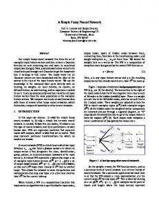
interpreted these findings as further support for the notion that sharp, angular objects may signal danger. In light of recent evidence, we also expected to see activation in visual sensory pathways in response to the downward V-shape. In a recent review, Vuilleumier and Driver (2007) emphasized that affective, not just perceptual, properties of visual stimuli can be potent modulators of the visual cortex. Consistent with this premise, emotional, particularly unpleasant emotional, faces activate the face-sensitive region of the fusiform gyrus more strongly than neutral faces (Vuilleumier et al., 2001). Similarly, unpleasant scenes elicit more robust activation of the extrastriate cortex than neutral scenes (Sabatinelli, Lang, Keil, & Bradley, 2007). This enhanced sensory processing is likely a function of the heightened salience of these stimuli, which necessitates increased recruitment of attentional resources, including the sensory cortex. The amygdala has been shown to be crucial to facilitate this enhanced processing in the ventral visual pathway (Vuilleumier, Richardson, Armony, Driver, & Dolan, 2004), and thus, seems to be the key structure initiating preferential processing of salient, biologically relevant visual stimuli. We sought to determine whether the amygdala and related circuitry is recruited in response to a very simple geometric shape which is devoid of contextual affective cues, but has been shown to depict threat. Specifically, we predicted that shapes with downward-pointing V’s would elicit greater amygdala activation than the identical shape whose vertex pointed upward. Consistent with the work of Vuilleumier and Driver (2007), we also predicted greater activation in ventral visual processing regions, as well as increased connectivity between the amygdala and these areas. Whole-brain fMRI scans were conducted while participants viewed a large set of images of three simple geometric shapes: downward-pointing triangles, upward-pointing triangles, and circles that varied in number, size, and location (see Figure 1) in a block design. To maintain attention, participants made simple judgments about the number of shapes depicted in each image. As with our previous behavioral work, triangles were used rather than an open V to avoid the possible
Figure 1. fMRI paradigm and examples of shape stimuli. Across four runs, 12 blocks of each of the three stimulus types, downward-pointing triangles, upward-pointing triangles, and circles, were presented in a random order. Each block was 25 sec long and consisted of 10 different stimuli of the same shape, each presented for 2.5 sec. Each stimulus consisted of one to seven identical shapes. Shapes of four different sizes were presented, but size was held constant within any given image. The number, size, and location of the shapes were counterbalanced within a condition and were equated across the three different shape conditions. To ensure that participants maintained attention to the stimulus presentation, they were asked to press a button indicating whether greater or fewer than four shapes were presented in each image (no images contained four shapes). Each block was followed by a 15-sec rest period during which a fixation cross was displayed.
Larson et al.
1525
confound of V being a letter, and thus, being more salient. Upward-pointing triangles were used as a comparison condition in order to test the effects of the same shape when inverted. Circles provided another control condition and were selected to test the general effects of angular compared to nonangular stimuli.
METHODS Participants Twenty right-handed, healthy college students participated in this study and signed consent forms approved by the Michigan State University Institutional Review Board. Data from three subjects were discarded, one due to incorrect positioning of the equipment, another due to irregular anatomy, and a third due to lack of activation in primary visual areas suggesting lack of attention during the study. For the same reason, data from two functional runs were discarded in one of the remaining subjects, and data from one functional run were discarded in another subject. Seventeen subjects (10 men, mean age = 20.6 years, range = 18–26 years) were included in the data analysis.
Stimuli Stimuli included 120 unique pictures for each of the three conditions: circles, upward triangles, and downward triangles (Figure 1). Each of the 120 pictures per condition was unique in either number of objects, object size, or object positions. Each picture contained one, two, three, five, six, or seven outlines of each shape on a white background. Shapes were presented in four different sizes, but for each picture the size was held constant. Finally, the positions were randomized within condition across all 120 images. Stimuli were matched across conditions such that, for each shape condition, there was an image that exactly matched the other conditions on number of objects, object size, and object position. Stimuli were displayed in color on a 640 � 480 LCD monitor mounted on top of the RF head coil. The LCD subtended 128 � 168 of visual angle.
paradigm so that they became familiar with the task, and they were asked to pay close attention to the shape of the objects in each picture. Participants were told that the study aimed to understand how the visual system responds to different geometric shapes. The experiment was divided into four functional runs each lasting 6 min 15 sec. In each run, subjects were presented nine blocks of visual stimulation after an initial 15-sec ‘‘resting’’ period. In each block, 10 unique pictures from one condition were presented. Within a block, each picture was presented for 2.5 sec with no interstimulus interval. A 15-sec baseline condition (a white screen with a black fixation cross at the center) followed each block. Each condition was shown in three blocks per run. Both the order of conditions within each run and the order of pictures within a block were randomly determined. The four functional runs were presented to eight subjects in a forward order and others in a reverse order.
Image Acquisition Data were collected on a 3-T GE Signa EXCITE scanner (GE Healthcare, Milwaukee, WI) with an eight-channel head coil. During each session, images were first acquired for the purpose of localization, followed by first and higher-order shimming procedures to improve magnetic field homogeneity (Kim, Adalsteinsson, Glover, & Spielman, 2002). To study brain function, echo-planar images, starting from the most inferior regions of the brain, were then acquired with the following parameters: 34 contiguous 3-mm axial slices in an interleaved order, TE = 27.7 msec, TR = 2500 msec, flip angle = 808, FOV = 22 cm, matrix size = 64 � 64, ramp sampling, and with the first four data points discarded. Each volume of slices was acquired 146 times during each of the four functional runs while subjects viewed the pictures, resulting in a total of 584 volumes of images over the course of the entire experiment. After functional data acquisition, high-resolution volumetric T1-weighted spoiled gradient-recalled images with cerebrospinal fluid suppressed were obtained to cover the whole brain with one hundred twenty 1.5-mm sagittal slices, 88 flip angle, and 24 cm FOV. These images were used to identify anatomical locations.
Procedure A block-design paradigm was controlled by an IFIS-SA system (Invivo, Gainesville, FL). Two ergonomic keypads were placed under the hands of each subject. The subject was requested to press the right index finger button when there were more than four objects on a picture, and to press the left index finger button when there were less than four objects (no image contained exactly four objects). Accuracy for this judgment was 99.7% and there were no reaction time differences between the three shape conditions.1 Before entering the scanner, all subjects were trained by viewing a 2-min practice
1526
Journal of Cognitive Neuroscience
fMRI Data Preprocessing and Analysis All fMRI data preprocessing and analysis was conducted with AFNI software (Cox, 1996). For each subject, acquisition timing difference was first corrected for different slice locations. With the first functional image as the reference, rigid-body motion correction was done in three translational and three rotational directions. The amount of motion in these directions was estimated and then the estimations were used in data analysis. For each subject, spatial blurring with a full width half
Volume 21, Number 8
maximum of 4 mm was applied to reduce random noise (Parrish, Gitelman, LaBar, & Mesulam, 2000), and also to reduce the issue of intersubject anatomical variation and Talairach transformation variation during group analysis. For the group analysis, all images were converted to Talairach and Tournoux (1988) coordinate space with an interpolation to 1 mm3 voxels. For analysis of each individual subject, the reference function throughout all functional runs for each picture category was generated on the basis of the convolution of the stimulus input and a gamma function, modeled as the impulse response when each picture was presented. The acquired functional data were compared with the reference functions using the 3dDeconvolve software for multiple linear regression analysis and general linear tests (Ward, 2002). Multiple linear regressions were applied on a voxelwise basis for t-statistic tests and to find the magnitude change when each picture condition was presented, compared to the reference functions. BOLD percent signal change relative to the baseline state was then calculated. General linear tests were also applied on a voxelwise basis to find the statistical significance of pairwise comparisons for all the picture conditions. For the above analysis, in addition to applying the reference functions for the three picture conditions, MRI signal modeling also included the subject motion estimations in the three translational and the three rotational directions, and the constant, linear, and quadratic trends for each of the four functional runs. Monte Carlo simulation of the effect of matrix and voxel sizes of the imaging volume, spatial correlation of voxels, voxel intensity thresholding, masking, and cluster identification was applied to estimate overall statistical significance with respect to the whole brain (Ward, 2000). Because the anterior cingulate cortex (ACC) was a specific region of interest (ROI), a similar procedure was carried to estimate the overall statistical significance with respect to this ROI.
Whole-brain Group Analysis After the percent signal change was estimated with respect to each picture condition for each subject, an ANOVA was performed for group analysis with a mixedeffect two-factor model, with picture condition (three levels) modeled as a fixed effect and subject modeled as the second factor as a random effect. The ANOVA results were used to extract the activated voxels for all pairwise condition contrasts (voxel-based p value