Journal of Theoretical and Applied Information Technology 28th February 2015. Vol.72 No.3 © 2005 - 2015 JATIT & LLS. All rights reserved.
ISSN: 1992-8645
www.jatit.org
E-ISSN: 1817-3195
AUTOMATIC GLAUCOMA DETECTION BASED ON THE TYPE OF FEATURES USED: A REVIEW 1
ANINDITA SEPTIARINI, 2AGUS HARJOKO Department of Computer Science, Mulawarman University, Samarinda, Indonesia 2 Department of Computer Science and Electronics, Gadjah Mada University, Yogyakarta, Indonesia E-mail:
[email protected],
[email protected] 1
ABSTRACT Glaucoma is an eye disease that is the second most common cause of blindness in worldwide. The characteristic of glaucoma are high eye pressure, loss of vision gradually which can cause blindness and damage to the structure of retina. The damages which may occur for example are structural form changes of the Optic Nerve Head (ONH) and Retinal Nerve Fiber Layer (RNFL) thickness. The observable part of ONH which is the features of glaucoma such as disc, cup, neuroretinal rim, Parapapillary atrophy and blood vessels. The structure of the retina can be observed through a retinal image, where the image is produced from several types of equipment such funduscopy, Confocal Scanning Laser Ophthalmoscopy (CSLO), Heidelberg Retina Tomograph (HRT) and Optical Coherence Tomography (OCT). This paper discusses about the automatic feature extraction technique in retinal fundus images which can be used for detection or classification of glaucoma. The technique is divided into two groups, namely morphological and non-morphological based on the type of features used. This grouping aims to determine what type of features extraction technique can be used to represent the glaucoma characteristic. Keywords: Glaucoma, Detection, Classification, Feature Extraction, Retinal Image allow any divergence accuracy of examination result due to the dependence on the domain knowledge of a different expert.
1. INTRODUCTION Glaucoma is the second leading cause of blindness worldwide [1]. It is characterized by a gradual loss of visual function to lead to blindness. Another feature associated with glaucoma is high pressure in the eye, changes in the structure of the Optic Nerve Head (ONH) and Retinal Nerve Fiber Layer (RNFL) thickness [2]. In ONH structural changes may occur in some parts such as discs, cup, neuroretinal rim, blood vessels and parapapillary atrophy (PPA). The examination of ONH can be done directly by using the direct ophthalmoscope, indirect ophthalmoscope or use the posterior pole lens is equipped with a slit lamp [3]. When an expert does the examination of ONH directly, it can make the patient feel uncomfortable. To overcome this, the examination of ONH can be observed through a retinal image, where the image produced by some types of equipment, such as funduscopy, Confocal Scanning Laser Ophthalmoscopy (CSLO), Heidelberg Retina Tomograph (HRT) and Optical Coherence Tomography (OCT) [4]. Almost all examinations performed manually by an expert,
Early detection of glaucoma is needed because the early stages of the disease symptoms are not felt by most of the patients and slow progression of the disease. It causes, damage is already severe in the ONH when the disease is detected. Oleh sebab itu dibutuhkan adanya pendeteksian glaucoma secara otomatis. The current research about automatic detection of glaucoma has been developed. The study was developed in a different way, where the difference can be viewed from a number of areas including the use of the features, methods of segmentation, feature extraction techniques and methods of classification. This paper are review about the automatic feature extraction technique in retinal images and provide an overview of the recent developments related to research automatic glaucoma detection. Our Contribution in this paper is to provide classification results feature extraction technique based on the type of the features. The type of feature is divided into two groups namely morphological and non-morphological. Papers
366
Journal of Theoretical and Applied Information Technology 28th February 2015. Vol.72 No.3 © 2005 - 2015 JATIT & LLS. All rights reserved.
ISSN: 1992-8645
www.jatit.org
were reviewed in this paper have been selected from the papers related to automatic glaucoma detection that disccused about automatic feature extraction technique with input data were retinal images. In the next section (2) explain about glaucoma, the type of features used described in section (3), then free database that can be used to glaucoma research, the open research issues and the conclusion sequentially described in section (4), (5) and (6).
E-ISSN: 1817-3195
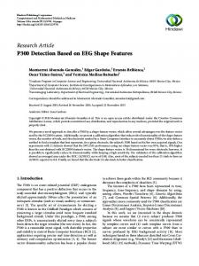
This section will explain about the structure of ONH, diagnosis of glaucoma based on the type of examination performed and the characteristics of glaucoma. 2.1 Structure of ONH ONH is an elliptical-shaped area located at the entrance of the optic nerve at the back of the eye. There are many parts of ONH that can be observed to distinguish between normal and glaucoma eyes such as disc, cup, neuroretinal rim, blood vessels and parapapillary atrophy (PPA). Anatomy of the eye and structure of ONH is shown in Figure 1 [5].
2. GLAUCOMA
Figure 1: Eye’s Anatomy And Structure Of ONH
Based on Figure 1 there are many part that related with glaucoma. ONH or also called as disc is an elliptical-shaped area. On the inside of the disc, there is another elliptical-shaped area with smaller size is called a cup. For an area that is between disc and cup called as neuroretinal rim. Parapapillary atrophy (PPA) is an area that does not always appear on the retina which is located on the outside of the ONH, where the PPA is directly adjacent to the disc. The part that looks like hair and emerged from the middle of the area referred to as the ONH blood vessels. Disc and cup are used to calculate the value of Cup-Disc Ratio (CDR). CDR is the value of a cup diameter divided by the diameter of the disc. CDR value can be calculated based on the diameter of the vertical or horizontal diameter of the disc and the cup. In 90% of normal eyes CDR-value of less than 0.5 [6]. The example of fundus image which present the diameter of disc and cup is shown in Figure 2 [3].
Figure 2: Disc And Cup In Fundus Image
Enlargement of cup size is an early change that could affect the value of the CDR, where the value of the CDR can be used as a parameter to detect glaucoma disease [6]. The example of cup size enlargement is shown in Figure 3 [3].
367
Journal of Theoretical and Applied Information Technology 28th February 2015. Vol.72 No.3 © 2005 - 2015 JATIT & LLS. All rights reserved.
ISSN: 1992-8645
www.jatit.org
E-ISSN: 1817-3195
does not have the effect of myopia and glaucoma disease. A second type of PPA is beta-zone. It occurred more frequently in patients with glaucoma, which has the characteristic white color. Both types of PPA are located outside the area of the disc. Beta-zone is directly bordering the disc on the temporal side, while alpha-zone directly adjacent to the beta-zone [6]. The areas of alphazone and beta-zone are shown in Figure 5.
Figure 3: Enlargment Of Cup Size
The neuroretinal rim which is the area between the disc and the cup in the normal eye looks pink-orange and has a broad estimate of between 1.4 to 2.0 mm 2. It is divided into four parts: Inferior, Superior, Nasal and Temporal (ISNT) [6]. Inferior rim is the distance between the disc and the cup on the top, the superior rim is the distance between the disc and the cup on the bottom, nasal rim is the distance between the disc and the cup in the section close to the nose while the temporal rim is the distance between the disc and the cup opposite the nasal rim. The location of the ISNT for more details is shown in Figure 4. Cup in ONH is not positioned at the center, so that the thickness of each section neuroretinal rim area is different. Sequentially based on the thickest part is inferior, superior, nasal and temporal. Inferior is the thickest part while the temporal thinnest part. The order is based on the thickness of the basis ISNT rule.
Figure 5: PPA With Alpha-Zone And Beta-Zone On The Right Eye
RNFL looks like a bunch of scratches that colored light is distributed evenly on the normal eye. In normal eye RNFL mostly seen in the inferior temporal area, followed in the area of the superior temporal, superior nasal and inferior nasal [7]. RNFL can be observed by ophthalmoscopy and wide angle photos without the red color. The structure of the RNFL is shown in Figure 6.
Figure 6: Structure Of RNFL
Figure 4: ISNT Rule on the Right Eye
PPA is a part with crescent-shaped, which consists of two types of alpha-zone and beta-zone. Alpha-zone is an area of hypopigmentation and hyperpigmentation. This part looks at the patient
2.2 Glaucoma Diagnosis Diagnosing glaucoma may be based on several type of examination. The examination of glaucoma can be done by looking at the patient's medical history, Intraocular Pressure (IOP), visual ability test and observation of ONH manually using ophthalmoscopy or by observing the retinal image
368
Journal of Theoretical and Applied Information Technology 28th February 2015. Vol.72 No.3 © 2005 - 2015 JATIT & LLS. All rights reserved.
ISSN: 1992-8645
www.jatit.org
structures. Qualitative examination of the ONH can be done by using the ONH Stereo Photographs (ONHSPs), Confocal Scanning Laser Ophthalmoscopy (CSLO), Scanning laser polarimetry (SLP) and Optical Coherence Tomography (OCT) to distinguish normal eyes and glaucoma [8]. Almost all examinations performed manually by an expert. The examination result has allowed difference accuracy due to the limitations and dependence domain knowledge from an expert. 2.3 Characteristics of Glaucoma Normal eyes and glaucoma can be distinguished based on the changes in the ONH and RNFL. Enlargement of cup size can lead to changes the value of CDR and neuroretinal rim area becomes smaller. Area neuroretinal rim thinning (notching) make void the ISNT rule. The changes in the initial stages of neuroretinal rim are located on the inferior and superior rim. Figure 7 shows the thinning of neuroretinal rim in the inferior part.
Figure 7: Notching
Nerve fiber layer hemorrhage appears as a red line that is parallel or close to the surface area dics. Hemorrhage is possible contained in the neuroretinal rim area or PPA. The example of hemorrhage is shown in Figure 8.
E-ISSN: 1817-3195
Patients which is has asymmetry disc conditions between the two eyes can be a sign of the possibility that the patient suffering from glaucoma. In general, the normal eye has a different value of CDR between right and left eyes, it value is not more than 0.2. When research on glaucoma utilize computer-based image analysis can be seen that 30% of patients with glaucoma have a cup size of areas in which asymmetry [7]. The next feature is the emergence of beta-zone PPA is also one of the characteristics of the disease glaucoma. Nerve fiber layer in normal eyes look like scratches are formed from a collection of axons in the retina, which is located around the neuroretinal rim to PPA. In the eyes of glaucoma nerve fiber layer thinning and become unclear.
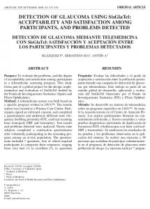
3. TYPE OF FEATURES Research about detection or classification of glaucoma using retinal image is influenced by the selection of features and feature extraction techniques. Feature extraction technique in retinal images are classified based on the type of the features. The type of feature is divided into two groups namely morphological and nonmorphological. In this research grouping type of features are presented as diagram and can be seen in Figure 9. To get the morphological feature we needs segmentation process before measuring the geometric parameters. The examples of the morphological features are RNFL, PPA and the part of ONH such as disc and cup. After segmentation process produce the disc and cup boundary we can use them to calculate the values of disc and cup diameter, disc and cup area, CDR and neuroretinal rim area. Non-Morphological feature is a feature that is no need segmentation process or extracted from the existing image (Image-based featured). Color, shape and texture are type of features that captured from the existing image. So it can be represented the characteristic of glaucoma disease. Color feature can be used to extract such as cup, neuroretinal rim, PPA to represent the characteristics. The blood vessels, neuroretinal rim and PPA can be extracted to represent characteristic based on shape feature, while RNFL can be extracted by texture feature.
Figure 8: Hemorrhage Of ONH On The Right Eye
369
Journal of Theoretical and Applied Information Technology 28th February 2015. Vol.72 No.3 © 2005 - 2015 JATIT & LLS. All rights reserved.
ISSN: 1992-8645
www.jatit.org
E-ISSN: 1817-3195
Figure 9: Group Research Based On Type Of Features
3.1 Morphological Features Research about glaucoma detection with morphological features has been done by Chrastek et al. (2005). It used several features that were CDR, Neuroretinal rim and PPA to detect glaucoma. To extract these features were used morphological method for localizing ONH, Hough transform to limit the search space and active contour models to find the end of the boundary line. The experiment was performed on 159 numbers of images (normal and glaucoma). The accuracy achieved in this study was 73.2% using the Linear Discriminant Analysis (LDA), 75.8% using Ctree and bagging 77.8% [5]. Several studies using CDR features to detect glaucoma was performed by Xu et al. [9], Muramatsu et al. [10], Joshi et al. [11], Yin et al. [12], Pruthi and Mukherjee [13] and Cheng et al. [14]. To obtain the value of CDR was require disc and cup segmentation process. In the study by Xu et al. (2007) the segmentation process performed by detecting the cup and disc boundary with free-form deformable model (snake) technique. Boundary was extracted based on combination of some of the information that is smoothness, gradient, depth and other information. The algorithm used is to modify the original snake technique. The method is applied to 100 retinal fundus images having both normal and glaucoma taken from the National University Hospital. Accuracy for boundary detection in this research was 94% [9]. In the research by Muramatsu et al. (2009) to detacted glaucoma proposed the several stages to
outline the boundary of disc [10]. The fundus images used for input data, in the early stages the image through filtering process with median filter and pixel averaging filter to reduce noise. Next, the ONH area estimated use p-tile thresholding method, followed by identifying the Region of Interest (ROI) in the form of ONH area using high pixel value in the red channel of RGB color image. After getting the expected ROI image the next stage is converted it to grayscale images, followed by improving contrast area of ONH and area of retina with Otsu thresholding method. In the last stage, the ONH edge detection is done by canny method. Then, the boundary of a disc used Snakes active contour method with spline operation. The experiment of this research used fundus images. The 80 pairs of experimental data consist of 25 pairs of glaucoma (24 pairs training data and a pairs testing data) and 55 normal pairs (44 pairs training data and 11 pairs testing data). The results for the accuracy of the segmentation disc and cup are respectively 83% and 51%. Optic disc segmentation process was done with the information integration of the local image that was around a point of interest into a multidimensional feature space. Next, the cup segmentation was done by anatomical features such as vessel bends at the cup boundary. Through this stage the resulting accuracy of two things were 91% for cup-to-disc vertical diameter ratio and 87% for cup-to-disc area ratio. The dataset consist of 33 normal and 105 glaucoma (total of 138) images [11].
370
Journal of Theoretical and Applied Information Technology 28th February 2015. Vol.72 No.3 © 2005 - 2015 JATIT & LLS. All rights reserved.
ISSN: 1992-8645
www.jatit.org
In [12] have used the dataset from database ORIGA light consisting of 625 retinal fundus images, where the data consists of 168 glaucoma images and 457 normal images. The dataset was randomly divided into two sections each of 325 data is used for training and the rest for the testing process. Selection of training and testing data randomly produced accuracy of 92% for segmentation disc and 81% for segmentation cup. The optic disc and cup segmentation is done based on the statistical model-based methods. Segmentation optic disc and cup performed by merging knowledge-based Circular Hough Transform and optimal channel selection. Another method that can be applied to disc and cup segmentation process consists of three stages. First, applied K-means clustering method, next stage was boundary process using Multi thresholding, Active Contour Method, Fuzzy C Means Clustering and ANN then performed morphological operations to fill the holes and a small part in the cluster of optic disc and cup. Third, Ellipse Fitting is used to mark the optic disc and cup boundary. This study used fundus image as input data. The method was tasted on 10 images consisting of normal and glaucoma fundus image. The accuracy reached 97.77% using ANFIS, 98.12% and 97.35% using SVM using Backpropagation [13]. The superpixel classification methods can be used to segment the optic disc and cup [14]. Optic disc segmentation process for classifying each superpixel including disc / non-disc used histograms and statistical surround center (CSC). This method produced accuracy reached 90.5%. The cup segmentation also used histograms and CSC, the location information is also included as features and the accuracy result reached 85.9%. The dataset was used consist of 2326 retinal fundus images. The data taken from two different place, first from Singapore Malay Eye Study (Simes) with 3072x2048 pixel size as 650 data (168 glaucoma and 482 normal). Second, from the Singapore Chinese Eye Study (SCES) 1676 data (46 glaucoma and normal 1630). Narasimhan and Vijayarekha [15] uses the combination of CDR and ISNT ratio features to detect glaucoma. The extraction progress of disc and cup were measured using a K-means clustering, while the ratio ISNT generated by calculating the area of blood vessel in ISNT quadrant. For the detection of blood vessel performed using local entropy thresholding. In the dataset 16 images used for training (consisting of 7 normal and 9
E-ISSN: 1817-3195
glaucoma) and 36 images used for testing (consisting of 15 normal and 21 glaucoma). The image data derived from Aravind Eye Hospital, Madurai Tamilnadu India. Research conducted by Mary and Marri focus on the localization part of ONH or disc, where the results can be used for segmentation process of disc and cup from fundus retinal image. ONH localization is done by combining the Hough space method then divides the area into n × n part. For ONH automated segmentation use Pyramidal Decompotition method. This research used 100 retinal images from DRIVE and DIARET0 database [16]. 3.2 Non-Morphological Features Research using non-morphological features have been carried out by Bock et al. [17] using some feature extraction methods such as the values of pixels intensity, Gabor Filter (textures), the coefficient of Fourier Transform (FFT) and the histogram model. The result of each features extraction methods then reduced using Principal Component Analysis (PCA). Next, merged the features using two way feature merging (970 dim) and a 2-stage classification (2 dim). From 200 total of dataset consist of 100 training data (50 Normal and 50 Glaucoma) and 100 testing the data (50 Normal and 50 Glaucoma). The best result of accuracy reached 86% with 2-stage classification and SVM classification methods. In 2010, Bock et al. developed the previous research by modifying the feature extraction method used. The feature extraction methods were used such as the value of intensity pixel rows, FFT and Bspline methods, then reduced the size of the features dimensions using PCA. This research made the Glaucoma Risk Index (GRI) system using twostage classification scheme to combined different features (fraw, ffft, fspline) in the first stage classification, then in the next stage the features normalized become one feature as fp. This research used 575 experimental data (239 glaucoma and 336 normal images). The accuracy of Area Under Convergence (AUC) reached 80% and GRI reached 88% [18]. The blood vessels in fundus image of ONH structure used as a feature to detect glaucoma by Matsopoulos et al. [19]. Selection of feature extraction method based on the shape of blood vessel. The steps being taken in this research was detected the vessel central axis, made automatic retinal image registration module with selforganizing maps (SOMS) to find attributes such as:
371
Journal of Theoretical and Applied Information Technology 28th February 2015. Vol.72 No.3 © 2005 - 2015 JATIT & LLS. All rights reserved.
ISSN: 1992-8645
www.jatit.org
point-related, the shape of vessel and the classification data. Total of dataset were 127 images. The accuracy reached 87.5% using multilayered Artificial Neural Network (ANN) method for classification. Features based on texture by fractal dimension (FD) method can be used to detect RNFL loss. These methods include particularly simple box counting method, maximum likelihood estimators, and spectral-based methods. This model uses two types of spectral parameters of 1D and 2D.1D model is a modification of spectrum to the size of a two-dimensional image. This research used fundus images as data set, consist of 16 glaucoma and 14 normal images. For testing the data are divided into three classes: class A, B and C. Class A has 310 data from the patient's glaucoma with tissues containing in RNFL. Class B has 176 data from patients of glaucoma without RNFL. Class C has 310 data from the normal patients. The accuracy of result test reached 74% using data from class AC, while the accuracy of result test reached 93% using data from class BC. It showed that the loss of RNFL can be used as feature to detect glaucoma [20]. The features to detected glaucoma was formed using HOS and Discrete Wavelet Transform (DWT) method [8]. Both of methods were needed to create integration index which called Glaucoma Risk Index (GRI). This research was used 60 fundus images as experimental data (30 normal and 30 glaucoma), which taken from Kasturba Medical College, Manipal India. The accuracy of classification reached 95% by using Support Vector Machine (SVM) method with polynomial kernel of order 2. The research by Lamani et al. [21] attempted to show that fractal dimension (FD) of image feature could be adopted as alternative diagnostic parameter to detect glaucoma. In this research extracted FD feature from fundus images using two algorithms: Semi-variance and Box counting. Total of 80 images as experimental data consisted of 50 glaucoma and 30 normal images. The accuracy of glaucoma detection using statistical analysis reached 95%. The research about glaucomatous image classification using texture features within images proposed by Annu and Justin [22]. Energy distribution over wavelet sub bands and Principal Component Analysis (PCA) were applied to find these important texture features. This research formed the features using Discrete Wavelet (DWT)
E-ISSN: 1817-3195
with three wavelet filters, namely: filter Daubechies (db3), the symlets (sym3) and biorthogonal (bio3.3, bio3.5, and bio3 .7). For comparison, the features were also formed using PCA method to show that features which formed by the proposed method was better. The classification is done by using the Probabilistic Neural Network (PNN) with experimental data of 20 retinal fundus images is divided into two types 10 normal and 10 glaucoma image, the data of which 15 are used for training process. Accuracy by using DWT features reached 95% while 90% using PCA. The used of texture feature extraction has done by Karthikeyan and Rengarajan [23] using two methods: histogram and Gray Level Cooccurrence Matrix (GLCM). The results of feature extraction were combined using the Sequential Forward Floating Selection (SFFS). Experimental data in fundus images were generated using the fundus camera Topcon TRC50 EX with image size 1900x1600 pixels derived from the Aravind eye hospital, Madurai. The results of the sensitivity, specificity and accuracy reached respectively 96%, 94% and 95% with 32 GLCM quantisasi level. Detection of glaucoma using a modified method of spatial fuzzy C-means applied by Shoba and Theresa [24]. This research applied two types of learning techniques to detect glaucoma by retinal image. Thresholding technique is used to classify the intensity of the pixels into two classes and it is not suitable for spatial image processing. Therefore the used of K-Means clustering and C-Means clustering has the results more accurate although it more complex. Experimental data formed as fundus images, which taken from egmore Eye Hospital. This research used normal retina as training data, so if the testing data is not included in the same cluster with data training then these data became part of the glaucoma group. Experimental data in the form of image Heidelberg retina tomograph (HRT) used in the research [25]. This research made a framework for detecting the progress of the glaucoma disease. The output is a status progress (if there was disease progression) or no-progressed (if there was no disease progression), by looking at the condition of the ONH. The ONH features formed using the algorithm Variational Expectation Maximization (VEM). The experimental data used were 36 eyes of 33 participants (progress) and 210 eyes of 148 participants (no-progress), the result of sensitivity reached 86 % and specificity reached 88%.
372
Journal of Theoretical and Applied Information Technology 28th February 2015. Vol.72 No.3 © 2005 - 2015 JATIT & LLS. All rights reserved.
ISSN: 1992-8645
www.jatit.org
In study of Noronha et al. [26] the focus of research was on the classification of glaucoma which is divided into three classes: normal, mild glaucoma and moderate/severe glaucoma. In this research, the features generated using Higher Order Spectra (HOS) cumulants and Naïve Bayesian methods for the classification, so the accuracy reached up to 92.65%. The total of dataset were 272 images. The data are divided into three classes: 100 normal images, 72 images and 100 images glaucoma mild moderate/severe glaucoma. 4.
PUBLIC DATABASES FOR GLAUCOMA EXPERIMENTATION
This section explains the several public databases retinal fundus images which can be used for research related to glaucoma briefly. Available databases can be used to automatically extract the features for glaucoma detection. Here are some examples of public database were constructed particularly for automatic feature extraction of glaucoma. • STARE (Structured Analysis of the Retina) STARE database is created with the aim to develop research related to the diagnosis automatically on the human eye. This database provides 81 images consisting of 31 images and 50 with normal retinal images which suggest some diseases of the retina, such as exudate and haemorrhages that occur in the ONH [27]. • DRIVE (Digital Retina Images for Vessel Extraction) DRIVE is a retinal fundus image database created to develop research on the segmentation of blood vessels in retinal image. It was created for the needs of the program diabetic retinopathy screening in the Netherlands. This database provides 40 images as training data and 20 images as a data testin [28]. • ORIGAlight (An Online Retinal Fundus Image Database for Glaucoma Analysis and Research) ORIGAlight is retinal fundus images database were made for research that related with developing of glaucoma. This database consists of 650 images (168 glaucoma and 482 non-glaucoma images). Each image data in the database is equipped with grading information for traits related to glaucoma. The images in the database are from the Singapore Malay Eye Study (Simes) [29]. • RIM-ONE (An Open Retinal Image Database for Optic Nerve Evaluation)
E-ISSN: 1817-3195
RIM-ONE is a fundus image database that can be used for research that related to glaucoma disease and focused on ONH segmentation. This database consists of 169 images for each image of the ONH and provided examples of the manual segmentation performed by five experts. Overall, the image is divided into several classes. It divide as follows Normal 118 images, early glaucoma 12 images, Moderate glaucoma 14 images, deep glaucoma 14 images and ocular hypertension (OHT) 11 images. This database was developed from three hospitals namely Hospital Universitario de Canarias, Hospital Clínico San Carlos and Hospital Universitario Miguel Servet. The main purpose of this database is to design an automated software system that supports to glaucoma diagnosing [30]. 5.
OPEN RESEARCH ISSUES
Researches related with automatic glaucoma detection have been done, but the open research issues are still potential in this fields. The following are some potential research issues that can still be developed, such as : • Use a combination of different feautures for automatic glaucoma detection. • Use a combination of different feauture extraction technique : non-morphology (imagebased feature) and morphology (need segmentation process). • Making the classification of glaucoma using different feature or type of feature extraction technique, which the result is not only distinguish normal or glaucoma but several stage of glaucoma (early, mild and advance). 6.
CONCLUSION
In this paper are discussed about detection of glaucoma. The type of features extracted that are used can be divided into two groups: morphological and non-morphology feature extraction techniques. Morphological feature extraction technique can be used to produce some features such as diameter of disc, diameter of cup, the value of CDR and area neuroretinal rim based on the rules of ISNT. The features that can be formed with non-morphological feature extraction technique such PPA, RNFL and blood vessels. Base on the previous researches, it can be seen that the detection of glaucoma by using non-morphological features yields the accuracy higher than using morphological features. This grouping aims to determine what type of features can be used to detect glaucoma easily.
373
Journal of Theoretical and Applied Information Technology 28th February 2015. Vol.72 No.3 © 2005 - 2015 JATIT & LLS. All rights reserved.
ISSN: 1992-8645
www.jatit.org
[12]
REFRENCES: [1] [2]
[3]
[4]
[5]
[6]
[7]
[8]
[9]
[10]
[11]
H. A. Quigley, “Number Of People With Glaucoma Worldwide” ,1996, pp. 389–393. G. . Krieglstein and R. . Weinreb, Glaucoma Progress III (Essentials In Ophthalmology). Berlin Heidelberg: Springer-Verlag, 2009. R. R. A. Bourne, “GLOSSARY The Optic Nerve Head In Glaucoma”, COMMUNITY EYE Heal., 2006, Vol. 19, no. 59, pp. 44– 45. E. M. Hoffmann, L. M. Zangwill, J. G. Crowston, and R. N. Weinreb, “Optic Disk Size and Glaucoma”, 2007, Vol. 52, no. 1. R. Chrastek, M. Wolf, K. Donath, H. Niemann, D. Paulus, T. Hothorn, B. Lausen, R. Lammer, C. . Mardin, and G. Michelson, “Automated Segmentation of The Optic Nerve Head for Diagnosis of Glaucoma”, Medical Image Analysis, 2005, Vol. 9, pp. 297–314. N. T. Choplin and D. C. Lundy, Eds., “Atlas of Glaucoma”, Second Edition London: Informa Healthcare, 2007. C. J. Morrison and P. I. Pollack, “Glaucoma Science and Practice”, Thieme Medical Publishers, Inc, 2003. M. R. K. Mookiah, U. R. Acharya, C. M. Lim, A. Petznick, and J. S. Suri, “Data Mining Technique for Automated Diagnosis of Glaucoma using Higher Order Spectra and Wavelet Energy Features”, Knowledge-Based System, 2012, Vol. 33, pp. 73–82. J. Xu, O. Chutatape, E. Sung, C. Zheng, and P. C. T. Kuan, “Optic Disk Feature Extraction Via Modified Deformable Model Technique for Glaucoma Analysis”, Pattern Recognition, 2007, Vol. 40, pp. 2063–2076. C. Muramatsu, T. Nakagawa, A. Sawada, Y. Hatanaka, T. Hara, T. Yamamoto, and H. Fujita, “Determination of Cup and Disc Ratio of Optical Nerve Head for Diagnosis of Glaucoma on Stereo Retinal Fundus Image Pairs”, Proc. SPIE, 2009, Vol. 7260, pp. 1–8. G. D. Joshi, J. Sivaswamy, and S. R. Krishnadas, “Optic Disk and Cup Segmentation from Monocular Colour Retinal Images for Glaucoma Assessment”, IEEE Transactions On Medical Imaging, 2010, pp. 1–14.
[13]
[14]
[15]
[16]
[17]
[18]
[19]
[20]
374
E-ISSN: 1817-3195
F. Yin, J. Liu, D. W. K. Wong, N. M. Tan, C. Cheung, M. Baskaran, T. Aung, and T. Y. Wong, “Automated Segmentation of Optic Disc and Optic Cup in Fundus Images for Glaucoma Diagnosis”, IEEE, 2012. J. Pruthi and S. Mukherjee, “Computer Based Early Diagnosis of Glaucoma in Biomedical Data Using Image Processing and Automated Early Nerve Fiber Layer Defects Detection using Feature Extraction in Retinal Colored Stereo Fundus Images”, International Journal of Scientific and Engineering Research, Vol. 4, no. 4, 2013, pp. 1822–1828. J. Cheng, J. Liu, Y. Xu, F. Yin, D. Wing, K. Wong, N. Tan, and D. Tao, “Superpixel Classification Based Optic Disc and Optic Cup Segmentation for Glaucoma Screening”, IEEE Transactions On Medical Imaging, Vol. 32, no. 6, 2013, pp. 1019– 1032. K. Narasimhan and K. Vijayarekha, “An Efficient Automated System For Glaucoma Detection using Fundus Image”, Journal of Theoretical and Applied Information Technology, Vol. 33, no. 1, 2011, pp. 104– 110. M. C. V. S. Mary and B. J. S. Marri, “Automatic Optic Nerve Head Segmentation for Glaucomatous Detection using Hough Transform and Pyramidal Decomposition”, International Journal of Computer Applications, 2012, pp. 33–37. R. Bock, J. Meier, G. Michelson, L. . Nyul, and J. Hornegger, “Classifying Glaucoma with Image-Based Features from Fundus Photographs”, Springer-Verlag, 2007, pp. 355–364. R. Bock, J. Meier, L. G. Nyúl, J. Hornegger, and G. Michelson, “Glaucoma risk index : Automated Glaucoma Detection from Color Fundus Iimages”, Medical Image Analysis, Vol. 14, no. 3, 2010, pp. 471–481. G. K. Matsopoulos, P. A. Asvestas, K. K. Delibasis, N. A. Mouravliansky, and T. G. Zeyen, “Detection of Glaucomatous Change based on Vessel Shape Analysis”, Computerized Medical Imaging and Graphics, Vol. 32, 2008, pp. 183–192 R. Kolar and J. Jan, “Detection of Glaucomatous Eye via Color Fundus Images Using Fractal Dimensions”,
Journal of Theoretical and Applied Information Technology 28th February 2015. Vol.72 No.3 © 2005 - 2015 JATIT & LLS. All rights reserved.
ISSN: 1992-8645
[21]
[22]
[23]
[24]
[25]
[26]
[27]
[28]
[29]
www.jatit.org
RADIOENGINEERING, Vol. 17, no. 3, 2008, pp. 109–114. D. Lamani, Ramegowda, and T. . Manjunath, “Fractal Dimension as Diagnostic Parameter to Detect Glaucoma”, International Journal of Innovations in Engineering and Technology, Vol. 2, no. 1, 2013, pp. 63–69. N. Annu and J. Justin, “Classification of Glaucoma Images using Wavelet based Energy Features and PCA”, International Journal of Scientific and Engineering Research, Vol. 4, no. 5, 2013, pp. 1369– 1374. S. Karthikeyan and N. Rengarajan, “Performance Analysis of Gray Level CoOccurrence Matrix Texture Features for Glaucoma Diagnosis”, American Journal of Applied Sciences, Vol. 11, no. 2, 2014, pp. 248–257. S. J. G. Shoba and A. B. Therese, “A Modified Spatial Fuzzy C-Means Clustering Algorithm for Detecting Glaucoma in Retinal Fundus Images”, Journal of Computer Science, Vol. 10, no. 8, 2014, pp. 1362–1372. A. Belghith, M. Balasubramanian, C. Bowd, R. N. Weinreb, and L. M. Zangwill, “A Unified Framework for Glaucoma Progression Detection using Heidelberg Retina Tomograph Images”, Computerized Medical Imaging and Graphics, Vol. 38, no. 5, 2014, pp. 411–420. K. P. Noronha, U. R. Acharya, K. P. Nayak, R. Joy, and S. V Bhandary, “Automated Classification of Glaucoma Stages Using Higher Order Cumulant Features”, Biomedical Signal Processing and Control, Vol. 10, 2014, pp. 174–183. M. Goldbaum, Structured Analysis of the Retina (Stare), 2000. URL http://www.ces.clemson.edu/ ahoover/stare/ J. Staal, M. Abramoff, M. Niemeijer, M. Viergever, B. V Ginneken, and ., “Ridge Based Vessel Segmentation in Color Images of the Retina”, IEEE Transactions On Medical Imaging, Vol. 23. Z. Zhang, F. S. Yin, J. Liu, W. K. Wong, N. M. Tan, B. H. Lee, J. Cheng, and T. Y. Wong, “An Online Retinal Fundus Image Database for Glaucoma Analysis and Research”, 2010, pp. 3065–3068.
[30]
375
E-ISSN: 1817-3195
F. Fumero, S. Alayon, J. L. Sanchez, J. Sigut, and G. M. Hernandez, “RIM-ONE : An Open Retinal Image Database for Optic Nerve Evaluation”, Proc. 24th Int. Symp. Comput. Med. Syst., 2011, pp. 2–7.